

We use cookies to ensure our website works properly and to personalise your experience. Cookies policy
S. D. Patil Institute of Pharmacy, S. D. Patil Nagar, Takari Road, Y. C. College Campus, Urun-Islampur, Tal. Walwa, Dist. Sangli (MS), India. 415409.
Nanoparticulate drug delivery systems have diverse advantages over traditional pharmaceutical drug carriers, and include advantages such as the stabilization of drugs, the controlled release of drugs, increasing the bioavailability of drugs, and the improved targeting of drugs at their specific site of action. Within this framework, polymeric nanoparticles consist of nanospheres and nanocapsules, and are some of the most advanced systems available for the encapsulation and delivery of drugs. This review is intended to describe some of the advanced principles of polymeric nanoparticle formulation, including solvent evaporation, spontaneous emulsification or solvent diffusion, polymerization, coacervation or ionic gelation, supercritical fluid technology, and particle replication in non-wetting templates. Particular emphasis is given to the characteristics of polymeric nanoparticles, including drug release, and their bio-distribution characteristics. Finally, this review highlights the use of PEGylation and ligand-mediated modifications to reduce the uptake of nanospheres and nanocapsules by cells, increase the circulation time of nanoparticles, and to optimize targeting to receptors.
Particulate dispersions or solid particles between 10nm and 1000nm in length are known as nanoparticles. For example, drugs are able to be dissolved, entrapped, encapsulated or chemically bound to a nanoparticle matrix. Based on how they are synthesized, nanoparticles can be of the nanospheres or nanocapsules variety. Nanocapsules consist of a cavity formed by a polymer membrane that can be used to confine the drug. By contrast, nanospheres are systems where the drug is physically and uniformly dispersed within the matrix. The use of biodegradable polymeric nanoparticles, especially poly (ethylene glycol)-coated long-circulating nanoparticles, has increased in the last few years as a means to deliver drugs, target specific organs, and be used as nucleic acid delivery vehicles in gene therapy, and to transport and express protein, peptide, and gene cargoes.
Designing nanoparticles as a drug delivery system requires control over particle size, surface properties, and the release of pharmacologically active agents, all of which allow for site-specific action of the drug at the therapeutically optimal rate and within the optimal dose regimen. Liposomes are potential carriers that provide important advantages, including protecting drug from degradation, delivering drugs to target sites while decreasing toxicity and side effects. However, their applications are very limited due to important issues such as low encapsulation of drugs, rapid leakage of water-soluble drugs due to the presence of blood components, and rapid leakage. Meanwhile, polymeric nanoparticles have some advantages over liposomes and increase the drug/protein stability and facilitate control over their release. Nanoparticles also have the ability to increase drug delivery.
1. The size and surface characteristics of nanoparticles can be modified to improve both passive and active drug targeting following paredntral administration.
2. They control and sustain release of the drug, thus altering organ distribution of the drug and subsequent drug clearance in a way that increases the drug's therapeutic value and decreases side effects.
3. Characteristics of controlled release and particle degradation can easily be tuned by choice of matrix components. Drug loading is high and drugs can be added to the systems without any chemical change; this is an important factor to preserve the effectiveness of the drug.
4. Site-specific targeting can be achieved by attaching targeting ligands to the surface of particles or using magnetic guidance.
5. The system can be used for various routes of administration including oral, nasal, parenteral, intra-ocular etc.
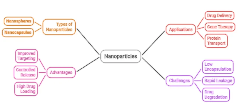
Figure 1: Nanoparticles in Drug Delivery
Despite benefits, there are drawbacks to working with nanoparticles. Due to their surface area to volume ratio, aggregation of particles is a real concern. This makes solid or liquid form handling very difficult. Small size and a large surface area can also lead to insufficient drug loading, or a burst release of the drug. These issues must be resolved in order for nanoparticles to be used clinically or marketed. This paper looks at recently published articles and discusses the development of nanoparticle-based drug delivery systems as well as the control of drug release.
Preparation of Nanoparticles
Nanoparticles made of proteins, polysaccharides, and synthetic polymers, etc., can be made in a variety of ways. The selection of the matrix materials will depend on a number of factors, which include the size of nanoparticles to be prepared, the characteristics of the drug (e.g., aqueous solubility and stability), the surface characteristics (e.g., charge and permeability), the levels of biodegradability, biocompatibility, and toxicity, the desired drug release profile, and the antigenicity of the final product.
Nanoparticles have been synthesized most frequently using three approaches: [1] the dispersing of preformed polymers; [2] polymerization of monomers; and [3] gelation or coacervation of hydrophilic polymers. Other approaches including supercritical fluids technology and the particle replication in non-wetting templates (PRINT) method have also been reported in the literature. PRINT, among the other methods, has absolute control over particle size, shape and composition, which could show an example for mass production of nanoparticles in industry.
Dispersion of preformed polymers: Dispersion of preformed polymers is a technique to fabricate biodegradable nanoparticles from poly (lactic acid) (PLA), poly (D, L-glycolide) (PLG), poly (D, L-lactide-co-glycolide) (PLGA), and poly (cyanoacrylate) (PCA). This technique has many variations and some are described below. The solvent evaporation method: The polymer is dissolved in an organic solvent e.g. dichloromethane, chloroform or ethyl acetate, which is also a solvent for the dissolution of a hydrophobic drug. A polymer-drug solution is then emulsified in aqueous solution of a surfactant resulting in the formation of an oil-water (o/w) emulsion. The emulsion is stabilized and run to dryness with either a vacuum or by continuously stirring. The particle size was affected by the type and concentration of the stabilizer, the speed of the homogenizer, and the polymer concentration. [13] To obtain small particle size, a high-speed homogenizer or ultrasonication is commonly implemented.
Spontaneous emulsification or solvent diffusion method: This approach is derived from the existing solvent evaporation method. In this method, the oil phase (water-immiscible organic solvent) is mixed with a water-miscible solvent. Because of the spontaneous diffusion of solvents, interfacial turbulence forms and smaller droplets are formed. Higher concentration of water-miscible solvents results in smaller particle size.
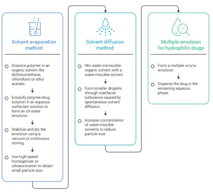
Figure 2: Preparation of Nanoparticles via Preformed Polymers
Solvent evaporation and solvent diffusion methods can be used for both hydrophilic and hydrophobic drugs. For hydrophilic drugs, a multiple w/o/w emulsion is formed having the drug dispersed in the remaining aqueous phase.
Polymerization method
This method polymerizes monomers into nanoparticles in an aqueous phase. Drug can be included by either being mixed with the monomers during polymerization, or can adsorb onto the nanoparticles post polymerization. The suspension of nanoparticles is then purified to remove the stabilizers and surfactants by centrifugation and resuspension of the particles in an isotonic medium. This is reported for the synthesis of polybutylcyanoacrylate or poly (alkylcyanoacrylate) nanoparticles [16,17]. Nanoparticle size and the formation of nanocapsules are also dependent on the concentration of the surfactants and stabilizers. [18]
Coacervation or ionic gelation method
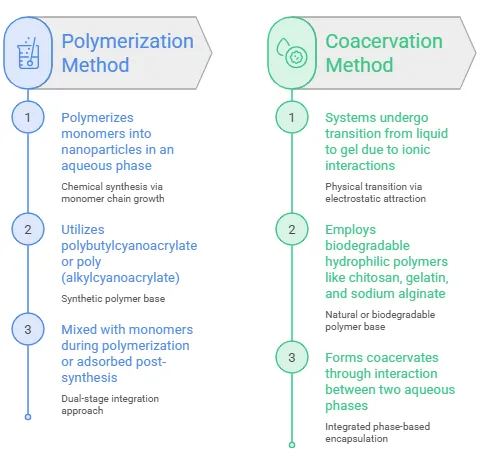
Figure 3: Which Nanoparticle Fabrication Method Should Be Utilized?
There has been considerable interest in the fabrication of nanoparticles using biodegradable hydrophilic polymers such as chitosan, gelatin and sodium alginate. Calvo and coworkers reported a method for the preparation of hydrophilic chitosan nanoparticles by ionic gelation.[19, 20] the process includes a mixture divided into two aqueous phases, of which one is polymer chitosan, a di-block copolymer of ethylene oxide or propylene oxide (PEO-PPO) and the other is a polyanion sodium tripolyphosphate. In this process, positively charged chitosan amino groups interact with negatively charged sodium tripolyphosphate and form coacervates in the nanometer size range. Formation of coacervates occurs due to electrostatic interactions between two aqueous phases, whereas, ionic gelation is a process whereby systems undergo transition from liquid to gel due to ionic interactions at room temperature..
Production of nanoparticles using supercritical fluid technology
Conventional techniques like solvent extraction-evaporation, and solvent diffusion or organic phase separation methods use unsafe organic solvents. For this reason, supercritical fluid technology can be used to prepare biodegradable micro- and nanoparticles, as supercritical fluids are non-hazardous [21].
A supercritical fluid is a solvent that is above its critical temperature and remains a single phase no matter the pressure [21]. Among supercritical fluids, supercritical CO2 (SC CO2) is mostly used because of its relatively low and safe critical conditions (Tc = 31.1 °C, Pc = 73.8 bars), and its low cost. The most common supercritical fluid technologies are the supercritical anti-solvent (SAS) and rapid expansion of a supercritical solution (RESS) methods. In SAS, a liquid solvent (usually, methanol) dissolves the solute to be micronized, and at the process conditions, due to the solute’s insolubility in the supercritical fluid, the extraction of the liquid solvent by the supercritical fluid causes the instantaneous precipitation of the solute and the formation of nanoparticles [8]. Thote and Gupta (2005) used a modified SAS method to produce the hydrophilic drug dexamethasone phosphate drug nanoparticles for microencapsulation [22].
Unlike the SAS process, RESP involves dissolving a solute in a supercritical fluid (e.g. supercritical methanol) that is then rapid expanded via a small nozzle into a region of lower pressure. This leads to the solvent power of the supercritical fluid decreasing and the solute precipitating. This process is considered clean due to the fact that the solute that precipitates is almost completely solvent free. RESS and the subsequent modifications of this process have been successful in the preparation of polymeric nanoparticles. Supercritical fluid technology is subsequently an eco-friendly method that lends itself to mass production, but is still fairly expensive due to specifically designed equipment.
Effect of Characteristics of Nanoparticles on Drug Delivery
Particle size
The size, as well as the size distribution of nanoparticles, are the most important aspects of a system. Size and size distribution affect the distribution and behavior of a system in a living organism. They can also affect the targeting ability and toxicity of a system, in addition to drug loading and release as well as system stability. It has been evaluated that nanoparticles may offer enhanced drug delivery over microparticles, and several advantages have been noted over microparticles. Nanoparticles have high intracellular uptake and move to a variety of biological areas. Desai et al. studied nanoparticles of 100 nm vs. microparticles of 1 µm and 10 µm in a Caco-2 cell line, and found that 100 nm nanoparticles had a 2.5 and 6 fold, respectively, higher uptake than microparticles [25]. It was later reported that nanoparticles penetrated throughout the submucosal regions in an intestinal loop model, while microparticles remained in the epithelium [26]. It was reported that nanoparticles are able to cross the blood brain barrier after opening of tight junctions using hyperosmotic mannitol. From this, it may be concluded that sustained delivery of therapeutic agents for brain tumors and other similar difficult-to-treat diseases may be possible. Tween 80 coated nanoparticles have been reported to cross the blood brain barrier [28]. In some cell types, only submicron sized nanoparticles are able to be taken up, while larger microparticles are not able to be taken up.
Particle size has an impact on drug release. Because smaller particles have a bigger surface area, the majority of the drug would be at or close to the particle surface, resulting in rapid drug release.bigger particles, on the other hand, have bigger cores that enable more medication to be progressively encased. Diffuse out thirty. Additionally, during the storage and transportation of nanoparticle dispersion, smaller particles are more likely to aggregate. Creating nanoparticles with the smallest size while maintaining the highest level of stability is always difficult.
Particle size can also have an impact on polymer breakdown. For example, it was discovered that the rate of PLGA polymer breakdown increased as particle size increased in vitro [31]. It was believed that PLGA degradation products in smaller particles could easily diffuse out of the particles, whereas in larger particles, the degradation products were more likely to stay in the polymer matrix for a longer amount of time, causing the polymer material to degrade autocatalytically. Larger particles were therefore thought to accelerate both drug release and polymer breakdown.Nevertheless, Panyam et al. synthesized PLGA particles in a variety of size ranges and discovered that the rates of polymer breakdown in vitro did not significantly change for different size particles [32].
Nowadays, dynamic light scattering or photon-correlation spectroscopy are the quickest and most common ways to measure particle size.Photon-correlation spectroscopy uses Brownian motion and light scattering characteristics to calculate the particle's diameter, but it also requires knowledge of the medium's viscosity [33]. Scanning or transmission electron microscopy (SEM or TEM) is typically used to confirm the findings of photon-correlation spectroscopy.
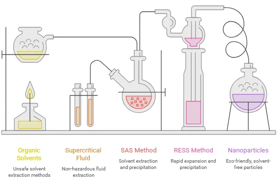
Figure 4: Nanoparticle Production and Drug Delivery
Surface properties of nanoparticles
When nanoparticles are injected intravenously, the body's immune systems quickly identify them, and phagocytes remove them from the bloodstream [34]. The amount of adsorbed blood components, mostly proteins (opsonins), is determined by the hydrophobicity of the nanoparticles' surface in addition to their size. This in turn affects the nanoparticles' in vivo destiny [34, 35]. Opsonization, the process by which these opsonins bind to the surface of nanoparticles, serves as a link between phagocytes and nanoparticles. When a medication is linked to traditional carriers, it results in alteration in the drug's biodistribution profile because it mostly reaches the mononuclear phagocyte system (MPS), which includes the bone marrow, liver, spleen, and lungs. In fact, once in the bloodstream, the macrophages of MPS-rich organs quickly opsonize and eliminate surface non-modified nanoparticles (conventional nanoparticles) [36]. IgG and complement C3 components are typically utilized for the identification of foreign compounds, particularly foreign macromolecules.
Hence, to increase the likelihood of the success in drug targeting by nanoparticles, it is necessary to minimize the opsonization and to prolong the circulation of nanoparticles in vivo. This can be achieved by (a) surface coating of nanoparticles with hydrophilic polymers/surfactants; (b) formulation of nanoparticles with biodegradable copolymers with hydrophilic segments such as polyethylene glycol (PEG), polyethylene oxide, polyoxamer, poloxamine and polysorbate 80 (Tween 80).
Studies show that PEG conformation at the nanoparticle surface is of utmost importance for the opsonin repelling function of the PEG layer. PEG surfaces in brush-like and intermediate configurations reduced phagocytosis and complement activation whereas PEG surfaces in mushroom-like configuration were potent complement activators and favoured phagocytosis [2, 37].
The zeta potential of a nanoparticle is commonly used to characterise the surface charge property of nanoparticles [38]. It reflects the electrical potential of particles and is influenced by the composition of the particle and the medium in which it is dispersed. Nanoparticles with a zeta potential above (+/-) 30 mV have been shown to be stable in suspension, as the surface charge prevents aggregation of the particles. The zeta potential can also be used to determine whether a charged active material is encapsulated within the centre of the nanocapsule or adsorbed onto the surface.
Drug loading
A good nanoparticulate system should ideally have a high drug-loading capacity, which will lower the amount of matrix materials needed for administration. There are two ways to load drugs: • Incorporating during the synthesis of nanoparticles (incorporation technique) • Using an adsorption/absorption approach, the drug is absorbed after the nanoparticles are formed by incubating the carrier with a concentrated drug solution.
The solid-state drug solubility in matrix material or polymer (solid dissolution or dispersion), which is correlated with the polymer composition, molecular weight, drug-polymer interaction, and the presence of end functional groups (ester or carboxyl), greatly influences drug loading and entrapment efficiency [39, 40, 41]. The PEG moiety has minimal to no impact on medication loading 42.When a macromolecule or protein is loaded at or close to its isoelectric point, where it has maximum adsorption and minimum solubility, it exhibits the highest loading efficiency [19]. Ionic interaction between the drug and matrix materials can be a highly efficient strategy to improve the drug loading for small compounds, according to research [43, 44].
Drug release
Drug release and polymer biodegradation are crucial elements to take into account when creating a successful nanoparticulate system. Drug solubility, desorption of the surface-bound or adsorbed drug, drug diffusion through the nanoparticle matrix, erosion/degradation of the nanoparticle matrix, and the combination of erosion/diffusion processes are the general factors that determine the drug release rate. Therefore, the release process is controlled by the matrix materials' solubility, diffusion, and biodegradation.
In the case of nanospheres, where the drug is uniformly distributed, the release occurs by diffusion or erosion of the matrix under sink conditions. If the diffusion of the drug is faster than matrix erosion, the mechanism of release is largely controlled by a diffusion process. The rapid initial release or ‘burst’ is mainly attributed to weakly bound or adsorbed drug to the large surface of nanoparticles [45]. It is evident that the method of incorporation has an effect on release profile. If the drug is loaded by incorporation method, the system has a relatively small burst effect and better sustained release characteristics [46]. If the nanoparticle is coated by polymer, the release is then controlled by diffusion of the drug from the core across the polymeric membrane. The membrane coating acts as a barrier to release, therefore, the solubility and diffusivity of drug in polymer membrane becomes determining factor in drug release. Furthermore release rate can also be affected by ionic interaction between the drug and addition of auxillary ingredients. When the drug is involved in interaction with auxillary ingredients to form a less water soluble complex, then the drug release can be very slow with almost no burst release effect 43; whereas if the addition of auxillary ingredients e.g., addition of ethylene oxide-propylene oxide block copolymer (PEO-PPO) to chitosan, reduces the interaction of the model drug bovine serum albumin (BSA) with the matrix material (chitosan) due to competitive electrostatic interaction of PEO-PPO with chitosan, then an increase in drug release could be observed [20].
Various methods which can be used to study the in vitro release of the drug are: (1) side-by-side diffusion cells with artificial or biological membranes; (2) dialysis bag diffusion technique; (3) reverse dialysis bag technique; (4) agitation followed by ultracentrifugation/centrifugation; (5) Ultra-filtration or centrifugal ultra-filtration techniques. Usually the release study is carried out by controlled agitation followed by centrifugation. Due to the time-consuming nature and technical difficulties encountered in the separation of nanoparticles from release media, the dialysis technique is generally preferred. Applications of Nanoparticulate Delivery Systems
Tumor targeting using nanoparticulate delivery systems
The use of nanoparticles for tumor targeting is justified by two factors: 1) the enhanced permeability and retention effect or active targeting by ligands on the surface of nanoparticles will enable nanoparticles to deliver a concentrated dose of drug in the vicinity of the tumor targets; and 2) the nanoparticles will limit drug distribution to the target organ, thereby reducing drug exposure of health tissues.
Verdun et al. showed that doxorubicin concentrations in the liver, spleen, and lungs were greater in mice treated with doxorubicin integrated into poly (isohexylcyanoacrylate) nanopsheres than in animals treated with pure doxorubicin [47]. Research indicates that the drug distribution pattern in vivo is significantly influenced by the polymeric composition of nanoparticles, including the type, hydrophobicity, and biodegradation profile of the polymer, as well as the molecular weight of the associated drug, its location within the nanospheres, and the mode of incorporation technique, adsorption or incorporation. Although the precise underlying mechanism is yet unknown, the biodistribution of nanoparticles occurs quickly—between ½ and 3 hours—and most likely includes MPS and the endocytosis/phagocytosis process [48].
The biodistribution and pharmacokinetics (PK) of a cyclic RGDdoxorubicin-nanoparticle formulation in tumor-bearing mice were recently described by Bibby et al. 49. Their biodistribution tests showed rising drug concentrations in the liver, spleen, and tumor and falling drug concentrations with time in the heart, lung, kidney, and plasma. At 48 hours after injection, the bulk of the administered dosage (56%) showed up in the liver and only 1.6% in the tumor, demonstrating the liver's strong propensity to absorb nanoparticles. This suggests that preventing particle absorption by the mononuclear phagocytic system (MPS) in the liver and spleen is the biggest obstacle to employing nanoparticles for tumor targeting.
uch propensity of MPS for endocytosis/phagocytosis of nanoparticles provides an opportunity to effectively deliver therapeutic agents to these cells. This biodistribution can be of benefit for the chemotherapeutic treatment of MPS- rich organs/tissues localized tumors like hepatocarcinoma, hepatic metastasis arising from igestive tract or gynaecological cancers, brochopulmonary tumors, primitive tumors and metastasis, small cell tumors, myeloma and leukemia. It has been proved that using doxorubicin loaded conventional nanoparticles was effective against hepatic metastasis model in mice. It was found there was greater reduction in the degree of metastasis than when free drug was used. The underlying mechanism responsible for the increased therapeutic efficacy of the formulation was transfer of doxorubicin from healthy tissue, acting as a drug reservoir to the malignant tissues [50]. Histological examination showed a considerable accumulation of nanoparticles in the lysosomal vesicles of Kupffer cells, whereas nanoparticles could not be clearly identified in tumoral cells [50]. Thus Kupffer cells, after a massive uptake of nanoparticles by phagocytosis, were able to induce the release of doxorubicin, leading to a gradient of drug concentration, favorable for a prolonged diffusion of the free and still active drug towards the neighboring metastatic cells [50].
It is possible to anticipate some cytotoxicity against Kupffer cells when using conventional nanoparticles as carriers in chemotherapy. This would lead to a Kupffer cell deficiency, which would naturally result in decreased liver uptake and a decreased therapeutic effect with intervals of less than two weeks administration [51]. Additionally, conventional nanoparticles can target bone marrow (MPS tissue), which is a crucial yet unpleasant site of action for the majority of anticancer medications since chemotherapy using such carriers may intensify the myelosuppressive impact. Therefore, only cancers at the level of MPS-rich organs may be targeted by conventional nanoparticles to increase the efficacy of anticancer treatments.
Additionally, if fast nanoparticle clearance happens soon after intravenous delivery, it is not practicable to route anticancer drug-loaded nanoparticles to different tumoral locations.
Long circulating nanoparticles
Nanoparticles must be able to target cancers that are located outside of MPS-rich tissues in order to be effective as a medication delivery device. Over the last ten years, a lot of effort has gone into creating so-called "stealth." Nanoparticles must be able to target cancers that are located outside of MPS-rich tissues in order to be effective as a medication delivery device. Over the last ten years, a lot of effort has gone into creating so-called "stealth." particles or PEGylated nanoparticles, which phagocytes and macrophages cannot see [52]. The use of hydrophilic polymers (such as polyethylene glycol, poloxamines, poloxamers, and polysaccharides) to effectively coat the surface of typical nanoparticles had an opposite impact to the absorption by the MPS [52, 53] which marked a significant advancement in the field.
At the particle surface, these coatings create a dynamic "cloud" of neutral and hydrophilic chains that resist plasma proteins [54, 55]. Consequently, those coated nanoparticles stay in the bloodstream for a longer amount of time since they are invisible to MPS. There are two approaches to introduce hydrophilic polymers to the surface: either by using block or branched copolymers to produce nanoparticles or by adsorbing surfactants [51, 52].
According to studies, PEG-coated nanoparticles can selectively extravasate at diseased places such tumors or inflammatory areas with leaky blood vessels, in addition to having a longer half-life in the blood compartment [51]. Consequently, the ability to directly target cancers outside of MPS-rich locations has been enhanced by such long-circulating nanoparticles [51]. The biological destiny of nanoparticles depends critically on the size and surface properties of the colloidal carriers. To achieve the decrease of opsonization reactions and subsequent clearance by macrophages, a hydrophilic surface and a size less than 100 nm are crucial [52].
Ovarian carcinoma while specific peptides or carbohydrates may be used to target integrins and selectins [56]. Oyewumi et al demonstrated that the benefits of folate ligand coating were to facilitate tumor cell internalization and retention of Gd-nanoparticles in the tumor tissue [57]. Targeting with small ligands appears more likely to succeed since they are easier to handle and manufacture. Furthermore, it could be advantageous when the active targeting ligands are used in combination with the long-circulating nanoparticles to maximize the likelihood of the success in active targeting of nanoparticles.
Reversion of multidrug resistance in tumour cells
Anticancer drugs, even if they are located in the tumour interstitium, can turn out to be of limited efficacy against numerous solid tumour types, because cancer cells are able to develop mechanisms of resistance [58]. These mechanisms allow tumours to evade chemotherapy. Multidrug resistance (MDR) is one of the most serious problems in chemotherapy. MDR occurs mainly due to the over expression of the plasma membrane pglycoprotein (Pgp), which is capable of extruding various positively charged xenobiotics, including some anticancer drugs, out of cells [58]. In order to restore the tumoral cells’ sensitivity to anticancer drugs by circumventing Pgp-mediated MDR, several strategies including the use of colloidal carriers have been applied. The rationale behind the association of drugs with colloidal carriers, such as nanoparticles, against drug resistance derives from the fact that Pgp probably recognizes the drug to be effluxed out of the tumoral cells only when this drug is present in the plasma membrane, and not when it is located in the cytoplasm or lysosomes after endocytosis [59 60].
Nanoparticles for oral delivery of peptides and proteins
Numerous bioactive compounds and vaccines based on peptides and proteins have been discovered as a result of significant developments in biotechnology and biochemistry. The development of appropriate carriers is still difficult since these compounds' bioavailability is restricted by the gastrointestinal tract's epithelial barriers and their vulnerability to digestive enzyme breakdown. Bioactive compounds can be encapsulated and shielded from hydrolytic and enzymatic breakdown by polymeric nanoparticles. For example, insulin-loaded nanoparticles have been shown to reduce blood glucose and maintain insulin action in diabetic rats for up to 14 days after oral delivery [61].
Human mucosa has a surface area that is 200 times larger than that of skin [62]. Proteolytic enzymes in the gut lumen, such as pepsin, trypsin, and chymotrypsin; proteolytic enzymes at the brush border membrane (endopeptidases); bacterial gut flora; and the mucus layer and epithelial cell lining itself are just a few of the physiological and morphological barriers that the gastrointestinal tract offers against the delivery of proteins or peptides [63].
The mucosa's histological structure is made to effectively stop the absorption of environmental particles. Delivering the medication in a colloidal carrier system, such nanoparticles, which can improve the interactions between the drug delivery system and the GI tract's epithelia cells, is a crucial tactic to get beyond the gastrointestinal barrier.
Targeting of nanoparticles to epithelial cells in the GI tract using ligands
Strategies designed to enhance the interaction of nanoparticles with adsorptive enterocytes and M-cells in the Peyer's patches located in the GI tract can be divided into strategies based on specific binding to ligands or receptors or on the mechanism of nonspecific adsorption. The entero- and mucus cells possess specific carbohydrates on their surfaces that could serve as ligand-binding sites. Certain glycoproteins and lectins bind specifically to that type of surface structure by receptor-mediated recognition. Both bean lectin and tomato lectin have been studied to increase oral peptide adsorption. [64, 65]. Vitamin B12 is absorbed in the intact small intestine via receptor-mediated endocytosis. The potential exists to increase oral bioavailability of different peptides (e.g., granulocyte colony stimulating factor, erythropoietin) by means of covalent binding to vitamin B12. This intrinsic process requires mucus membrane mucoprotein, which binds to cobalamin and reaches the ileum.
Absorption enhancement using non-specific interactions
In general, to be absorbed through the gastrointestinal tract, macromolecules and particles travel through either the paracellular route or the endocytotic pathway. For nanoparticles, absorption through the paracellular route, uses less than 1% of the mucosal surface area. The paracellular absorption of macromolecules can be enhanced with polymers such as chitosan [68], starch [69] and poly (acrylates) [70], amongst others. The endocytotic pathway for absorption of nanoparticles occurs through either receptor-mediated endocytosis (active targeting) or adsorptive endocytosis (which does not involve any ligands). This pathway is initiated by an unspecific physical adsorption of the material to the cell surface by hydrogen bonding or hydrophobic interactions [71]. Adsorptive endocytosis is extremely dependent upon the size and surface properties of the material. If the surface charge of the nanoparticles is either positive or uncharged, it will provide adsorptive selectivity to enterocytes, whereas a negative charge and hydrophilic surface will favor adsorptive Mcells [73]. It is clear that all the (nano) particles' surface properties such as charge and hydrophilicity have an important impact on affinity. This is demonstrated with poly (styrene) nanoparticles, which upon carboxylation [72], have a different effect.
Nanoparticles for gene delivery
Polynucleotide vaccines deliver antigen encoding genes, which are expressed by host cells. Antigenic proteins are thus synthesized locally and are recognized by Antigen Presenting Cells (APCs) to initiate an appropriate immune response. Polynucleotide based vaccines give humoral and cell-mediated immunity, as the synthesis of antigenic proteins intracellularly stimulates both arms of the immune system. Another advantage of polynucleotide based vaccines is that the price of DNA production is significantly low compared to other protein based vaccines. In addition, the storage and handling of DNA is easier than the other vaccine components. This makes polynucleotide based vaccines an ideal replacement for most conventional vaccines for application in immunotherapy. However, there are many challenges associated with the delivery of polynucleotides, which will need to be solved for their application in this field to be feasible. These challenges are related to the delivery of the polynucleotides to the target cells, the transfer of polynucleotides to the nucleus of the target cells, and the maintenance of the stability of the polynucleotides during their transport to the target cells.
Due to their rapid escape to the cytoplasm, nanoparticles loaded with plasmid DNA examined in this study might be useful for the sustained release of genes. Plasmid DNA-loaded nanoparticles have been found to be rapidly released from the lysosomes. It was reported that after endolysosomal escape, nanoparticles could be used for the sustained release of DNA and thus sustained gene expression [75]. The use of PLGA nanoparticles loaded with the therapeutic genes of bone morphogenic protein could be an effective way of using nanoparticles as a means of healing bone healing.
Nanoparticles for drug delivery into the brain
The blood-brain barrier (BBB) is the biggest challenge to central nervous system (CNS) drug development. The endothelial cells that form the barrier have tight junctions, active transport systems, and metabolic enzymes. Because of this, water and lipid-soluble substances have a hard time traversing the barrier. The barrier also moves lipid-soluble molecules out of the brain using enzymatic and transport systems. Because of these mechanisms, the BBB only allows a select group of molecules to enter the brain.
Targeting of nanoparticles to the brain is achieved by the presence of and the interaction of nanoparticles to specific receptor-mediated transport systems at the BBB. It has been shown in the literature that self-non-transportable drugs can be delivered across the BBB, using a chimeric construct, capable of undergoing receptor-mediated transcytosis, including polysorbate 80/LDL, OX26, lactoferrin, cell penetrating peptides, melanotransferrin and others. It has been shown that poly (butylcyanoacrylate) nanoparticles are capable of crossing the BBB, and have delivered dalargin and doxorubicin to the brain. It also delivered other agents, which demonstrates the importance of this system since the barrier is so difficult to cross. Polysorbate coated nanoparticles (80), although some success has been reported, have shown rapid degradation and are toxic as the concentration of polysorbate increases. OX26 MAbs (anti-transferrin receptor MAbs) are the most studied targeting antibodies and have been used in studies to test the ability of lipsosomes to cross the BBB, an important barrier that manifests difficulty to overcome.
But recently, Ji et al. showed that the brain absorbs lactoferrin, an iron-binding glycoprotein from the transferrin (Tf) family, twice as much as OX26 and transferrrin in vivo [79]. These BBB-specific compounds may soon be utilized to direct nanoparticles to the brain.
CONCLUSION
The aforementioned demonstrate the enormous potential of nanoparticulate systems, which may transform physiologically active substances that are poorly soluble, poorly absorbed, and labile into viable deliverable medications. Because of its hydrophilic coating, which keeps the reticular-endothelial system from recognizing it, the core of this system has a lengthy circulation time and may include a range of medications, enzymes, and genes.
Particle engineering and a deeper comprehension of the many biological interaction pathways are still needed to enhance this medication delivery technology. To make the idea of nanoparticle technology a workable practical application as the next generation of medication delivery systems, more developments are required.
REFERENCES

Tanvi Patil, Rutuja Mahind, Neha Patil, Tejaswini Pawar, Anjali Patil, Suprita Mane, Advanced Polymeric Nanoparticulate Drug Delivery Systems: Formulation Strategies, Controlled Release, Surface Engineering, and Site-Specific Therapeutic Targeting, Int. J. of Pharm. Sci., 2026, Vol 4, Issue 10, 4813-4827. https://doi.org/10.5281/zenodo.23793418
 10.5281/zenodo.23793418