

We use cookies to ensure our website works properly and to personalise your experience. Cookies policy
Department of Pharmacology, Sarojini Naidu Vanita Pharmacy Maha Vidyalaya (Co-Ed.), Tarnaka, Hyderabad, Telangana, India-500017.
Background: Polyendocrine metabolic ovarian syndrome (PMOS), formerly designated as polycystic ovary syndrome (PCOS), is the most prevalent endocrine and metabolic disorder affecting women of reproductive age worldwide. It comprises complex, interconnected reproductive, endocrine, metabolic, inflammatory, and neuroendocrine abnormalities whose precise mechanistic drivers remain incompletely defined. Because human PMOS is inherently heterogeneous and profoundly influenced by genetic predisposition, dietary habits, and environmental factors, preclinical animal models are indispensable for dissecting disease etiology, validating predictive biomarkers, and screening novel therapeutic interventions.Main Body: High-fat, high-fructose, or Western-style diets (WSD) reliably induce obesity, dyslipidemia, insulin resistance, chronic systemic inflammation, and oxidative stress; however, dietary manipulation alone frequently yields mild or inconsistent ovarian morphological and reproductive disruptions. Conversely, combined diet–chemical or endocrine-disruption models—utilizing aromatase inhibitors (letrozole), androgen precursors (dehydroepiandrosterone [DHEA]), direct androgens (testosterone propionate [TP]), synthetic estrogens (estradiol valerate [EV]), or prenatal androgenization (dihydrotestosterone [DHT]) alongside obesogenic feeding—synergistically recapitulate the complete PMOS phenotype. These dual-induction models establish robust hyperandrogenemia, estrous cyclicity arrest, polycystic follicular morphology, severe insulin resistance, gut microbial dysbiosis, and chronic ovarian inflammation.Conclusion: Combined diet and hormonal protocols provide superior translational fidelity for mimicking human PMOS. Standardizing interventional windows, diet compositions, and endocrine dosages alongside multi-omics, single-cell transcriptomics, and artificial intelligence frameworks is essential for accelerating targeted preclinical pharmacotherapy
Polyendocrine metabolic ovarian syndrome (PMOS) is the updated consensus terminology representing the clinical spectrum historically recognized as polycystic ovary syndrome (PCOS) [1]. This updated nomenclature highlights the multifaceted pathology of the disorder, moving beyond an isolated gynecological focus to emphasize three cardinal disease axes: polyendocrine dysfunction, systemic metabolic dysregulation, and ovarian morphological alterations [1, 2].
First characterized by Stein and Leventhal in 1935, PMOS remains incompletely understood despite decades of research. Diagnostic criteria have evolved from focusing solely on menstrual irregularities, hyperandrogenism, and ultrasound-detected ovarian morphology to acknowledging the pivotal contributions of intrinsic insulin resistance, visceral adiposity, disrupted hypothalamic-pituitary-ovarian (HPO) neuroendocrine signaling, chronic low-grade inflammation, and accelerated long-term cardiometabolic risk [3–5].
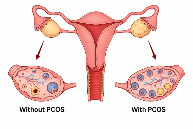
Figure 1: Pathophysiological and morphological differences between a normal ovary and polyendocrine metabolic ovarian syndrome.
Epidemiology and Global Burden
PMOS represents the leading endocrine disorder among women of reproductive age globally [6]. Contemporary systematic reviews and Global Burden of Disease (GBD) analyses estimate a pooled global prevalence of 9.2% to 11.5% under the Rotterdam consensus, with substantial regional variation [7–9]. The highest burdens are documented in the Eastern Mediterranean (15.1%) and South-East Asian (14.3%) regions, followed by Europe (11.7%) and the Americas (10.5%) [9]. GBD metrics indicate that prevalent cases expanded from ~35 million in 1990 to over 65.8 million, with the sharpest increases noted in adolescent and young adult cohorts [7, 8, 10].
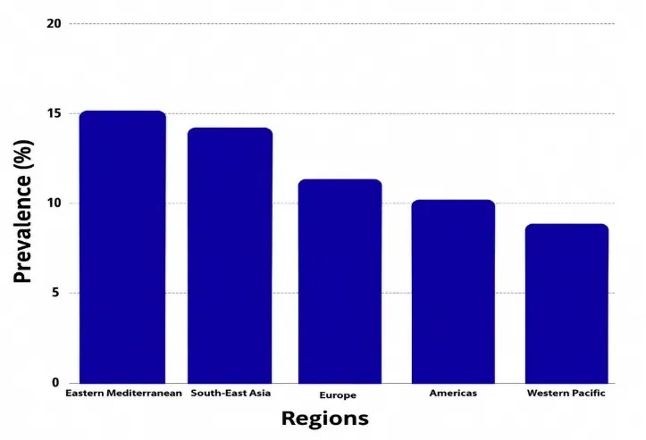
Figure 2: Global prevalence of PMOS across WHO regions
Etiology and Pathophysiological Framework
PMOS emerges from a complex interaction among multiple pathogenic drivers:
1. Genetic Predisposition: Genome-wide association and familial studies demonstrate high heritability involving loci responsible for steroidogenesis, insulin receptor signaling, and gonadotropin feedback regulation [11].
2. Insulin Resistance and Compensatory Hyperinsulinemia: Present in 50% to 70% of affected women across lean and obese phenotypes, hyperinsulinemia directly stimulates theca cell androgen biosynthesis while downregulating hepatic sex hormone-binding globulin (SHBG) synthesis [3, 12].
3. Hyperandrogenism & HPO Axis Alterations: Accelerated GnRH pulsatility drives excessive luteinizing hormone (LH) secretion over follicle-stimulating hormone (FSH), promoting theca cell hyperplasia and arrest of follicular maturation [12, 13].
4. Chronic Low-Grade Inflammation & Oxidative Stress: Elevated circulating TNF-α, IL-6, reactive oxygen species (ROS), and lipid peroxidation products perpetuate insulin resistance, disrupt granulosa cell function, and drive microvascular endothelial damage [2, 5, 14].
Current Clinical and Pharmacological Management
Management of PMOS centers on lifestyle intervention as first-line therapy, combined with targeted pharmacological therapies to alleviate specific reproductive, metabolic, or dermatological symptoms [14–17].
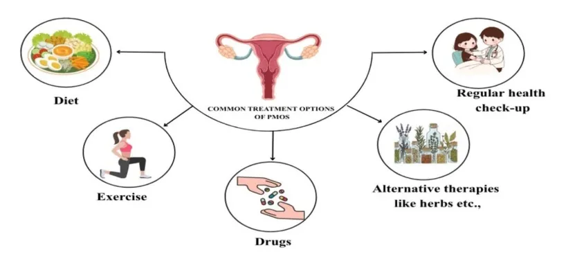
Figure 4: Overview of clinical management options and lifestyle interventions for PMOS.
Table 1: Symptomatic Relief and Evidence Levels for Current PMOS Interventions
| Treatment Category | Intervention | Benefits | Evidence Level |
|---|---|---|---|
| Pharmacological | Metformin (1500-2550 mg/day) | Menstrual regulation (91% success), improved insulin sensitivity | High [18,19] |
| GLP-1RAs | Menstrual regulation, metabolic improvement | Moderate [18] | |
| SPIOMET Regimen | Multi-domain efficacy: ovulatory, anti-androgenic, metabolic | Moderate [19] | |
| Spironolactone | Reduced hirsutism (50% Ferriman-Gallwey reduction) | High [19] | |
| Combined Oral Contraceptives (COCs) | Endometrial protection, cycle regularity, suppressed ovarian androgen secretion | High [19, 20] | |
| Clomiphene Citrate / Letrozole | Ovulation induction, improved live birth rates in anovulatory infertility | Moderate [20] | |
| Supplements | Myo-Inositol | Improved ovulation, insulin sensitivity | Moderate [21] |
| Omega-3, Antioxidants | Reduced inflammation, improved metabolic markers | Moderate [21,22] |
Preclinical Models of PMOS
Because no single experimental paradigm completely captures the full spectrum of human PMOS, animal models are selected according to the specific pathogenic component under investigation—ranging from neuroendocrine hyperandrogenemia to metabolic lipotoxicity.
1. Diet-Induced PMOS Models
Diet-only protocols rely on chronic nutrient excess to generate obesity, visceral adiposity, compensatory hyperinsulinemia, and lipotoxicity [23–27].
Table 2: Preclinical Animal Models, Strains, Diets, and Durations in Diet-Induced Obesity/Metabolic Studies.
| Model | Strain | Diet Composition / Dose | Route | Duration | References |
|---|---|---|---|---|---|
| High-fat diet (HFD) alone | Wistar rats | HFD 40–45% energy from fat | Oral (dietary) | 8–12 weeks | 24 |
| Wistar rats | HFD (5.24 kcal/g, energy supply ratio: protein 20%, carbohydrate 20%, fat 60%) | Oral (dietary) | 8–12 weeks | 25 | |
| Wistar rats | HFD 40% calories from fat | Oral (dietary) | 20–30 days to 8 weeks | 26 | |
| Wistar rats | HFD 45% kcal from fat | Oral (ad libitum feeding) | 21 consecutive days (3 weeks) | 27 | |
| C57BL/6J mice (21-day-old (3-week-old) female mice) | HFD containing 60% kcal from fat | Oral (dietary) | 20 Days | 28 | |
| High-fat, high-fructose diet (HF/HFr) | Prepubertal C57BL/6J female mice | HFD 60% fat + 10% fructose in drinking water | Oral (dietary + drinking water) | 5 weeks | 29 |
| C57BL/6J mice | HFD 60% fat + 20% fructose with time-restricted feeding | Oral (dietary) | 4–8 weeks | 30 | |
| Western-style diet (WSD) / High-fat high-sugar diet (HFHS) |
C57BL/6J female mice | HFHS: 45% kcal fat, 35% kcal carbohydrate | Oral (dietary) | 12 weeks | 31 |
| Wistar rats | HFD (obesogenic diet containing approximately 40% kcal from fat with added sugar/sucrose) | Oral (dietary) | 24 weeks | 32 | |
| C57BL/6J mice | Varied macronutrient diets (high fat + high sugar combinations) | Oral (dietary) | 8–12 weeks | 33 | |
| Sprague–Dawley rats | HFHS containing increased fat and sucrose content (obesogenic diet; high saturated fat with added sugar/sucrose) | Oral feeding (ad libitum) | Approximately 26 weeks | 34 |
High-fat diet (HFD) alone model
This model uses female Wistar rats or C57BL/6J mice fed a high-fat diet (40–45% energy from fat) via oral feeding for 8–12 weeks. In letrozole combination studies, HFD alone serves as a control group with diet composition but without letrozole administration [24,25].
Although many HFD studies are embedded within combination protocols, several primary studies use HFD alone as a comparator to evaluate the metabolic contribution to PMOS phenotypes when combined with hormonal agents [26,27]. Chronic high-fat feeding increases adiposity, insulin resistance, and inflammatory signaling, which can secondarily affect ovarian function [25]. In these studies, HFD alone induces obesity and metabolic changes but typically does not fully replicate the reproductive PMOS phenotype.These models are simple, noninvasive for studying metabolic dysfunction and used as a control or baseline for combination models. However, in the absence of direct endocrine manipulation, HFD alone often fails to induce consistent hyperandrogenism or anovulation, limiting its utility as a stand-alone PMOS model [27,28].
High-fat, high-fructose diet (HF/HFr) model in prepubertal mice
This model uses prepubertal female mice (C57BL/6J, 3–4 weeks old) fed a high-fat, high-fructose diet (60% kcal from fat + 20% fructose in drinking water) via oral feeding for 4–8 weeks. In combination studies, letrozole (1 mg/kg/day, oral gavage) is added for 5 weeks alongside HF/HFr feeding [29,30]. It is primarily a mechanistic rather than a classic PMOS model [30].
This model uses a HF/HFr diet in prepubertal female mice to study the impact of severe dietary stress on metabolic and endocrine dysregulation relevant to PMOS. It is designed to examine how early-life metabolic insults affect later reproductive and endocrine outcomes and is often combined with letrozole or other inducers to intensify phenotypes [29].
HF/HFr diet intake drives adiposity, insulin resistance, and hepatic metabolic stress, which can alter hypothalamic-pituitary-ovarian signaling and ovarian steroidogenesis [30].
Western-style diet (WSD) / high-fat high-sugar diet (HFHS) model
This model uses female mice or rats fed a WSD or HFHS diet (typically 40–60% energy from fat with added sucrose/fructose) via oral feeding for 8–16 weeks. In 2024, C57BL/6J female mice were fed a HFHS (45% kcal fat, 35% kcal carbohydrate) for 12 weeks, while HFHS rat studies used Wistar rats fed 45% fat + 17% sucrose diet for 12–16 weeks [31,33]. This model mimics human obesogenic dietary patterns.
This model exposes female rodents to a WSD or HFHS diet enriched in fat and simple sugars to mimic obesogenic dietary patterns associated with PMOS [34]. Chronic WSD/HFHS feeding increases adiposity, insulin resistance, and inflammatory signaling, which can amplify pre-existing endocrine dysfunction and worsen ovarian phenotypes. In PMOS-susceptible or hyperandrogenic models, WSD/HFHS intensifies anovulation, ovarian cyst formation, and metabolic abnormalities, demonstrating a synergistic effect of diet and endocrine stress [32].
It is useful for studying diet–gene and diet–hormone interactions in PMOS that reproduces both metabolic and reproductive PMOS features without hormonal induction [31,32].
2. Synergistic Combination Models: Diet Coupled with Endocrine Disruptors
Combining obesogenic diets with pharmacological or endocrine inducers reliably yields the complete, severe PMOS phenotype, bringing together hyperandrogenism, cystic folliculogenesis, and metabolic dysfunction [35–52].
Table 3: Preclinical Combination Models of PMOS (Hormonal Disruption + Obesogenic Diets)
| Model | Strain | Diet Composition / Dose | Route | Duration | Reference |
|---|---|---|---|---|---|
| Letrozole + high-fat diet (LET + HFD) | Wistar rats | Letrozole 1 mg/kg/day + HFD 40–45% fat | Oral gavage + diet | 12 weeks (Letrozole + HFD) | 24 |
| Wistar rats (180–220 g) | Letrozole (1 mg/kg/day) + New HFD (Approximately 60% kcal from fat) | Oral gavage + Oral feeding (ad libitum) | 21 days | 48 | |
| Wistar rats | Letrozole 1 mg/kg/day (prepared in 1% carboxymethyl cellulose sodium) + High-fat diet (used concurrently with letrozole; compared with standard chow) | Oral gavage + diet | 21 days + 5 additional days | 35 | |
| Sprague Dawley rats | Letrozole 1 mg/kg/day + HFD 40% fat | Oral gavage + diet | 27–30 days | 37 | |
| Sprague Dawley rats | Letrozole 1 mg/kg/day + HFD 45% fat | Oral gavage + diet | Approximately 5 weeks | 38 | |
| C57BL/6N mice | Letrozole pellet implantation + High-fat diet (60% kcal from fat) | Letrozole: subcutaneous pellet + HFD: oral feeding | 4 weeks | 39 | |
| C57BL/6J mice | Letrozole 1 mg/kg/day + HFD 60% fat | Oral gavage + diet | 5 weeks | 36 | |
| Letrozole + high-fat, high-fructose diet (HF/HFr) | Female C57BL/6 mice (prepubertal) | Letrozole 50 µg/day (3 mg slow-release pellet) + HFD combined with fructose supplementation (obesogenic diet) | Subcutaneous implantation of continuous-release pellet + Oral feeding (ad libitum) | 5 Weeks | 29 |
| Female C57BL/6 mice (prepubertal) | Letrozole 50 µg/day (3 mg slow-release pellet) + HFD combined with fructose supplementation (obesogenic diet) | Subcutaneous implantation of continuous-release pellet + Oral feeding (ad libitum) | 5 Weeks | 40 | |
| Testosterone propionate + high-fat diet (TP + HFD) | Sprague–Dawley rats | TP 1.5 mg/kg + HFD 40–45% fat | Subcutaneous + oral diet | 3 times/week for 8–12 weeks | 41 |
| Dehydroepiandrosterone + high-fat diet (DHEA + HFD) model | C57BL/6J female mice (prepubertal, 25 days old) | DHEA 6 mg/100 g body weight + HFD (60% kcal from fat) | Subcutaneous injection + oral diet | 20 days (DHEA) + 8–12 weeks (HFD) | 42 |
| C57BL/6J female mice | DHEA 6 mg/100 g body weight + HFD (composition not specified) | Subcutaneous injection + oral diet | 20–30 days (DHEA) + HFD continuation | 43 | |
| C57BL/6J female mice | DHEA 6 mg/100 g body weight + HFD 40–45% fat + antibiotic cocktail (ampicillin, metronidazole, neomycin, vancomycin) | Subcutaneous injection + oral diet + oral gavage | 20–30 days (DHEA) + HFD duration + antibiotic treatment | 44 | |
| Female C57BL/6J mice | DHEA+ High-fat diet (60% fat) | Subcutaneous DHEA injection + oral HFD | 20 days induction followed by continuation/withdrawal evaluation | 45 | |
| C57BL/6J female mice | Low-dose DHEA (dose not specified) + HFD 40–45% fat | Subcutaneous injection + oral diet | 20–30 days | 46 | |
| Estradiol valerate + high-fat diet (EV + HFD) model | Wistar albino rats (150–170 g) | Estradiol valerate (4 mg/rat) + HFD (45% kcal from fat) | Subcutaneous injection + Oral feeding (ad libitum) | 60 days | 47 |
| Wistar rats (21-day-old (prepubertal)) | Estradiol valerate (4 mg/rat) + HFD | Subcutaneous injection + Oral feeding (ad libitum) | 84 days | 49 | |
| Prenatal androgenization (DHT) + high-fat diet | Female C57BL/6J mice | DHT (250 µg DHT/day administered to pregnant dams) + HFD (60% kcal from fat) | Subcutaneous injection to pregnant dams + Oral feeding (ad libitum) | From the prepubertal period after weaning until adulthood | 50 |
| Female Long-Evans rats (21-day-old (prepubertal)) | DHT (7.5 mg, 90-day continuous-release pellet) + HFD (60% kcal from fat) | Subcutaneous implantation + Oral feeding (ad libitum) | 90 days | 51 | |
| Western-style diet + hyperandrogenemia | Adult female rhesus macaques (Macaca mulatta) | Testosterone (testosterone delivered via subcutaneous Silastic implant) + Western-style diet (WSD) | Subcutaneous testosterone implant + High-calorie obesogenic diet enriched in animal fat, cholesterol and simple sugars | 5 years | 52 |
Letrozole + high-fat diet (LET + HFD) model
Letrozole inhibits aromatase, reducing estradiol and increasing androgens, thereby disrupting the HPO axis and causing anovulation and polycystic ovaries in addition to high-fat feeding induced changes which exacerbate metabolic dysfunction and mimic comorbidities of human PMOS [48]. In mice, Letrozole + HFD for 5 weeks produces elevated testosterone, anovulation, adipocyte and hepatocyte hypertrophy, and more severe ovarian pathology [36,39].
This model uses female Wistar rats (6–8 weeks old) or C57BL/6J mice fed a high-fat diet (45% energy from fat) via oral feeding for 12 weeks, combined with letrozole administration (1 mg/kg/day, oral gavage or 0.5 mg/kg/day in diet) for 5–12 weeks [24,39].This model combines letrozole with a high-fat diet to induce both reproductive and metabolic PMOS features in female rodents. It was developed to overcome the limitation of letrozole-alone models, which reproduce hyperandrogenism and anovulation but lack significant metabolic abnormalities [35,36].
Strong reproductive phenotype with added metabolic abnormalities; superior to letrozole alone for modeling obese/insulin-resistant PMOS; useful for studying metabolic, inflammatory, and gut–PMOS interactions; validated across multiple primary studies [37,38]. This model is more complex than single-agent models; still may not fully capture human phenotypic heterogeneity; outcomes depend on diet composition, treatment duration, and letrozole dose [35,38].
Letrozole + high-fat, high-fructose diet (LET + HF/HFr) model in mice
This model uses prepubertal female C57BL/6J mice (3–4 weeks old) Letrozole 50 µg/day (3 mg slow-release pellet) and HFD combined with fructose supplementation (obesogenic diet) in Oral feeding (ad libitum) for 5 weeks [29]. Combination of letrozole with a high-fat, high-fructose (HF/HFr) diet in prepubertal female mice to study how severe dietary stress modulates letrozole-induced endocrine and metabolic dysregulation relevant to PMOS. It is used to examine interactions between metabolic stress and aromatase inhibition in a developmental context [40].
The HF/HFr diet exacerbates adiposity, insulin resistance, and hepatic metabolic stress, which amplify endocrine dysregulation and may intensify PMOS-like phenotypes. Letrozole and HF/HFr combination allows investigation of how early-life metabolic stress interacts with endocrine disruption. This model enables study of early-life dietary effects on letrozole-induced PMOS-like phenotypes which is useful for mechanistic studies of metabolic-endocrine interactions. It also allows testing of dietary interventions [29, 40]. Limitations include over represent extreme phenotypes, varying composition and timing which may change across studies. It is primarily a mechanistic rather than a classic PMOS model.
Testosterone propionate + high-fat diet (TP + HFD) model
Combination of testosterone propionate with a high-fat diet in rats to induce both reproductive and metabolic PMOS features. Testosterone propionate elevates circulating androgens, disrupting the HPO axis, impairing follicular development, and promoting anovulation and polycystic ovarian morphology in addition to high-fat feeding induced changes intensifying metabolic dysfunction and altering the intestinal microenvironment. In prenatally androgenized mice, HFD further exacerbates uterine and metabolic phenotypes.This model uses female Wistar rats (6–8 weeks old) or C57BL/6J mice receiving testosterone propionate (1.5 mg/kg, subcutaneous injection, 3 times/week) combined with a high-fat diet (40–45% energy from fat) via oral feeding for 8–12 weeks. It was compared directly with letrozole-based models to evaluate differences in pituitary-ovarian axis disruption, metabolic changes, and gut microenvironment alterations [41].
This is a strong integrated reproductive and metabolic phenotype model, useful for studying androgen-driven PMOS with obesity and insulin resistance and suitable for investigating gut–brain–ovary axis and uterine effects.It is a complex model requires dual manipulation (hormone plus diet), hence interpretation must account for independent contributions of androgen excess and metabolic stress.
Dehydroepiandrosterone + high-fat diet (DHEA + HFD) model
Combination of subcutaneous DHEA administration with high-fat diet in female rodents to rapidly induce both metabolic and reproductive PMOS features [48]. It was designed to overcome the limitations of DHEA-alone or HFD-alone models, which each reproduce only partial aspects of PMOS. DHEA increases androgen precursors, leading to hyperandrogenism, anovulation, and polycystic ovarian morphology [45, 46].
This model uses female Wistar rats (6–8 weeks old) receiving DHEA (6 mg/100 g body weight, subcutaneous injection, daily) combined with a high-fat diet (40% calories from fat) via oral feeding for 20–30 days [42]. Rapid induction (20–30 days) of both metabolic and reproductive PMOS features; strong phenotypic resemblance to human PMOS; useful for studying combined endocrine-metabolic dysfunction [43, 44].
Estradiol valerate + high-fat diet (EV + HFD) model
This model uses female Wistar rats (8–10 weeks old) receiving estradiol valerate (4 mg/100 g body weight, single intramuscular injection) combined with a high-fat diet (40–45% energy from fat) via oral feeding for 8–12 weeks [47]. This model combines a single or repeated injection of estradiol valerate with high-fat diet in female rats to produce PMOS-like manifestations that include both reproductive and metabolic disturbances, along with oxidative and inflammatory changes.
Estradiol valerate exerts negative feedback on the HPO axis, suppressing gonadotropin release and causing anovulation and polycystic ovarian morphology [49]. High-fat feeding induces adiposity, insulin resistance, and oxidative stress, which further impair ovarian function and amplify inflammatory and oxidative pathways implicated in PMOS pathogenesis. This model reproduces minimizes repeated handling compared with some hormonal protocols, hence suitable for studying oxidative stress, inflammation, and metabolic-reproductive crosstalk. Estrogen-induced model may not fully mimic human hyperandrogenic PMOS, Estradiol valerate dose and timing are critical; outcomes influenced by diet composition and duration [47, 49].
Prenatal androgenization (DHT) + high-fat diet
This model uses C57BL/6J female mice prenatally exposed to dihydrotestosterone (DHT) 250 µg/day via subcutaneous injection to pregnant dams for 3 days (gestational days E16.5–E18.5), followed by high-fat diet (45% fat) feeding from weaning to adulthood (8–12 weeks) [50]. Major phenotypes include ovary-independent uterine dysfunction, impaired decidualization, placental inflammation, fetal growth restriction, subfertility, irregular estrous cycles, hyperandrogenism, glucose intolerance, and increased adiposity. High-fat diet exacerbates all defects beyond prenatal androgenization alone [51].
This model developmental origin of PCOS, captures ovary-independent uterine pathology (novel finding), useful for studying uterine/placental dysfunction and pregnancy complications, allows diet–gene interaction studies. It requires breeding and prenatal manipulation (high complexity), long study duration, HFD may overrepresent extreme phenotypes, not suitable for rapid screening [50, 51].
Western-style diet + hyperandrogenemia
Prenatally androgenized female rhesus macaques provide a non-rodent PCOS model in which chronic mild hyperandrogenemia is combined with an obesogenic WSD from puberty. In this model, macaques receive subcutaneous testosterone implants (mean serum T ≈1.35 ng/ml vs 0.27 ng/ml in cholesterol-treated controls) and are fed either standard chow (≈15% calories from fat) or WSD (≈36% calories from fat) for 5 years, with a fertility trial in year 4.
The T+WSD group develops increased body weight, BMI, and body fat, elevated fasting insulin, higher HOMA-IR, impaired glucose clearance and greater glucose/insulin AUC on intravenous glucose tolerance testing, along with delayed estradiol surge, reduced luteal progesterone, lower AMH, more antral follicles, and reduced fertility compared with controls or single-factor treatments. Mechanistically, prolonged hyperandrogenemia accelerates and amplifies WSD-induced metabolic dysfunction, closely mirroring the higher insulin resistance seen in obese women with PCOS. Advantages include high translational relevance, integration of reproductive and metabolic phenotypes over a long exposure window, and spontaneous conceptions without assisted reproduction, while limitations are small sample size, heterogeneity in metabolic responses, progressive obesity even in controls due to ad libitum feeding, long duration, and the need for specialized primate facilities [52].
Biomarkers and Histopathological Endpoints in PMOS Validation
Accurate validation of preclinical models requires a multi-parametric panel comprising endocrine, metabolic, inflammatory, oxidative, and ovarian morphological endpoints [24, 28, 43, 47, 50].
Table 4: Diagnostic Parameters, Biomarkers, and Histopathological Alterations in Preclinical PMOS Models
| Category | Biomarker/Parameter | Method | Normal Range | Alteration in PMOS | Reference |
|---|---|---|---|---|---|
| Hormonal | Total / Free Testosterone | ELISA / Chemiluminescence immunoassay | ~0.2–1 ng/mL | ↑ Increased (hyperandrogenemia) | [28, 43, 44, 51] |
| Dehydroepiandrosterone sulfate (DHEA-S) | ELISA | ~0.5–5 µg/dL (species dependent) | ↑ Increased Adrenal androgen excess | [28, 46] | |
| Estradiol (E2) | ELISA | ~20–80 pg/mL (cycle dependent) | ↑/↓ Model-dependent dysregulation | [47, 49] | |
| Progesterone | ELISA | ~5–30 ng/mL (diestrus/metestrus dependent) | ↓ Reduced due to anovulation | [24, 25] | |
| Luteinizing hormone (LH) | ELISA | ~0.5–5 ng/mL | ↑ Increased LH secretion and LH/FSH imbalance | [25, 26] | |
| Follicle-stimulating hormone (FSH) | ELISA | ~1–10 ng/mL | ↓ or unchanged | [25] | |
| LH/FSH ratio | Calculated | ~0.5–2 | ↑ Increased ratio | [25] | |
| Insulin | ELISA | ~0.5–2 ng/mL | ↑ Hyperinsulinemia | [28, 37] | |
| Metabolic | Body weight | Digital weighing balance | Depends on strain (~180–250 g rats; 18–25 g mice) | ↑ Increased body weight | [28, 31, 51] |
| Visceral fat weight | Dissection and weighing | Low adiposity | ↑ Increased adiposity | [28] | |
| Fasting glucose | Glucometer | ~80–120 mg/dL | ↑ Hyperglycemia | [37, 48] | |
| HOMA-IR | Formula: glucose × insulin/constant | <2 | ↑ Insulin resistance | [28, 30] | |
| Glucose tolerance test (GTT) | Oral glucose tolerance test | Normal glucose clearance | Delayed glucose clearance | [28] | |
| Total cholesterol | Biochemical assay | ~50–100 mg/dL | ↑ Increased | [24] | |
| Triglycerides | Biochemical assay | ~50–150 mg/dL | ↑ Increased | [28] | |
| LDL cholesterol | Biochemical assay | Low level | ↑ Increased | [30] | |
| HDL cholesterol | Biochemical assay | Higher protective level | ↓ Reduced | [24] | |
| Reproductive | Estrous cycle | Vaginal smear cytology | Regular 4–5 day cycle | Prolonged diestrus / irregular cycle | [24, 32, 34] |
| Ovulation rate | Oocyte counting after stimulation | Normal ovulation | ↓ Reduced ovulation | [51] | |
| Fertility index | Mating study | Normal pregnancy rate | ↓ Reduced fertility | [50] | |
| Ovarian weight | Analytical balance | ~50–100 mg (rat ovary) | Altered ovarian weight | [28, 47] | |
| Inflammatory | TNF-α | ELISA / qRT-PCR | Low basal expression | ↑ Increased | [43, 48] |
| IL-6 | ELISA / qRT-PCR | Low basal expression | ↑ Increased | [43, 44] | |
| IL-1β | ELISA / qRT-PCR | Low basal expression | ↑ Increased | [48] | |
| NF-κB pathway | Western blot / qPCR | Normal activity | ↑ Activated inflammatory signaling | [43] | |
| Oxidative Stress | MDA (malondialdehyde) | TBARS assay | ~2–5 nmol/mg protein | ↑ Increased lipid peroxidation | [47, 48] |
| ROS (Reactive Oxygen Species) | Fluorescence assay | Basal ROS production | ↑ Increased | [47] | |
| GSH (Glutathione) | Biochemical assay | Normal antioxidant level | ↓ Reduced | [48] | |
| SOD (Superoxide Dismutase) | Enzyme assay | Normal antioxidant activity | ↓ Reduced | [47] | |
| Catalase | Enzyme assay | Normal antioxidant activity | ↓ Reduced | [47] | |
| Histopathological Parameters (Ovarian morphology) | Follicle count | H&E staining | Normal follicular development | ↑ Atretic/cystic follicles | [28, 32, 36, 44, 50] |
| Corpus luteum | Histology | Presence of corpora lutea | ↓ Reduced/absent | [25, 46] | |
| Granulosa cell layer | Histology | Normal thickness | Altered thickness | [32] | |
| Theca cell layer | Histology | Normal thickness | ↑ Hyperplasia | [50, 51] | |
| Uterine morphology | Histology | Normal endometrium | Altered decidualization | [50] |
CONCLUSIONS AND FUTURE PROSPECTS
While current animal models have contributed considerably to defining the biology of PMOS, significant challenges remain regarding translational reproducibility and the representation of clinical sub-phenotypes.
1. Standardization of Synergistic Protocols: Achieving consistent and reproducible outcomes across laboratories requires harmonizing dietary parameters—including fat source, carbohydrate composition, induction duration, and precise hormonal doses.
2. Integration of Multi-Omics and AI Approaches: Integrating spatial transcriptomics, single-cell RNA sequencing, metabolomics, and gut metagenomics will help characterize cell-specific alterations within the ovarian stroma and the gut–brain–ovary axis. Applying machine learning frameworks to these high-dimensional datasets will aid in classifying endotypes and predicting therapeutic responses.
3. Exploration of Novel Molecular Targets: Preclinical research should focus on resolving chronic low-grade inflammation, mitochondrial oxidative stress, and theca cell hyperactivation. Promising areas for investigation include dual GLP-1/GIP incretin mimetics, selective non-steroidal androgen receptor modulators, mitochondrial-targeted antioxidants, and microbiome-derived postbiotics.
Declarations
Ethics Approval and Consent to Participate
Not applicable (Review manuscript).
Consent for Publication
Not applicable.
Availability of Data and Materials
Not applicable. Data sharing is not applicable to this article as no new datasets were generated during the current review.
Competing Interests
The authors declare that they have no competing financial or non-financial interests.
Funding
This work received no specific grant from any funding agency in the public, commercial, or not-for-profit sectors.
Authors' Contributions
VT conceptualized the review, conducted literature searches, compiled the preclinical model data, drafted the manuscript, and prepared the final tables. All authors read and approved the final manuscript.
Acknowledgements
The authors thank the Department of Pharmacology, Sarojini Naidu Vanita Pharmacy Maha Vidyalaya, for institutional infrastructure and support.
REFERENCES

Udutha Bharathi, Kukkali Sneha, Dr. Venu Talla, Advancements in Modeling Polyendocrine Metabolic Ovarian Syndrome: Focus on Dietary and Chemical Synergism, Int. J. of Pharm. Sci., 2026, Vol 4, Issue 11, 3661-3674. https://doi.org/10.5281/zenodo.24413023
 10.5281/zenodo.24413023