

We use cookies to ensure our website works properly and to personalise your experience. Cookies policy
Department of Pharmacy, Siddhis Institute of Pharmacy, Nandgaon, Murbad, Maharashtra 421401, India.
Bioadhesive drug delivery systems are designed to establish intimate contact with biological tissues and, in many applications, with the mucus layer covering mucosal surfaces. By increasing the residence time of a dosage form at its intended site, these systems may improve local drug availability, support sustained release, reduce dosing frequency, and in selected cases enhance systemic absorption. Their performance is governed by the interaction between polymer chemistry, dosage-form design, tissue physiology, mucus turnover, hydration, and mechanical stresses. This narrative review summarizes the principles of bioadhesion and mucoadhesion, major theories of adhesion, conventional and emerging polymer classes, formulation platforms, and methods used to evaluate adhesive performance and drug release. Natural polymers such as chitosan, alginate, pectin, hyaluronic acid, and plant-derived gums offer useful biocompatibility and formulation flexibility, while cellulose derivatives, poly (acrylic acid)-based polymers, thiolated polymers, catechol-functional materials, and other functionalized systems provide opportunities to tune adhesion, swelling, permeability, and retention. Evaluation should combine complementary in vitro, ex vivo, and, where justified, in vivo approaches rather than relying on a single force measurement. Texture-analyser tests, tensile detachment, flow-through wash-off, residence-time studies, rheology, swelling, permeation, and release testing each measure different aspects of performance and have limitations. Translation remains challenging because mucus composition and turnover vary by anatomical site and between individuals; strong adhesion can also impede drug release or irritate tissue. A rational development strategy should therefore balance adhesion, hydration, drug diffusion, tissue compatibility, manufacturability, and reproducibility. The review concludes with research priorities for standardized methods, physiologically relevant models, quality-by-design development, and more convincing links between
Conventional dosage forms may be cleared rapidly from mucosal surfaces by saliva, tears, nasal secretions, gastrointestinal motility, mucus renewal, or fluid flow. This short contact time can limit the duration of local treatment and reduce the fraction of a drug available for absorption. Bioadhesive systems seek to address this limitation by promoting contact between a formulation and a biological surface. The term bioadhesion is used broadly for adhesion to biological material, whereas mucoadhesion usually refers to interaction with mucus or mucosal tissue. In pharmaceutical literature, the terms sometimes overlap; the anatomical target and the experimental model should therefore be stated clearly (Andrews et al., 2009; Kulkarni et al., 2023; Kumar et al., 2020).
Mucosal delivery is attractive because it can be non-invasive and can support local therapy at oral, buccal, nasal, ocular, vaginal, rectal, and gastrointestinal sites. Depending on the drug and route, delivery may be intended to treat disease locally, protect a labile drug, or facilitate absorption across an epithelial barrier. Prolonged residence does not automatically guarantee improved bioavailability: the drug must also be released in an appropriate form, remain stable, and cross the relevant biological barrier when systemic action is intended (Kulkarni et al., 2023; Kumar et al., 2020).
The central design problem is therefore a balance. A formulation needs enough adhesion to resist clearance, but it should not damage tissue, cause unacceptable discomfort, trap the drug too strongly, or prevent normal physiological clearance. Polymer selection and evaluation methods must be matched to the intended site and dosage form. This review brings together foundational concepts and emerging polymer approaches, with particular attention to how adhesive properties should be measured and interpreted.
2. Review Scope and Literature Approach
This manuscript is structured as a narrative review rather than a registered systematic review. Foundational papers and reviews on mucoadhesion theories, polymer design, and evaluation methods were used to frame the discussion, including work on mucoadhesive platforms, functionalized polymers, and methodological limitations (Andrews et al., 2009; Asane et al., 2008; Khutoryanskiy, 2011; Kulkarni et al., 2023; Kumar et al., 2020; Smart, 2005; Surendranath et al., 2022). A submission-ready version should document a reproducible search of databases such as PubMed, Scopus, Web of Science, and Google Scholar, including the date searched, exact search strings, inclusion criteria, language limits, and the process used to screen records.
Suggested search terms include: (mucoadhesive OR bioadhesive) AND (drug delivery OR polymer) AND (residence time OR adhesion strength OR evaluation OR thiomer OR catechol). Include original experimental studies, relevant reviews, and regulatory or pharmacopeial material where directly applicable. Exclude papers that do not report enough information to interpret the material, model, or test conditions. Because this is a narrative draft, no claim is made that all available studies have been identified or that a formal risk-of-bias assessment has been completed.
3. Terminology, Mucosal Biology, and Residence Time
3.1 Bioadhesion and mucoadhesion
Bioadhesion describes attachment of a material to a biological substrate. Mucoadhesion is commonly used when the target is mucus, mucin, or a mucus-covered epithelial surface. A formulation may interact with mucus, epithelial tissue, or both; these interactions are not identical. Mucus is a hydrated, viscoelastic network rich in mucins, salts, lipids, proteins, and water. Its composition and thickness differ between sites and can change with disease, hydration, medication, and physiological state (Khutoryanskiy, 2011; Kulkarni et al., 2023; Kumar et al., 2020).
3.2 Why residence time matters
Residence time is the period a formulation remains at the intended site before being removed, dislodged, dissolved, or transported away. Increasing residence time can extend the period over which drug is released near a target tissue. It can also improve convenience where a formulation otherwise requires frequent administration. Nevertheless, residence time should be interpreted alongside drug-release rate, drug permeation, local tolerance, and clinical outcomes. A formulation that remains attached but releases little drug may not provide a therapeutic advantage.
3.3 Site-specific physiology
The buccal cavity is exposed to saliva, tongue movement, chewing, and swallowing. The eye is exposed to blinking and tear turnover. The nasal cavity has mucociliary clearance, while the gastrointestinal tract experiences fluid movement, pH variation, digestive enzymes, and motility. Vaginal and rectal environments have their own secretion patterns, microbial communities, and mechanical conditions. Consequently, an adhesive material that performs well on one tissue cannot be assumed to perform similarly at another site (Asane et al., 2008; Kulkarni et al., 2023; Kumar et al., 2020).
4. Mechanisms and Theories of Mucoadhesion
Mucoadhesion is a multistep interfacial process rather than a single chemical event. The initial stage generally involves contact and wetting, followed by polymer hydration and, for suitable polymers, interpenetration of polymer chains with components of the mucus layer. Interfacial interactions and mechanical resistance then contribute to measured adhesion. The relative importance of each process depends on polymer chemistry, molecular weight, charge, flexibility, cross-link density, hydration, contact time, and the properties of the biological substrate (Andrews et al., 2009; Khutoryanskiy, 2011; Kulkarni et al., 2023).
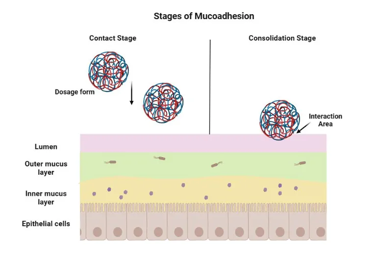
Figure 1: Mechanism of Mucoadhesion
4.1 Wetting theory
Wetting theory considers how well a liquid or hydrated polymer spreads over a biological surface. Good contact increases the area available for interaction. Contact angle and surface-energy measurements can help describe wetting, although a low contact angle alone does not establish strong mucoadhesion under physiological conditions.
4.2 Diffusion or interpenetration theory
This theory proposes that polymer chains and mucin chains interpenetrate across the interface. Chain mobility, molecular weight, contact time, and hydration affect the extent of interpenetration. Excessive cross-linking can restrict chain mobility, whereas excessive hydration may dilute the interface or weaken the dosage form.
4.3 Adsorption theory
Adsorption theory attributes adhesion to interfacial interactions such as hydrogen bonding, electrostatic attraction, van der Waals forces, and hydrophobic interactions. Functional groups including carboxyl, hydroxyl, amino, and thiol groups can influence these interactions. The effect of a functional group is context-dependent: pH, ionic strength, and competing molecules may strengthen or weaken the interaction.
4.4 Electronic theory
Electronic theory proposes that differences in electronic structure between two surfaces can lead to charge transfer and an electrical double layer at the interface. Although this theory can contribute to a conceptual description, it is difficult to isolate as the sole explanation for complex polymer–mucus adhesion.
4.5 Fracture theory
Fracture theory relates the force required to separate two joined surfaces to the strength of the adhesive interface and the geometry of the test. Tensile or detachment tests often report peak force or work of adhesion. These values depend on specimen dimensions, loading rate, contact time, hydration, and the tissue used; they should not be compared across studies without considering test conditions.
4.6 Mechanical interlocking
A hydrated formulation may enter surface irregularities and create mechanical interlocking. This mechanism can matter for rough biological surfaces or porous dosage forms, but it should not be assumed to dominate on all mucosal tissues. In practice, several theories may operate simultaneously, and no single theory explains all mucoadhesive systems (Andrews et al., 2009; Kulkarni et al., 2023; Serra et al., 2009).
5. Polymer Classes for Bioadhesive Drug Delivery
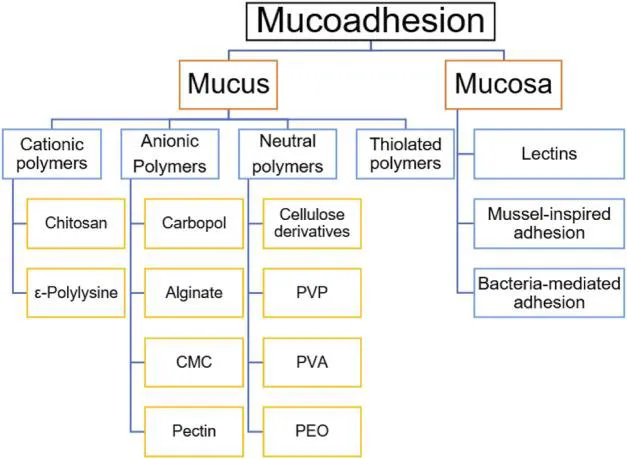
Figure 2: Classification of Bioadhesive Polymers
5.1 Natural polymers
Natural polymers are attractive because many are biodegradable or biocompatible and can be sourced from renewable materials. Their composition, molecular weight, purity, and performance can vary between batches, however, so raw-material specifications are important.
5.1.1 Chitosan
Chitosan is a cationic polysaccharide derived from chitin. Its protonated amino groups can interact electrostatically with negatively charged mucins, while its hydroxyl and amino groups contribute to hydrogen bonding. Chitosan has been investigated in films, gels, nanoparticles, and particulate systems. Its solubility and charge depend on pH and degree of deacetylation; therefore, adhesion observed under acidic conditions may not persist in a different physiological environment. Chemical modification can improve solubility or add functional groups, but the modification must be assessed for safety and reproducibility (Jawadi et al., 2022; Khutoryanskiy, 2011; Surendranath et al., 2022).
5.1.2 Alginate
Alginate is an anionic polysaccharide that forms gels in the presence of suitable multivalent ions. It can provide a hydrated matrix for controlled release and can be combined with cationic polymers to alter mechanical strength and swelling. Because alginate and mucin may both carry negative charges under some conditions, adhesion can depend strongly on formulation composition, ionic environment, and the use of complementary polymers.
5.1.3 Pectin, hyaluronic acid, and plant gums
Pectin, hyaluronic acid, guar gum, xanthan gum, gellan gum, and related materials have been explored as viscosity modifiers, gel-forming agents, or components of mucoadhesive matrices. Their performance depends on source, grade, esterification or substitution, molecular weight, and processing history. Natural origin alone does not establish safety or superiority; impurities, microbial quality, and batch variability require control.
Table 1: Comparison of Bioadhesive Polymers Used in Drug Delivery Systems
| Polymers | Source/ type |
|---|---|
| Chitosan | Natural |
| Alginate | Natural |
| Pectin | Natural |
| HPMC | Semi-synthetic |
| Carbopol | Synthetic |
| Thiolated polymers | Modified polymers |
5.2 Semi-synthetic cellulose derivatives
HPMC, hydroxypropyl cellulose, methylcellulose, and carboxymethyl cellulose are widely used pharmaceutical polymers. They can control viscosity, hydration, film formation, swelling, and drug release. CMC includes ionizable carboxyl groups, while HPMC is non-ionic. These materials may be used alone or blended with stronger adhesive polymers to balance manufacturability, adhesion, and release.
5.3 Synthetic polymers
Poly (acrylic acid)-based materials, including carbomer grades, are common mucoadhesive excipients because carboxyl groups can form hydrogen bonds with mucin and the polymers can swell substantially. Their behavior depends on neutralization, pH, ionic strength, cross-linking, and polymer grade. PVA, PEG, and other synthetic polymers may be used as film formers, plasticizers, or components of composite systems. Polymer selection should be based on the target site, intended exposure, route-specific safety, and the full formulation rather than adhesive strength alone (Asane et al., 2008; Smart, 2005).
5.4 Thiolated polymers (thiomers)
Thiomers are polymers modified with thiol-containing groups. These groups can form disulfide bonds with cysteine-rich domains in mucins, potentially improving adhesion and sometimes influencing permeability or drug release. The magnitude and persistence of the effect depend on the type and degree of modification, oxidation state, pH, and accessibility of the thiol groups. Characterization should include chemical confirmation of modification, free-thiol content, stability during storage, and appropriate safety testing (Bernkop-Schnürch, 2005; Surendranath et al., 2022).
5.5 Catechol-functional and bio-inspired polymers
Catechol chemistry, inspired by adhesive proteins found in mussels, has been investigated to improve adhesion in wet environments. Catechol groups can participate in hydrogen bonding, coordination, and oxidative coupling, depending on the local conditions. This approach is promising but introduces questions about oxidation, storage stability, synthesis reproducibility, and tissue compatibility. Results from non-mucosal wet adhesives should not be directly extrapolated to pharmaceutical mucosal applications without relevant testing (Jawadi et al., 2022).
5.6 Lectin-, peptide-, and ligand-functionalized systems
Ligands can be added to promote interactions with specific surface components. Such targeting may be useful when a defined biological receptor or carbohydrate structure is relevant. However, specificity, immunogenicity, mucosal penetration, and manufacturing complexity must be considered. Ligand binding is not equivalent to prolonged residence and should be measured separately from nonspecific adhesion.
Table 2: Representative polymer classes and formulation considerations
| Polymer/class | Useful features | Potential limitations | Common roles |
|---|---|---|---|
| Chitosan | Cationic; hydrogen bonding; modifiable | pH-dependent solubility; source variability | Films, gels, particles |
| Alginate | Gel formation; hydrated matrix | Adhesion may depend on charge and ions | Gels, beads, composite matrices |
| Pectin / gums | Renewable; viscosity and gel control | Batch variability; source and purity control | Gels, films, blends |
| HPMC / CMC | Established excipients; film and release control | Adhesion may require blending | Films, tablets, patches |
| Carbomer / PAA | Swelling; carboxyl-rich; strong interaction potential | pH/ionic-strength sensitivity; irritation must be assessed | Gels, tablets, films |
| Thiolated polymers | Potential covalent interaction with mucins | Synthesis and oxidation-state control | Advanced gels and particles |
| Catechol-functional polymers | Wet adhesion potential | Oxidation and long-term stability | Emerging adhesive platforms |
6. Dosage Forms and Routes of Administration
6.1 Buccal and sublingual systems
Buccal films, tablets, patches, and gels can provide local delivery or absorption through the oral mucosa. Films can conform to the surface and may be designed with a backing layer to direct drug release toward the mucosa. Key issues include saliva-induced dilution, taste, speech and eating interference, accidental swallowing, and the balance between adhesion and comfortable removal. Sublingual systems face rapid fluid exposure and must be designed for the specific absorption objective.
6.2 Nasal delivery
Nasal gels, powders, and particles may use mucoadhesion to resist mucociliary clearance and prolong contact. Longer retention can be beneficial for local treatment or absorption, but excessive adhesion or viscosity may impair ciliary function or cause discomfort. Formulation testing should consider pH, osmolality, sprayability where relevant, ciliary safety, and the effect of mucus on drug release.
6.3 Ocular delivery
Eye drops are rapidly diluted and cleared by blinking and tear turnover. In situ gels, inserts, and mucoadhesive particles are intended to increase contact time. Ocular formulations require particularly careful assessment of sterility, particulate matter, pH, osmolality, irritation, and visual effects. Strong adhesion should not compromise comfort or normal ocular surface function.
6.4 Gastrointestinal and gastroretentive systems
Gastrointestinal bioadhesive systems aim to increase contact with a selected region or prolong gastric residence. Performance is complicated by changing pH, food, motility, mucus renewal, and fluid volume. Adhesion to excised tissue in a static apparatus does not prove gastric retention in vivo. Studies should distinguish mucosal adhesion from floating, swelling, density-based, or other gastroretentive mechanisms.
6.5 Vaginal and rectal delivery
Gels, films, tablets, and inserts can provide localized delivery at vaginal or rectal sites. Formulation performance depends on secretions, tissue condition, pH, movement, and user acceptability. Compatibility with the local microbiome and mucosal barrier is important, particularly for repeated administration. Residence time should be measured under conditions that reflect the intended use.
6.6 Pulmonary and other mucosal applications
Mucoadhesive concepts have also been explored for respiratory delivery and other mucosal interfaces. In pulmonary applications, particle deposition, airway clearance, inhalation performance, and respiratory safety are central. The fact that a polymer is acceptable for oral use does not establish its suitability for inhalation or other specialized routes.
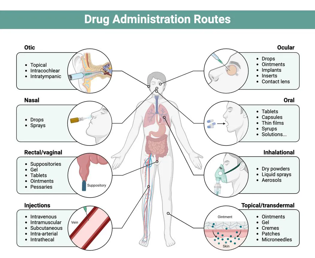
Figure 3: Different Dosage Forms and Routes of Administration
7. Formulation Design and Manufacturing
7.1 Defining the target product profile
Development should begin with the intended anatomical site, local or systemic therapeutic objective, target residence duration, dose, drug physicochemical properties, release profile, dosage-form dimensions, and patient-use requirements. The required adhesive performance should be defined in relation to physiological clearance rather than as an isolated maximum force.
7.2 Critical material attributes
Important polymer attributes include identity, molecular weight or viscosity grade, substitution or deacetylation level, charge density, functional-group content, moisture, particle size where relevant, and microbial or elemental impurity limits. For modified polymers, degree of substitution and residual reagents may be critical. Raw-material variation can change swelling, adhesion, and drug release.
7.3 Preparation methods
Common methods include solvent casting for films, direct compression or granulation for tablets, gel preparation by polymer hydration and neutralization, ionic gelation for selected particles, and spray-based methods for suitable powders. Process parameters such as mixing order, hydration time, shear, temperature, drying rate, compression force, and residual moisture can affect polymer structure and final performance.
7.4 Drug–polymer compatibility
Drug–polymer interactions may change drug solubility, crystallinity, stability, and release. Screening can include differential scanning calorimetry, infrared spectroscopy, X-ray diffraction, microscopy, and stability-indicating chromatography as appropriate. These techniques provide complementary evidence; a spectral shift or thermal change alone is not definitive proof of a specific interaction.
7.5 Quality by Design
A QbD approach identifies critical quality attributes, material attributes, and process parameters that influence performance. A design-of-experiments study can examine the effects of polymer ratio, plasticizer, pH, cross-linker, and drying conditions on adhesion, swelling, mechanical properties, and release. The design space should be supported by data and should include trade-offs, because maximizing one response can worsen another.
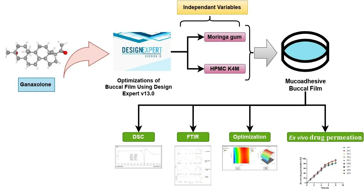
Figure 4: Formulation Development Process
8. Evaluation Strategies
No single test fully describes mucoadhesion. The evaluation plan should combine physicochemical characterization, adhesion testing, dosage-form performance, release testing, tissue compatibility, and—when scientifically justified—residence-time or pharmacokinetic studies. Methodological reviews have emphasized that results are difficult to compare when substrates, contact conditions, instruments, and data reporting differ (da Silva et al., 2017; Serra et al., 2009).
8.1 Basic physicochemical characterization
Depending on the dosage form, testing may include appearance, dimensions, mass variation, thickness, pH, drug content, content uniformity, hardness, friability, tensile strength, folding endurance, viscosity, spreadability, and moisture uptake. These measurements do not directly prove adhesion, but they help explain variation in adhesive performance and support quality control.
8.2 Swelling and hydration studies
Swelling is commonly measured by exposing a known mass or dosage form to a defined medium for specified intervals and recording mass or dimensional change. The medium, temperature, pH, ionic strength, and blotting procedure must be controlled. Swelling may increase contact and chain mobility, but excessive swelling can weaken the dosage form, alter release, or cause discomfort. Report the calculation, replicate number, variability, and endpoint.
8.3 Tensile detachment and texture-analyser methods
A texture analyser or universal testing instrument can bring a formulation into contact with mucosa or a mucin-coated substrate under a defined preload and contact time, then withdraw it at a controlled speed. Common outputs include peak detachment force and work of adhesion, calculated from the force–distance curve. The study should report probe geometry, tissue preparation, contact force, contact duration, withdrawal speed, temperature, hydration, and normalization method. Peak force is not an intrinsic material constant; it depends on the experimental setup (da Silva et al., 2017; Serra et al., 2009).
8.4 Wash-off and flow-through methods
Wash-off tests expose an attached formulation to a flowing liquid or moving apparatus and assess the amount remaining or time to detachment. These methods can approximate clearance better than a static test, but they remain simplified models. Flow rate, angle, substrate, fluid composition, temperature, and movement should be specified. For nasal or ocular applications, physiologically relevant clearance models are especially important.
8.5 Residence-time assessment
Residence time may be assessed in vitro, ex vivo, or in vivo. In vitro systems can provide controlled comparisons; ex vivo tissue may better represent surface chemistry but lacks active mucus renewal and normal physiology. In vivo imaging or labelled formulations may provide stronger evidence of retention, subject to suitable validation and ethical approval. The label must not substantially alter the formulation's properties. Distinguish formulation retention from drug retention, because the drug can leave the carrier while the carrier remains attached.
8.6 Rheology and viscoelastic properties
Rheology measures flow and deformation behavior. Viscosity, storage modulus, loss modulus, and yield stress can be relevant to gels and semisolids. A formulation that is too fluid may clear rapidly; one that is too stiff may spread poorly or cause discomfort. Rheology is complementary to adhesion testing and should not be used as a surrogate for mucoadhesive strength.
8.7 In vitro drug-release testing
Release tests should use an apparatus and medium appropriate for the dosage form and route, with justified sampling intervals, sink conditions where feasible, temperature control, and a validated analytical method. Release profiles may be described using zero-order, first-order, Higuchi, or Korsmeyer–Peppas models when the assumptions are appropriate. A high coefficient of determination alone does not prove a mechanism; model fit should be interpreted with residuals, parameter plausibility, and the physical structure of the formulation.
8.8 Permeation studies
Diffusion cells using appropriate biological tissue or validated membranes can help estimate drug permeation. Tissue source, thickness, integrity, receptor medium, temperature, exposed area, and sink conditions should be reported. Ex vivo permeation is not equivalent to clinical absorption; tissue variability and post-collection changes can influence results. Permeation enhancers require separate safety evaluation.
8.9 Mucoadhesive strength calculations
When detachment force is measured, adhesive stress may be calculated by dividing force by the nominal contact area, provided the area is known and the assumptions are stated. Work of adhesion is often obtained by integrating force over displacement. Studies should report raw or representative force–distance curves, replicate number, mean and dispersion, and a pre-defined method for excluding failed measurements. Avoid reporting only a single maximum value.
8.10 Biocompatibility and irritation
Biocompatibility assessment depends on route, exposure duration, and material composition. Appropriate approaches may include cytotoxicity tests, tissue irritation studies, histological evaluation, and assessment of barrier integrity. A polymer with a history of use in one route is not automatically suitable for another. Testing should follow relevant ethical and regulatory standards and should be designed to minimize unnecessary animal use.
8.11 Stability and packaging
Adhesive performance can change during storage because of moisture uptake, polymer oxidation, cross-linking, drug crystallization, or changes in mechanical strength. Stability programs should monitor appearance, drug content, degradation products, moisture, release, and relevant adhesive properties. Packaging should protect against moisture, light, oxygen, and contamination as appropriate. For thiolated or catechol-containing polymers, chemical-state stability may be particularly important.
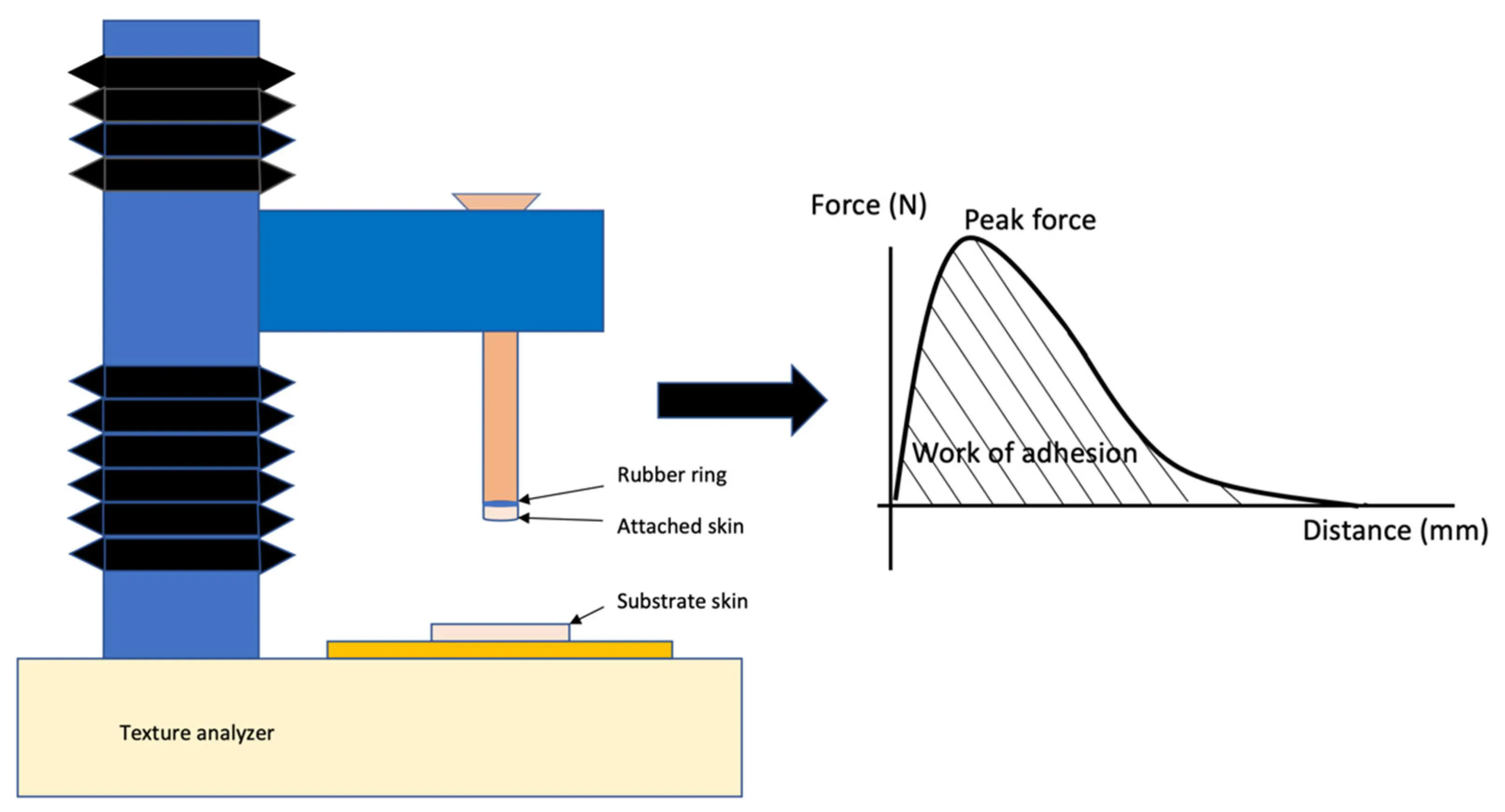
Figure 5: Evaluation Methods of Bioadhesive Drug Delivery Systems
Table 3: Evaluation methods: what they measure and key limitations
| Method | Main information | Important limitation |
|---|---|---|
| Texture analyser / tensile detachment | Peak force and work of adhesion | Highly dependent on substrate, geometry and test settings |
| Wash-off / flow-through | Resistance to fluid-driven removal | Simplified flow and tissue conditions |
| Swelling index | Hydration and dimensional/mass change | Does not directly quantify adhesion |
| Rheology | Viscosity and viscoelastic behavior | Cannot substitute for adhesion or residence testing |
| In vitro release | Drug-release profile and kinetics | May not predict tissue retention or clinical exposure |
| Ex vivo permeation | Drug transport through tissue | Tissue variability and limited physiological renewal |
Good reporting is essential for meaningful comparisons. At minimum, manuscripts should specify the polymer grade and supplier, formulation composition, substrate source and preparation, medium composition, temperature, contact time, loading and withdrawal settings, replicate number, statistical method, and whether values are normalized by area or mass. Biological tissues should be described in terms of source, storage, anatomical region, and integrity checks. Where multiple formulations are compared, controls should include an appropriate non-adhesive or reference formulation when feasible.
Statistical significance should not be confused with practical importance. A small increase in detachment force may not translate to longer residence in vivo. Report effect sizes and uncertainty, not only p-values. If a formulation is optimized across multiple responses, present the trade-offs between adhesion, release, mechanical integrity, comfort, and safety. Method standardization and better links between bench tests and clinical outcomes remain important needs in this field (da Silva et al., 2017; Serra et al., 2009).
9. Emerging Directions
9.1 Functional polymer modification
Functional modification aims to add or expose groups that improve interaction with mucin or tissue. Thiolation, catechol functionalization, hydrophobic modification, and ligand attachment are examples. The value of a modification should be demonstrated against a matched unmodified polymer, with chemical characterization and stability testing. Enhanced adhesion is not sufficient if the modification introduces toxicity, instability, or difficult manufacturing.
9.2 Stimuli-responsive and in situ gelling systems
Systems that respond to pH, temperature, ions, or other local signals can be administered as liquids and form a gel at the target site. This may improve conformity and retention. The transition must occur reliably under realistic physiological conditions and should not interfere with drug release. The response window can vary between patients and disease states, which may limit reproducibility.
9.3 Nanocarriers and hybrid platforms
Mucoadhesive nanoparticles, nanogels, liposomes, and hybrid carriers can combine adhesion with drug protection or controlled release. Their small size and surface chemistry influence mucus interaction: strong adhesion may keep particles near the surface, while mucus-penetrating designs may be preferable when access to underlying epithelium is needed. The optimal strategy depends on whether the goal is surface retention, tissue penetration, or systemic absorption.
9.4 Mucin-mimetic and bio-inspired materials
Mucin-mimetic materials and bio-inspired adhesive chemistries seek to reproduce useful aspects of natural mucus interactions. These approaches may improve understanding of interfacial behavior and provide more tunable systems. Key challenges include scalable synthesis, structural characterization, degradation products, storage stability, and demonstrating performance in relevant tissue models (Jawadi et al., 2022).
9.5 Translational development and regulatory considerations
Translation requires evidence of reproducible manufacture, appropriate quality controls, route-specific safety, stability, and clinically meaningful performance. A novel polymer may require additional characterization of residual monomers, reagents, impurities, degradation products, and biological effects. Developers should consult applicable national and international guidance for the intended product category and route; this review does not replace regulatory advice.
10. Challenges and Limitations
Several recurring limitations affect bioadhesive systems. First, mucus is dynamic and heterogeneous, and its properties vary across anatomical sites and disease states. Second, strong adhesion can conflict with rapid drug release, comfort, or normal tissue clearance. Third, in vitro and ex vivo tests do not fully reproduce active physiological processes. Fourth, studies often use different tissues, test fixtures, media, and reporting conventions, making direct comparison difficult (da Silva et al., 2017; Kulkarni et al., 2023; Serra et al., 2009).
Natural polymers can have variable composition, whereas chemically modified polymers may require more complex synthesis and safety characterization. Nanotechnology and targeting ligands can add manufacturing and analytical burdens. Finally, residence time is only one contributor to therapeutic performance: drug solubility, potency, stability, permeability, metabolism, and patient adherence also influence outcomes. Claims of improved efficacy or bioavailability should therefore be supported by relevant pharmacological or clinical evidence rather than inferred from adhesive strength alone.
11. Future Perspectives
Future research should prioritize standardized, physiologically relevant test methods and transparent reporting. Reference materials and inter-laboratory comparisons could help determine whether different instruments provide comparable measurements. Dynamic models that incorporate mucus renewal, fluid flow, temperature, and tissue movement may improve prediction. The development of synthetic mucus or mucin-based substrates with defined composition may aid early screening, but such models need validation against biological tissue.
A second priority is to link material attributes to clinical endpoints. For example, studies should test whether a measured increase in adhesion predicts a meaningful change in residence, drug exposure, symptom control, or dosing frequency. Patient-centered outcomes—comfort, ease of administration, taste, vision effects, and acceptability—should be considered early. Third, emerging polymers should be assessed for sustainability, scalable synthesis, batch consistency, and realistic storage conditions. Finally, advanced imaging and computational models may help describe interactions between polymer networks, mucus, and drug diffusion, provided predictions are validated experimentally.
CONCLUSION
Bioadhesive and mucoadhesive drug delivery systems offer a flexible approach to prolonging formulation residence at selected biological interfaces. Performance emerges from the combined effects of polymer chemistry, hydration, interfacial interactions, dosage-form structure, drug release, and site-specific physiology. Conventional materials such as chitosan, alginate, cellulose derivatives, and poly (acrylic acid)-based polymers remain useful, while thiolated, catechol-functional, ligand-modified, and hybrid systems offer additional design possibilities. Reliable evaluation requires complementary methods that distinguish adhesion strength, resistance to clearance, drug release, permeation, and tissue compatibility. Because no single laboratory test captures the complexity of mucosal environments, method details and limitations must be reported transparently. The most successful future systems will balance prolonged retention with safety, comfort, manufacturability, and clinically meaningful drug delivery.
Acknowledgements:
The authors have no acknowledgements to declare.
Funding Statement:
The authors received no specific funding for the preparation of this review article.
Conflict of Interest:
The authors declare that they have no competing interests.
Data Availability: No new datasets were generated or analysed for this review. The information discussed is based on previously published studies cited in the reference list.
Abbreviations
CMC, carboxymethyl cellulose; HPMC, hydroxypropyl methylcellulose; PAA, poly (acrylic acid); PEG, polyethylene glycol; PVA, poly (vinyl alcohol); QbD, quality by design; RH, relative humidity; USP, United States Pharmacopeia.
REFERENCES

Aditi Tarmale, Gouri Gavhane, Vinaya Vekhande, Manasi Padwal, Dhanashree Chandgir, Namrata Sabale, Bioadhesive Drug Delivery Systems for Prolonged Residence Time: Emerging Polymers and Evaluation Strategies, Int. J. of Pharm. Sci., 2026, Vol 4, Issue 11, 3552-3564. https://doi.org/10.5281/zenodo.24393797
 10.5281/zenodo.24393797