

We use cookies to ensure our website works properly and to personalise your experience. Cookies policy
Karnataka college of Pharmacy, Bengaluru 560064
Dermatophytosis is one of the most common superficial fungal infections affecting keratinized tissues such as the skin, hair, and nails. The condition is primarily caused by dermatophytes belonging to the genera Trichophyton, Microsporum, and Epidermophyton. Increasing incidence of chronic and recurrent dermatophytosis, particularly in tropical countries, has become a major therapeutic concern due to antifungal resistance, irrational corticosteroid use, and poor patient compliance. Conventional topical antifungal formulations such as creams, ointments, and lotions often demonstrate limited skin penetration, inadequate drug retention, frequent application requirements, and reduced therapeutic effectiveness. Nanoemulgel systems have emerged as advanced topical drug delivery platforms that combine the advantages of nanoemulsions and gel systems to overcome these limitations. The nanosized droplets enhance drug solubility, permeation, and bioavailability, while the gel matrix improves viscosity, spreadability, retention time, and sustained drug release. Nanoemulgels have shown promising outcomes in improving antifungal activity, skin permeation, formulation stability, and patient compliance compared with conventional topical systems. This review highlights the epidemiology, clinical presentation, diagnosis, and pathogenesis of dermatophytosis, together with current treatment approaches, and places special emphasis on nanoemulgel formulation strategies, components, preparation methods, mechanisms of skin penetration, evaluation parameters, recent patents, challenges, and future perspectives in antifungal therapy.
Dermatophytosis is one of the most prevalent superficial fungal infections affecting humans worldwide and primarily involves keratinized tissues such as the skin, hair, and nails [1,2]. The infection is caused by filamentous fungi known as dermatophytes, mainly belonging to the genera Trichophyton, Microsporum, and Epidermophyton, which can invade and utilise keratin as a nutrient source [1,3]. Clinically, it manifests as tinea infections such as tinea corporis, tinea cruris, tinea pedis, tinea capitis, and tinea unguium, associated with erythema, scaling, inflammation, and pruritus, leading to discomfort and reduced quality of life1,4.
In recent years, dermatophytosis has become a significant public health concern, particularly in tropical regions like India, where high humidity and temperature favors fungal growth 1,2. Factors such as overcrowding, poor hygiene, occlusive clothing, and urbanisation further contribute to its spread. Weitzman and Summerbell’s classic taxonomic account of the dermatophytes remains a useful reference point for the genera and species involved 5. An increase in chronic, recurrent, and treatment-resistant cases has been reported due to inappropriate antifungal use, incomplete therapy, irrational corticosteroid combinations, and emerging antifungal resistance 1,3.
Topical antifungal therapy remains the first-line treatment for localised infections due to targeted drug delivery and reduced systemic effects 3,6. Common agents include azoles, allylamines, benzylamines, and hydroxypyridones, which disrupt fungal cell membrane synthesis7,8. However, conventional formulations like creams and ointments show limitations such as poor skin penetration, low drug retention, frequent application, and reduced patient compliance 3,6.
The stratum corneum acts as a major barrier to drug delivery, especially for lipophilic drugs with low aqueous solubility 9. Inadequate drug levels at the infection site may lead to incomplete eradication and recurrence 3,9. Hence, advanced delivery systems are required to enhance penetration and retention.
Nano-based systems, particularly nanoemulsions, have shown potential in improving solubility and skin permeation due to their small droplet size and large surface area 10,11. However, their low viscosity and limited residence time restrict effectiveness in topical use.
Nanoemulgels, combining nanoemulsions with gel systems, offer improved stability, spreadability, bioadhesion, and ease of application while maintaining enhanced permeation 12,13,14. They provide controlled drug release, prolonged skin contact, and better patient compliance compared to conventional formulations 14,15.
Thus, nanoemulgel-based systems represent a promising approach for improving antifungal efficacy and reducing recurrence in dermatophytosis. This review focuses on recent advances in nanoemulgel-based antifungal systems, including formulation strategies, mechanisms, evaluation, and future prospects.
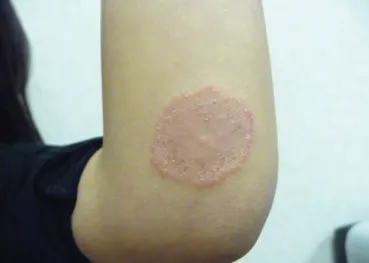
Fig 1: Dermatophytosis
Table 1. Classification of dermatophytosis by source of infection
|
Category |
Description |
|
Anthropophilic |
Human-adapted; spread by human contact 1 |
|
Zoophilic |
From animals; more inflammatory lesions 1 |
|
Geophilic |
Soil-derived; occasional human infection 1 |
Table 2. Classification of dermatophytosis by clinical site of infection
|
Type |
Site |
|
Tinea capitis |
Scalp and hair 2,3 |
|
Tinea faciei |
Non-bearded facial skin 2,3 |
|
Tinea barbae |
Beard and moustache region 2,3 |
|
Tinea corporis |
Trunk and glabrous skin 2,3 |
|
Tinea manuum |
Hands, palms, interdigital areas 2,3 |
|
Tinea cruris |
Groin region 2,3 |
|
Tinea pedis |
Feet, soles, interdigital spaces 2,3 |
|
Tinea unguium (onychomycosis) |
Nail infection with thickening/discoloration 2,3 |
Table 3. Other clinical variants of dermatophytosis
|
Variant |
Feature |
|
Tinea imbricata |
Concentric ring-like lesions 4 |
|
Tinea pseudobreccia |
Steroid-modified atypical lesions 4 |
According to multiple epidemiological studies, dermatophytosis affects approximately 20–25% of the global population at any given time, making it one of the most common superficial fungal infections worldwide 2,16,17. However, reported prevalence rates vary widely across regions and study populations, largely due to differences in climate, hygiene practices, socioeconomic status, and diagnostic criteria. Higher prevalence rates are consistently reported in tropical and subtropical regions, whereas lower rates are observed in temperate climates, suggesting a strong influence of environmental factors 2,18.
Marked variations in prevalence have been observed based on age, gender, and geographic location. Tinea capitis predominantly affects children, with prevalence estimates ranging from 5% to over 30% in school-aged populations in endemic regions, while tinea pedis and tinea unguium are more common in adolescents and adults 17,19. Several studies report a higher prevalence among males, with male-to-female ratios ranging from 1.5:1 to 3:1, which has been attributed to occupational exposure, excessive sweating, and use of occlusive footwear 18,20.
The burden of dermatophytosis is significantly higher in low- and middle-income countries, where overcrowding and limited access to healthcare facilitate transmission and recurrence. In recent years, an increasing proportion of cases have been described as chronic or recurrent, particularly in South Asia, highlighting evolving epidemiological trends and the growing challenge of disease persistence 20,21.
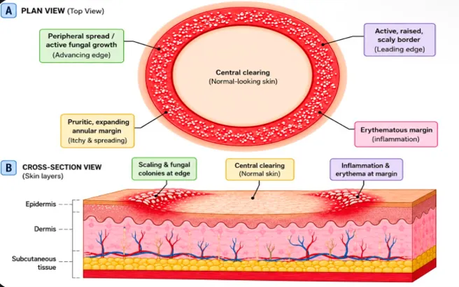
Figure 2: Schematic illustration of the classic annular dermatophytosis lesion
Dermatophytosis presents with a characteristic set of clinical features that vary by infection site, though most lesions share a common annular, scaly morphology. The classic presentation is an erythematous, well-demarcated plaque with an active, raised, scaly border and a tendency toward central clearing, producing the ring-like appearance from which the disease takes its common name 1,4. Pruritus is a near-universal complaint and is often the symptom that prompts patients to seek treatment 1,3.
Clinical features differ considerably by site. Tinea corporis and tinea cruris present as expanding annular plaques on the trunk, limbs, or groin, frequently accompanied by maceration and secondary excoriation from scratching 2,3. Tinea pedis typically manifests as interdigital scaling, fissuring, and maceration, or as a diffuse scaling pattern across the soles [2,4]. Tinea capitis in children can present with patchy hair loss, broken hair shafts close to the scalp surface, diffuse scaling, and, in more severe cases, a boggy, inflamed swelling (kerion) with regional lymphadenopathy 1,2. Tinea unguium (onychomycosis) presents more insidiously, with progressive nail plate thickening, yellow-brown discolouration, subungual hyperkeratosis, and eventual onycholysis 4.
In chronic or recurrent disease, which is increasingly reported in India, lesions may lose their classic annular outline and instead appear as widespread, poorly marginated, or atypical patches, particularly where topical corticosteroids have been misused (tinea incognito) [1,19]. This atypical presentation is a major reason accurate clinical recognition has become more difficult in recent years, a point returned to under Diagnostic Challenges below.
Clinical diagnosis of dermatophytosis is often presumptive, based on the morphology of the lesion, but laboratory confirmation is recommended wherever possible to avoid misdiagnosis and unnecessary or inappropriate treatment, particularly given the growing frequency of steroid-modified and atypical presentations 22.
Direct microscopy using a potassium hydroxide (KOH) wet mount of skin scrapings, hair, or nail clippings is the most widely used first-line diagnostic test, allowing visualisation of fungal hyphae or arthrospores within minutes of sample collection 23. Reported sensitivity varies considerably between studies and depends heavily on sampling technique, site, and observer experience 24.
Fungal culture on Sabouraud dextrose agar is traditionally regarded as the diagnostic gold standard, since it allows species-level identification of the causative organism, which can in turn guide antifungal selection. Its principal drawback is turnaround time, with growth and identification typically taking one to three weeks 23,24.
Dermoscopy has become a useful non-invasive adjunct for both initial diagnosis and monitoring of treatment response. Reported dermoscopic features of dermatophytosis include comma-shaped and corkscrew hairs, scattered black dots at broken hair shafts, and dotted or looped vessels against an erythematous background 24,25.
Molecular assays, including nested polymerase chain reaction (PCR) techniques targeting dermatophyte-specific gene sequences, offer substantially greater sensitivity than either KOH microscopy or culture and can return same-day results, making them valuable in high case-load or outbreak settings; cost and limited laboratory availability, however, still restrict their routine use in many regions 23.
In equivocal cases, or where nail involvement makes sampling difficult, periodic acid–Schiff (PAS) staining of tissue sections can help confirm the presence of fungal elements when other tests are inconclusive 25.
Dermatophytosis is a superficial fungal infection caused by keratinophilic fungi that target keratin-rich tissues such as the stratum corneum, hair, and nails. Its pathogenesis is a stepwise process involving interaction between fungal virulence factors and host tissues, beginning with exposure and leading to inflammation and colonization 26.
5.1 Initial contact and adhesion:
Infection begins when arthroconidia or hyphal fragments contact the skin surface. These fungal elements adhere to the keratinised stratum corneum, overcoming initial barriers. Studies show that spores of Trichophyton mentagrophytes attach rapidly and remain viable, indicating time-dependent adhesion and early colonization 20,27,28.
5.2 Germination and penetration:
After adhesion, spores germinate to form germ tubes that extend over the keratinised surface. These penetrate the stratum corneum by secreting proteolytic enzymes that degrade keratin. Studies show germ tube formation within 24 hours and significant hyphal penetration by 72 hours 27.
5.3 Keratin degradation and nutrient acquisition:
Dermatophytes utilize keratin as their main nutrient source. Studies on Trichophyton rubrum show growth only in the presence of keratin, while absence of keratin results in minimal growth. The fungi secrete keratinases and proteases that break keratin down into peptides and amino acids, supporting growth and facilitating deeper penetration 20,29,30.
5.4 Host immune response:
Fungal colonization activates the host immune system. Dermatophytes remain superficial, but their antigens trigger a T-cell-mediated delayed-type hypersensitivity response, which helps control infection; humoral immunity plays a comparatively limited role. Additional defences include intact skin, sebaceous secretions, and innate inhibitory factors 21.
5.5 Host-pathogen interaction:
The outcome depends on the balance between fungal virulence and host immunity. Fungal adhesion, germination, and keratin degradation promote infection, while host defences regulate inflammation and clearance. When fungal growth exceeds host control, infections may become chronic or recurrent 21,27,31.
Nanoemulsions are biphasic dispersions of two immiscible liquids, oil and water, stabilised by a surfactant or surfactant/co-surfactant system, with droplet diameters typically in the range of 10–100 nm 10,32. Unlike microemulsions, which form spontaneously and are thermodynamically stable, nanoemulsions are kinetically stable, non-equilibrium systems: they generally require energy input during formation and can, in principle, separate over sufficient time, although their small droplet size and resulting Brownian motion make this process very slow in practice 10,33.
Nanoemulsions are broadly classified into three types based on the arrangement of the dispersed and continuous phases:
The choice of nanoemulsion type depends on the physicochemical properties of the drug, the intended route of administration, and the desired release profile. For topical antifungal delivery, O/W nanoemulsions are strongly preferred, since their aqueous outer phase is directly compatible with hydrophilic gelling agents such as Carbopol, allowing straightforward conversion into a nanoemulgel.
A nanoemulsion typically consists of an oil phase, a surfactant/co-surfactant system, and an aqueous phase. The oil phase facilitates drug solubilisation, while surfactants reduce interfacial tension and promote the formation of nanosized droplets. Nanoemulsions can be prepared using high-energy techniques such as ultrasonication and high-pressure homogenisation, or low-energy methods including spontaneous emulsification and phase-inversion techniques 33.
The physicochemical properties of nanoemulsions are influenced by formulation variables such as oil concentration, surfactant-to-co-surfactant ratio, oil characteristics, and preparation conditions. Pseudo-ternary phase diagrams are commonly used for optimisation to identify suitable nanoemulsion regions and obtain formulations with desirable droplet size, polydispersity index, and stability 35.
Nanoemulsions have gained significant attention in topical drug delivery due to their ability to enhance the solubility, penetration, and therapeutic performance of poorly water-soluble drugs. The nanosized droplets provide a large surface area, which improves drug partitioning into the skin and facilitates enhanced permeation through the stratum corneum. The presence of surfactants and oils can also improve skin hydration and reduce the barrier properties of the outer skin layer, thereby promoting drug transport to deeper skin regions 36,37.
For topical antifungal therapy, nanoemulsions offer several advantages, including improved drug solubilisation, prolonged residence time at the application site, controlled drug release, and enhanced antifungal activity. These properties are particularly beneficial for lipophilic antifungal agents such as terbinafine hydrochloride, where limited aqueous solubility may affect conventional topical formulations; incorporating such drugs into nanoemulsion systems can improve drug availability at the site of infection and potentially enhance therapeutic outcomes 38.
Furthermore, nanoemulsions can be incorporated into gel systems to form nanoemulgels, combining the advantages of nanoemulsions with the improved viscosity, spreadability, and patient acceptability of gels. This approach provides better retention on the skin surface and supports sustained drug release, making nanoemulgels promising carriers for the management of dermatophytosis 39.
Nanoemulgel is an advanced hybrid drug delivery system combining nanoemulsions with gel-based topical formulations. It was developed to overcome limitations of conventional gels and nanoemulsions, especially for poorly water-soluble drugs. Nanoemulsions provide enhanced solubilisation and permeation of lipophilic drugs, but their low viscosity and poor retention limit effectiveness; incorporation into a gel matrix improves consistency, residence time, patient compliance, and controlled drug release, making the nanoemulgel a superior topical system 33,40.
Nanoemulgels consist of nanosized oil droplets (below 500 nm) dispersed in an aqueous phase and stabilised by surfactants and co-surfactants. These are incorporated into a three-dimensional gel network using polymers like Carbopol, HPMC, or natural gelling agents. This structure keeps the drug solubilised in the oil phase while providing the rheological and bioadhesive properties of the gel base 41.
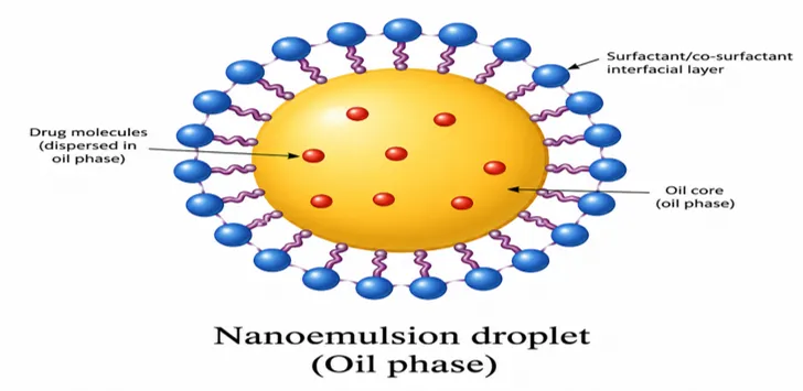
Figure 3: Schematic representation of nanoemulgel structure
Table 4. Comparison between conventional emulgel and nanoemulgel
|
Parameter |
Conventional emulgel |
Nanoemulgel |
|
Thermodynamic stability |
Less stable droplets coalesce, causing creaming or settling 46 |
More stable, very small droplet size and Brownian motion resist settling 33 |
|
Particle size |
Usually large (>500 nm) 45 |
Very small (<100 nm), uniformly distributed 47 |
|
Bioavailability |
Lower, less surface area, poor penetration 48 |
Higher — small size and large surface area 49 |
|
Skin permeation |
Low, due to larger droplet size 50 |
High, due to better interaction with skin layers 33,50 |
|
Method of preparation |
Needs high-energy methods 51 |
Can be prepared by high- or low-energy methods 44 |
|
Systemic absorption |
Very low |
Improved, due to nanosize and better surface properties 33 |
|
Ability to cross the blood–brain barrier |
Cannot cross 52 |
Can cross, due to very small particle size 53 |
Dermatophytosis is a common superficial fungal infection affecting skin and keratinised tissues. It is caused by dermatophytes that colonise the stratum corneum, leading to itching, scaling, and inflammation. Conventional topical antifungal therapies often show limited efficacy due to poor drug penetration, especially for lipophilic drugs, resulting in prolonged treatment and frequent recurrence 53,54.
Limitations of conventional topical therapy:
The stratum corneum acts as a major barrier to drug permeation due to its dense lipid matrix, restricting antifungal drugs with poor aqueous solubility. This limits drug diffusion and results in sub-therapeutic concentrations in deeper epidermal layers where dermatophytes persist [53,54]. Conventional formulations such as creams and ointments fail to maintain effective drug levels at the infection site, leading to incomplete eradication and longer treatment; frequent application and irritation from excipients reduce patient compliance, increasing relapse and the risk of drug resistance 53,54.
14. Current Treatment Options for Dermatophytosis:
Topical antifungal therapy is the first-line treatment for most uncomplicated dermatophytosis, as it delivers high drug concentrations at the site of infection with minimal systemic exposure. Common topical agents include azoles (e.g., clotrimazole, econazole, luliconazole, sertaconazole) and other antifungals such as amorolfine, which act against dermatophytes by disrupting ergosterol synthesis in the fungal cell membrane, leading to increased membrane permeability and fungal cell death. These agents are particularly effective for superficial infections such as tinea corporis, tinea cruris, and tinea pedis, with typical regimens applied once or twice daily for two to four weeks [7]. However, the effectiveness of topical therapy may be limited by poor penetration into deeper epidermal layers or nails and inconsistent patient application; expert guidance recommends continuing treatment for at least two to four weeks beyond clinical resolution to reduce relapse rates 1,3,8.
Systemic antifungal therapy is indicated in extensive, chronic, recurrent, or treatment-resistant dermatophytosis, as well as in infections involving the nails (onychomycosis) or scalp (tinea capitis). Widely used systemic antifungals include terbinafine, itraconazole, griseofulvin, and fluconazole, with terbinafine and itraconazole most commonly prescribed due to their fungicidal activity and effectiveness in clinical practice. These agents achieve therapeutic concentrations in keratinised tissues and are effective when topical agents fail; however, systemic administration carries a higher risk of adverse effects such as hepatotoxicity, gastrointestinal disturbance, and drug–drug interactions, and baseline as well as periodic liver function monitoring is recommended for prolonged therapy 2,3,4.
In more extensive or recurrent infections, clinicians often employ combination therapy using both topical and systemic antifungals. Combining agents can improve treatment outcomes by reducing fungal load more rapidly and helping overcome partial response to monotherapy. Although robust comparative studies are limited, clinical practice often supports combined use in extensive lesions or in those with a poor response to topical treatment alone.
Successful management also involves adjunctive supportive measures that reduce recurrence and transmission, including maintaining dry, clean skin, avoiding tight or occlusive clothing, and practising good personal hygiene. Patients should be counselled against self-medicating with over-the-counter combination creams containing corticosteroids, as inappropriate steroid use can alter lesion appearance, suppress local immunity, and contribute to persistent or atypical presentations such as tinea incognito. Education on hygiene and avoidance of steroid-containing formulations significantly reduces recurrence and household transmission 3,4,31.
15. Advantages of Nanoemulgel Systems:
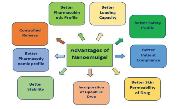
Fig 4: Advantages of Nanoemulgel
Nanoemulgel are hybrid systems that incorporate nanoemulsions, with droplet sizes in the nanometre range, into a gel base. The nanoscale droplets significantly enhance the solubility and skin uptake of hydrophobic antifungal agents by increasing surface area and improving interaction with skin lipids, facilitating deeper penetration through the stratum corneum 1,6.
The gel matrix provides increased viscosity and prolonged residence time at the application site, enabling sustained drug release and reducing the frequency of application compared with conventional gels or creams. These features are associated with enhanced therapeutic outcomes and improved patient adherence 1,6,9.
Numerous studies have demonstrated that nanoemulgels of antifungal drugs exhibit superior antifungal activity compared with conventional formulations. For example, a terbinafine-loaded nanoemulgel showed enhanced drug release and better skin permeation compared with marketed products, supporting its potential for more effective management of dermatophytosis 2. Similarly, miconazole nitrate nanoemulgels exhibited significantly higher antifungal activity against Candida species compared with standard topical creams 4. Nanoemulgel formulations also showed improved physicochemical stability and sustained drug release profiles, contributing to enhanced efficacy and reduced treatment durations 9,10.
Through increased drug delivery to target tissue, sustained release, and improved patient acceptability, nanoemulgels represent a promising topical strategy for overcoming the limitations of conventional antifungal therapies and improving clinical outcomes in dermatophytosis management 1,2,4,6,9,10.
16. Preparation Methods of Nanoemulgel:
Nanoemulgel formulations typically begin with the preparation of a drug-loaded nanoemulsion, followed by incorporation of the optimised nanoemulsion into a suitable gel matrix. Both high-energy methods, which use external forces to reduce droplet size, and low-energy methods, which exploit the physicochemical properties of the formulation components, have been reported for nanoemulsion formation prior to gel formation 33,41,55.
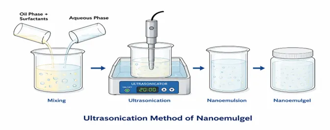
Figure 5: Ultrasonication method
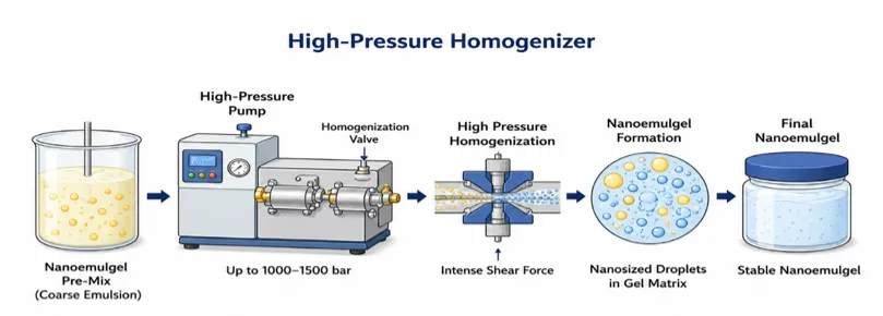
Figure 6: High-pressure homogenization method
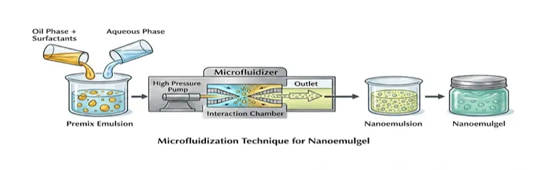
Figure 7. Microfluidisation technique
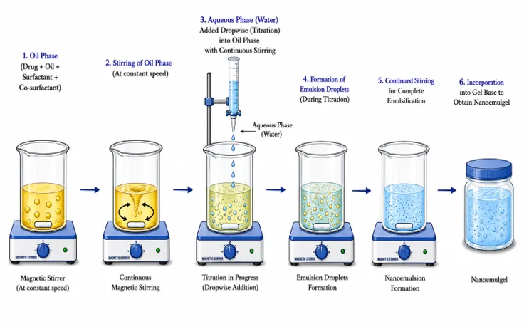
Figure 8. Aqueous titration method for nanoemulsion nanoemulgel preparation
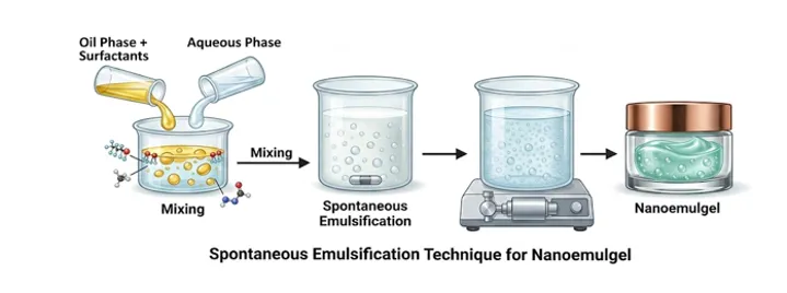
Figure 9: Spontaneous emulsification
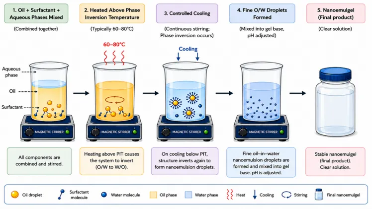
Figure 10: Phase inversion temperature (PIT) method
17. Recent Patents on Nanoemulgel:
Table 5: Recent Patents on Nanoemulgel
|
Patent Number |
API/Drug |
Title |
Disease/ Use |
Assignee/ Inventor |
Year |
|
US11185504B2 |
Aromatase inhibitors |
Nanoemulgel for transdermal delivery of aromatase inhibitors |
Breast cancer |
Qatar University [56] |
2021 |
|
CA3050535C |
Anti-inflammatory nutraceuticals (resveratrol, etc.) |
Nano-based formulation for treating inflammation |
Inflammatory disorders |
Nanosphere Health Sciences Inc. |
2021 |
|
CN107303263B |
Tripterygium glycosides |
Nanoemulsion gel preparation method |
Rheumatoid arthritis, psoriasis |
Second Military Medical University [57] |
2020 |
|
EP3099301B1 |
Besifloxacin |
Nanoemulgel for acne treatment |
Acne vulgaris |
Vyome Therapeutics Ltd. |
2019 |
|
WO2020240451A1 |
Brinzolamide |
In-situ nanoemulgel system |
Glaucoma |
Hemant Bhalerao, Sajeev Chandran |
2020 |
|
WO2020121329A1 |
Minoxidil + castor oil |
Nanoemulgel for hair growth |
Androgenic alopecia |
Sudha Suresh et al. |
2020 |
|
BR102019014044A2 |
Ketoconazole |
Nanoemulgel for nail fungal infection |
Onychomycosis |
Rayanne Rocha Pereira et al. |
2021 |
18. Components of Nanoemulgel:
Nanoemulgels are advanced topical drug delivery systems formed by incorporating nanoemulsions into a gel matrix. They combine the advantages of nanoemulsions, such as enhanced drug solubility and permeation, with the benefits of gels, including improved viscosity, spreadability, and patient acceptability. A typical nanoemulgel formulation consists of an oil phase, surfactants, co-surfactants, gelling agents, an aqueous phase, preservatives, and penetration enhancers 41,48,49.
The oil phase is a vital component of nanoemulgels because it solubilises lipophilic drugs and enhances skin permeation. Oil selection depends on drug solubility, compatibility, and permeation-enhancing ability; oils also improve drug diffusion through the stratum corneum by interacting with skin lipids 41,44. Commonly used oils include oleic acid, isopropyl myristate, castor oil, eucalyptus oil, peppermint oil, and clove oil 48,49
Functions: solubilization of hydrophobic drugs; enhancement of skin permeation; improvement of drug loading capacity; stabilisation of nanoemulsion droplets.
Surfactants reduce the interfacial tension between oil and water phases and stabilise nanosized droplets. Non-ionic surfactants are most commonly used because of their lower toxicity and better skin compatibility 44,45. Common surfactants include Tween 80, Tween 20, Span 80, and Cremophor RH40 43,48.
Functions: stabilization of the nanoemulsion; reduction of droplet size; improvement of formulation stability; enhancement of drug permeation.
Co-surfactants work together with surfactants to further reduce interfacial tension and improve the flexibility of the interfacial film, helping to form stable nanoemulsions with a smaller droplet size 44. Common co-surfactants include PEG 400, propylene glycol, ethanol, and Transcutol P 48.
Functions: improvement of nanoemulsion stability; reduction of interfacial tension; enhancement of drug solubilisation; facilitation of spontaneous emulsification.
Gelling agents convert nanoemulsions into nanoemulgels by increasing viscosity and improving consistency, enhancing spreadability, bioadhesion, retention time, and patient compliance 43,54. Commonly used gelling agents include Carbopol 934, Carbopol 940, HPMC, xanthan gum, and sodium alginate 48,54,58.
Functions: increase viscosity; improve topical retention; provide controlled drug release; enhance formulation stability.
The aqueous phase forms the continuous phase of oil-in-water nanoemulsions. Purified or distilled water is commonly used, as it provides compatibility and stability to the formulation 41.
Functions: formation of the external phase; drug dispersion medium; maintenance of formulation consistency.
Preservatives are added to prevent microbial contamination and increase the shelf life of nanoemulgels. Methylparaben and propylparaben are commonly used preservatives in topical formulations 54.
Functions: prevention of microbial growth; improvement of product stability; enhancement of shelf life.
Penetration enhancers improve drug permeation through the stratum corneum by altering skin lipid structure and increasing drug diffusion 49. Common penetration enhancers include oleic acid, ethanol, propylene glycol, and essential oils 41,48.
Functions: enhancement of skin permeation; improvement of bioavailability; increased drug retention at the target site.
19. Mechanism of Skin Penetration of Nanoemulgels:
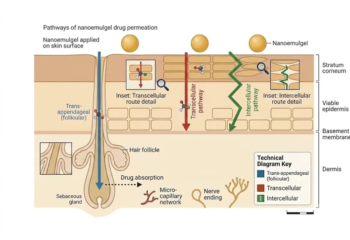
Figure 11: Pathways of nanoemulgel drug permeation through the skin
Nanoemulgels improve topical antifungal delivery through multiple mechanisms that enhance drug penetration across the stratum corneum, the primary barrier of the skin. The nanosized droplets present in nanoemulgels provide a larger surface area and intimate contact with the skin surface, facilitating enhanced diffusion of antifungal drugs into deeper epidermal layers. Because of their extremely small droplet size, nanoemulgels can penetrate more efficiently through intercellular lipid pathways and follicular routes compared with conventional creams and ointments 59,60.
Surfactants and co-surfactants incorporated into nanoemulgel systems additionally improve permeation by reducing interfacial tension and temporarily disturbing the highly ordered lipid arrangement of the stratum corneum, which enhances membrane fluidity and promotes deeper diffusion of antifungal agents into infected tissues 60,61. Certain oils commonly used in nanoemulgel formulations, including oleic acid and eucalyptus oil, also function as penetration enhancers by fluidising skin lipids and increasing membrane permeability 61.
Another important mechanism involves follicular targeting. Hair follicles and sebaceous glands can act as reservoirs for nanosized droplets, allowing prolonged drug retention and sustained release within infected skin layers — an advantage that is especially relevant in chronic dermatophytosis, where fungal persistence in deeper skin structures contributes to recurrence 62.
The gel matrix further enhances therapeutic performance by improving the viscosity and bioadhesion of the formulation. Increased residence time at the site of application promotes sustained drug release, reduces the frequency of application, and improves patient compliance 59,63. Overall, nanoemulgels enhance antifungal efficacy through improved solubilisation, enhanced permeation, prolonged retention, and controlled drug release.
20. Mechanism of Drug Permeation Through Skin:
The skin serves as an effective protective barrier against external substances, with the stratum corneum representing the primary obstacle to drug penetration. For a topically administered drug to produce a therapeutic effect, it must cross this barrier and reach the viable epidermis and dermis. Drug permeation through the skin generally occurs through three pathways: transcellular, intercellular, and trans-appendageal, illustrated in Figure 9 above. The transcellular pathway involves direct passage through corneocytes, whereas the intercellular pathway involves diffusion through the lipid matrix surrounding these cells; the trans-appendageal pathway allows drug transport through hair follicles and sweat glands 53.
Nanoemulgels enhance skin permeation by incorporating the drug into nanosized oil droplets dispersed within a gel matrix. The extremely small droplet size provides a larger surface area for drug absorption and facilitates closer contact with the skin surface. Surfactants and co-surfactants present in the formulation may temporarily modify the lipid arrangement of the stratum corneum, thereby improving drug diffusion. In addition, the gel matrix prolongs residence time at the application site, allowing sustained drug release and enhanced penetration into deeper skin layers 54,58.
21. Factors Affecting Nanoemulgel Performance:
The performance of a nanoemulgel is influenced by several formulation and processing variables. Droplet size is one of the most critical factors, as smaller droplets provide a larger interfacial surface area and improve drug release and skin permeation. The polydispersity index (PDI) reflects the uniformity of droplet size distribution, while zeta potential indicates the physical stability of the formulation by predicting the tendency of droplets to aggregate 54.
The type and concentration of the oil phase significantly influence drug solubilisation and loading capacity. Similarly, the surfactant and co-surfactant ratio determines the ability of the system to form stable nanosized droplets. The viscosity of the gel base affects spreadability, drug diffusion, and retention at the site of application. Furthermore, pH must remain within the physiological skin range to ensure patient comfort and formulation stability. Storage temperature and environmental conditions also play an important role in maintaining long-term stability and preventing phase separation or droplet growth 54,55,58,64.
22. Challenges and Limitations of Nanoemulgel Systems:
Despite their numerous advantages, nanoemulgels face several challenges that may limit their widespread commercial use. High concentrations of surfactants are often required to maintain nanoemulsion stability, which may increase the risk of skin irritation in sensitive individuals, and physical instability phenomena such as coalescence, flocculation, and Ostwald ripening can also affect formulation quality during storage 55,58,65.
Large-scale manufacturing remains a significant challenge because specialised equipment such as high-pressure homogenisers and microfluidisers is often required to achieve nanosized droplets, which increases production costs and complicates scale-up. Regulatory guidelines for nanotechnology-based pharmaceutical products are also still evolving, creating uncertainty in approval pathways, and further clinical studies are required to establish the long-term safety, efficacy, and therapeutic superiority of nanoemulgel formulations compared with conventional topical dosage forms 54,58,66,67.
23. Evaluation Parameters of Nanoemulgels:
Nanoemulgel formulations are extensively characterised to ensure stability, therapeutic efficacy, safety, and patient acceptability. Evaluation parameters such as particle size, zeta potential, viscosity, spreadability, drug release, and permeation studies play a major role in determining formulation quality and performance.
23.1 Evaluation parameters of Nanoemulgel:
Table 6. Evaluation parameters of nanoemulgel
|
Parameter |
Purpose |
Method/ Instrument |
|
pH determination |
Ensures skin compatibility |
Digital pH meter |
|
Viscosity |
Determines flow behavior of nanoemulgel |
Brookfield viscometer |
|
Spreadability |
Assesses ease of topical application |
Glass slide method |
|
Drug content |
Determines uniform drug distribution |
UV spectrophotometric method |
|
Entrapment efficiency |
Measures percentage of drug entrapped |
Centrifugation method |
|
In vitro drug release |
Evaluates release pattern of the drug |
Franz diffusion cell |
|
Ex vivo permeation study |
Determines permeation across excised skin |
Franz diffusion apparatus |
|
Antifungal activity |
Evaluates therapeutic effectiveness |
Zone of inhibition method |
|
Stability studies |
Determines physical and chemical stability |
ICH stability conditions |
23. Characterization of Terbinafine Hydrochloride Nanoemulsion:
Table 7. Characterization of terbinafine hydrochloride nanoemulsion
|
Technique |
Purpose |
Principle/ Application |
|
Dynamic Light Scattering (DLS) |
Particle size analysis |
Measures Brownian motion of nano-sized droplets |
|
Polydispersity Index (PDI) |
Assesses uniformity of droplet distribution |
Obtained from DLS using a Zetasizer |
|
Zeta potential analysis |
Evaluates formulation stability |
Determines surface charge of droplets |
|
Fourier Transform Infrared Spectroscopy (FTIR) |
Drug excipient compatibility |
Identifies functional groups and interactions |
|
Differential Scanning Calorimetry (DSC) |
Thermal behavior |
Detects crystallinity and drug excipient interactions |
24. Current Clinical Challenges in Dermatophytosis:
Dermatophytosis has emerged as a major public health concern, particularly in tropical and subtropical countries such as India. In recent years, clinicians have reported a substantial increase in chronic, recurrent, and treatment-resistant fungal infections, making disease management increasingly difficult. Several factors contribute to these challenges, including antifungal resistance, misuse of topical corticosteroids, poor patient compliance, and incomplete treatment courses.
25. Future Perspectives of Nanoemulgels in Dermatophytosis Therapy:
Recent progress in nanotechnology has created new opportunities for the development of highly efficient nanoemulgel systems for dermatophytosis treatment. Future research is expected to focus on targeted delivery systems capable of improving drug accumulation at infected tissues while minimising systemic exposure and adverse effects 68.
The incorporation of herbal bioactive compounds such as essential oils, phytoconstituents, and plant extracts into nanoemulgels is also gaining considerable attention, since many natural compounds possess antifungal, anti-inflammatory, and penetration-enhancing properties. Combination therapy involving synthetic antifungal agents and natural permeation enhancers may improve therapeutic outcomes and reduce antifungal resistance 69.
Modern pharmaceutical approaches such as Quality by Design (QbD) and computationally assisted formulation optimisation are additionally expected to improve reproducibility, stability, and industrial scalability, supporting the development of more efficient and commercially viable nanoemulgel systems 70.
Future advancements may also include stimuli-responsive or “smart” nanoemulgels capable of releasing drugs in response to pH, temperature, or fungal enzymes, which may further improve targeted delivery and therapeutic effectiveness in chronic dermatophytosis 68,70.
CONCLUSION:
Dermatophytosis continues to represent a major public health concern worldwide due to its increasing prevalence, recurrent nature, and emerging antifungal resistance. Conventional topical formulations often fail to provide adequate drug penetration and prolonged therapeutic action, leading to incomplete eradication and frequent relapse. Nanoemulgel systems have emerged as promising advanced topical delivery platforms capable of overcoming these limitations through enhanced drug solubilisation, improved skin permeation, sustained release, and prolonged retention at the site of infection.
The combination of nanoemulsion technology with gel-based systems provides superior physicochemical stability, better patient compliance, and enhanced antifungal efficacy compared with conventional formulations. Recent advances in nanotechnology, formulation optimisation, and the incorporation of natural bioactive compounds further highlight the growing potential of nanoemulgels in dermatophytosis management. Although challenges related to stability, large-scale production, regulatory approval, and long-term safety still exist, ongoing research is expected to support the successful clinical translation of nanoemulgel-based antifungal systems. Overall, nanoemulgels represent a highly promising and innovative strategy for the effective topical treatment of dermatophytosis.
REFERENCES

Navya N, Beny Baby, Bhuvana, Pallavi A, Tejashree K C, Advances in Nanoemulgel-Based Topical Antifungal Systems for the Treatment of Dermatophytosis, Int. J. of Pharm. Sci., 2026, Vol 4, Issue 9, 2530-2553. https://doi.org/10.5281/zenodo.22878057
 10.5281/zenodo.22878057