

We use cookies to ensure our website works properly and to personalise your experience. Cookies policy
Department of Pharmacy, Lokmanya Tilak Institute of Pharmacy, Kharghar.
Dermatophytosis is a common superficial fungal infection caused by dermatophytes belonging mainly to the genera Trichophyton, Microsporum and Epidermophyton, affecting keratinized tissues such as skin, hair and nails. Conventional topical antifungal formulations, including creams, gels, lotions and ointments, are widely used for the treatment of localized infections. However, limitations such as poor drug penetration, inadequate drug retention, frequent application and the emergence of antifungal resistance have created a need for advanced topical drug delivery systems. This review focuses on recent advancements in topical drug delivery approaches for the effective management of dermatophytosis. Various novel formulations, including polymeric nanohydrogels, liposomes, niosomes, microemulsions, nanoemulsions and nanostructured lipid carriers, are discussed for their potential to enhance skin penetration, improve drug solubility, provide controlled drug release and increase therapeutic efficacy. Different formulation strategies involving antifungal agents such as econazole, griseofulvin, terbinafine and luliconazole are also highlighted. Furthermore, important evaluation parameters, including pH, viscosity, droplet size distribution and in-vitro drug release studies, are discussed to assess formulation quality and performance. Despite their promising potential, challenges such as formulation instability, skin irritation, limited drug penetration and large-scale manufacturing remain significant concerns. Future research should focus on improving formulation safety, long-term stability and clinical effectiveness to develop more efficient and patient-friendly topical antifungal therapies. Overall, advanced topical drug delivery systems offer promising opportunities for improving the treatment outcomes of dermatophytosis.
1.1 Overview of Dermatophytosis:
Dermatophytosis is when a type of fungus called a dermatophytic fungus grows on the keratinised tissues. These tissues include nails, hair and the outer layer of the skin. The infection can be caused by members of three groups of fungi. These groups are called genera. The three groups are Trichophyton, Microsporum and Epidermophyton. Each of these groups has features, in their shape how they work and the way they look to the body’s immune system. This information was reported by Weitzman and Summerbell in 1995.[1]
1.2 Causative Agents/Organisms:
Fungi, like Trichophyton, Microsporum, Epidermophyton, Nannizzia, Paraphyton, Lophophyton, Arthroderma, Ctenomyces and Guarromyces live in humans and animals. Out of all these Trichophyton, Microsporum and Epidermophyton are the common. The fungi Trichophyton rubrum, Trichophyton interdigital, Trichophyton tonsurans, Epidermophyton floccosum and Trichophyton violaceum are called anthropophilic fungi. These mainly infect humans. Rarely infect animals. The fungi Trichophyton mentagrophytes, Microsporum canis and Trichophyton verrucosum are called fungi. These mainly affect animals. Can also infect humans. [2]
1.3 Common Infection:
Table 1.3: 1 Types of Infection [3]
| Sr. No. | Type of infection | Description |
|---|---|---|
| 1. | Tinea corporis (ringworm) | A red, ring-shaped, scaly, itchy patch with central clearing and an active rim |
| 2. | Tinea cruris (Jock itch) | Refers to the area of the upper thigh situated opposite the scrotum |
| 3. | Tinea pedis (Athlete’s foot) | This usually affects the skin between the toes, though it may spread to the sole, the sides, and the dorsum of the affected foot. Acute form: erythema and maceration between the toes, at times with painful vesicles. Chronic form: scaling, peeling, and erythema between the toes; nevertheless, it can extend to other areas of the foot. |
| 4. | Tinea unguium (Onychomycosis) | Commonly observed in people with dystrophic toenails, this condition is marked by nails that are thickened, brittle, and discoloured. |
| 5. | Tinea capitis | Ringworm of the scalp |
1.4 Current Treatment:
Treatment of dermatophytosis involves both nonpharmacological and pharmacological approaches. The skin should be fully dried before getting dressed, and wearing loose-fitting garments made of cotton or synthetic material is recommended. It is recommended that you avoid walking barefoot and refrain from sharing clothing or other items with family members.[3]
Topical treatment:
For the management of naive tinea cases or localized infections, topical therapy is typically advised. It works very well for localized infections and naive infections. A topical agent ought to have characteristics like broad-spectrum activity, a high mycological cure rate, ease of application, and a low rate of side effects. When choosing among the available topical antifungal drugs, the number of applications and the duration of treatment are the deciding factors.[3]
Based on Type of Infection:
Table 1.4: 1 Treatment of Dermatophytosis Based on Type of Infection [4]
| SR. NO. |
Type of infection | Current Therapeutic Regimen: | In Naïve Cases: | In Recalcitrant Cases: |
|---|---|---|---|---|
| 1 | Tinea pedis | Terbinafine -250mg for 2 weeks Itraconazole-200mg daily |
Terbinafine 250 mg/day | Itraconazole-200mg-400 mg/day, in divided dose |
| 2 | Tinea crusis and corpis | Terbinafine -250 mg once daily Itraconazole-100 mg– 200 mg/day |
Terbinafine -250 mg once daily Itraconazole-100 mg– 200 mg/day |
Itraconazole-200mg-400 mg/day, in divided dose |
| 3 | Tinea incognito | Itraconazole 200 mg– 400 mg daily, for 4–6 weeks or longer | Griseofulvin -250 mg– 500 mg twice daily fluconazole -150 mg–300 mg/week |
- |
Systemic or oral treatment:
The selection of an antifungal agent is guided by the drug's pharmacological properties, any previous exposure to antifungals, the location and size of the lesion, the skin surface affected, and the patient's age. Oral antifungal drugs are considered for treatment when repeated use of topical agents does not clear lesions covering a large body surface area. Systemic therapy is taken into consideration when vellus hair is involved.[3]
Patients who are elderly:
An elderly patient in good health may be managed according to the recommendations used for a young adult. In elderly patients, topical therapy should be preferred; systemic therapy is needed only when topical treatment fails, when lesions are extensive, or in recalcitrant cases. Because systemic triazole drugs (itraconazole, fluconazole) can cause multiple drug interactions, oral terbinafine should be the preferred option.[4]
Age group of paediatric patients:
Administration of fluconazole and terbinafine in the paediatric age group. Although fluconazole may be given during infancy, terbinafine is advised only for children over 2 years old.[4]
Females who are pregnant:
Topical antifungals are absorbed systemically only minimally or not at all, so they can be prescribed during any stage of pregnancy. As for systemic therapy, terbinafine belongs to pregnancy category B; nevertheless, there are no data on its use during pregnancy, and it is also unknown whether terbinafine crosses the placental barrier. It is recommended to use effective contraception for 2 months after taking oral itraconazole before conception. During pregnancy, other systemic antifungals should be avoided.[4]
Table 1.4: 2 Oral and Topical Drugs Used to Treat Dermatophytosis [3]
| Sr. No. |
Oral Drugs | Dose |
|---|---|---|
| 1 | Itraconazole | 100-200mg/day |
| 2 | Terbinafine | 250mg/day |
| 3 | Fluconazole | 150-300mg/day |
| 4 | Griseofulvin | 250-500mg/day |
| Sr. No. |
Topical Drugs | Dose |
| 1 | Luliconazole | Once or twice a day for 2-6 weeks |
| 2 | Sertaconazole | Once or twice a day for 2-6 weeks |
| 3 | Terbinafine | Twice daily for 2 weeks |
| 4 | Amorolfine | Twice daily for 4 weeks |
1.5. ANTIFUNGAL RESISTANCE AND ITS MECHANISM:
Dermatophytes can develop resistance to antifungal drugs through multiple mechanisms, and each of these mechanisms may greatly diminish how well standard treatments work. The main resistance mechanisms are listed below:
1.5.1. Genetic Mutations
When genes that encode key fungal enzymes carry mutations, the effectiveness of antifungal agents can be compromised. For example, alterations in the ERG11 gene, which encodes lanosterol demethylase, can lead to reduced binding of azoles—an antifungal class that acts on this enzyme. Such mutations disrupt ergosterol production, and ergosterol is an essential constituent of the fungal cell membrane.
1.5.2. Efflux Pumps:
A commonly documented mechanism of resistance involves the activation of membrane-embedded efflux pumps, which can identify and extrude a wide variety of compounds, thereby producing multidrug resistance (MDR). Dermatophytes are capable of developing or upregulating efflux pumps, including those in the ATP binding cassette (ABC) and major facilitator superfamily (MFS) families. These pumps actively move antifungal drugs out of the cell, which lowers the drug concentration inside the cell and consequently weakens the drugs' effectiveness. Higher expression of these pumps can lead to multidrug resistance.
1.5.3. Breakdown by Enzymes:
Certain dermatophytes generate enzymes capable of chemically altering or breaking down antifungal medications, which stops these drugs from arriving at their intended target. For instance, some fungi release esterases or oxidases that disable antifungals before those drugs can interfere with the workings of fungal cells.[5]
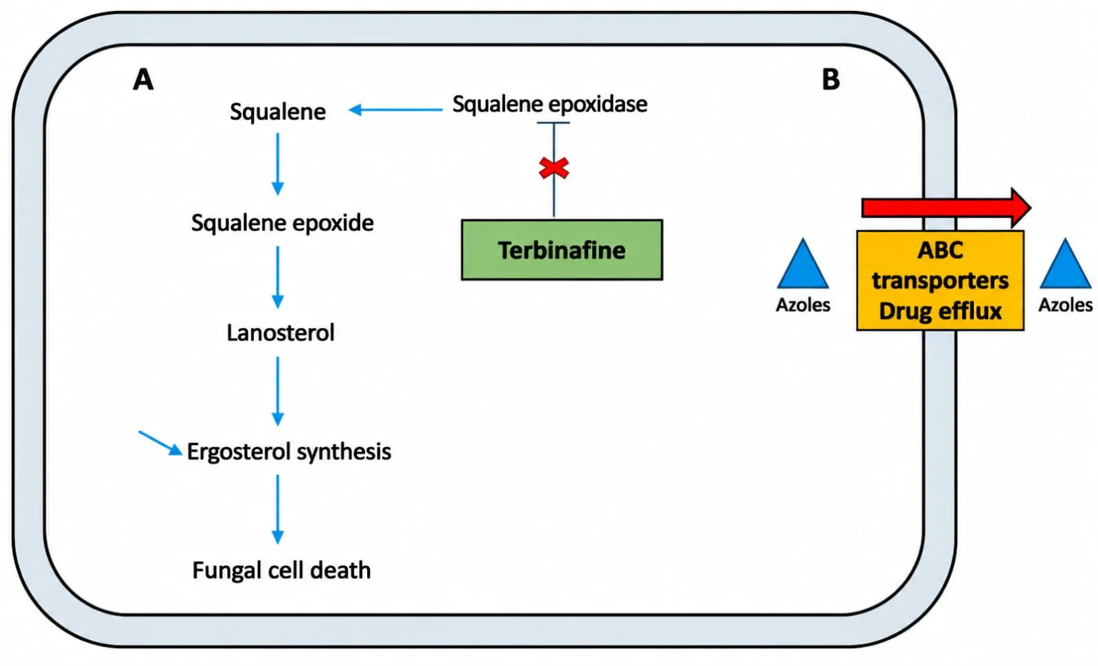
Figure 1.5: 1. Mechanism of Antifungal Resistance
1.6. NEED FOR ADVANCES IN TOPICAL TREATMENT:
Options for treating skin inflammation with topical medications are currently limited, so there is a need to create a drug delivery system that can release drugs in a controlled manner. This approach is intended to produce more effective outcomes in relieving pain, reducing inflammation, ensuring greater safety, and preventing adverse reactions.1 By means of such a delivery system, therapeutic agents are able to reach the deeper layers of the epidermis and dermis, thus maximizing their therapeutic efficacy. Researchers are investigating formulations that boost skin permeability and penetrate the skin via various mechanisms in order to enhance topical drug delivery. One potentially effective strategy involves the use of nanoparticle systems to increase skin permeability and promote the deposition of the encapsulated pharmacological agents. However, studies have shown that these nanoparticles frequently do not penetrate the stratum corneum (SC) but instead build up in the SC layer, gradually releasing the encapsulated drug into the upper epidermis. From that point, the drug diffuses passively into the skin layers beneath .Although recent research has demonstrated that nanoparticles can carry active pharmaceutical ingredients (APIs) through hair follicles and into the skin layers, the total amount that reaches the dermal sites is still limited .As the most recent drug delivery systems, a range of nanotechnology techniques have been introduced, including protein-based nanoparticles, lipid based nanoparticles, nanoemulsions, nanocrystals, nanodiamonds, carbon nanotubes, nanosuspensions, and NH. These technologies show a considerable significant effect in enhancing drug delivery through the skin and offer attractive avenues for further exploration and development in the field of topical drug delivery.[6]
2.ADVANCED TOPICAL DRUG DELIVERY SYSTEM:
Existing topical treatments for skin inflammation are restricted in their effectiveness, which makes it necessary to create a drug delivery system capable of releasing medication in a controlled manner. The goal of this approach is to produce better outcomes in relieving pain and inflammation, while also providing improved safety and avoiding unwanted side effects. Through the use of a delivery system of this kind, therapeutic agents are able to reach the deeper layers of the epidermis and dermis, thereby maximizing their therapeutic effectiveness. To improve topical drug delivery, researchers are investigating formulations that boost skin permeability and penetrate the skin via a range of mechanisms.[6] Novel drug delivery systems (NDDS) rank among the most extensively studied topical formulations in pharmaceutical research. Owing to its distinctive capacity to regulate the release kinetics of encapsulated drugs, to encapsulate a broad variety of drugs, and to increase disease-specific localization, NDDS lowers dosing frequency and improves clinical efficacy. Nevertheless, when an appropriate topical formulation is being implemented, one must grasp the detailed mechanism of antifungal therapeutics in order to maintain sufficient therapeutic performance.[7] One potentially effective approach involves employing nanoparticle systems to boost skin permeability and to facilitate deposition of the encapsulated pharmacological agents. Yet studies have shown that such nanoparticles frequently fail to penetrate the stratum corneum (SC) and instead build up within the SC layer, slowly releasing the encapsulated drug into the upper epidermis. From that point, the drug diffuses passively into the skin layers beneath.3 Although recent research indicates that nanoparticles are able to carry active pharmaceutical ingredients (APIs) through hair follicles and into the skin layers, the overall quantity that reaches the dermal sites is still limited. As the most recent class of drug delivery systems, a variety of nanotechnology techniques have been introduced, such as protein-based nanoparticles, lipid-based nanoparticles, nanoemulsions, nanocrystals, nanodiamonds, carbon nanotubes, nanosuspensions, and NH. These technologies show a considerably significant effect in enhancing drug delivery through the skin and constitute appealing avenues for further exploration and development within the field of topical drug delivery.[6]
3.ADVANCED TOPICAL FORMULATIONS:
3.1. Nanohydrogels Based on Polymers
For the creation of smart systems intended for skin applications, polymer-based NH have most frequently been identified as promising candidates. The polymeric channel network forming the core of a hydrogel can be produced through either physical or chemical cross-linking of homopolymers or copolymers; such cross-linking makes the hydrogel swell when exposed to an aqueous environment. Polymer NH are materials with viscoelastic properties and hydrophilic polymeric networks in the sub-micron size range, and they are used as nanocarriers in drug delivery.19 The type and amount of polymer building blocks employed in producing viscoplastic nanogels dictate the properties exhibited by the final product. NH are manufactured using raw materials derived from natural polymers, synthetic polymers, or a combination of both. Synthetic polymers are a class of polymers that may be produced by processing natural materials or by synthesizing primary components from oil, gas, and other sources. These polymers are distinguished by their long molecular chains and organic linkages. NH composed of synthetic polymers offer an alternative to conventional polymer networks, along with several advantages.[6] [22]
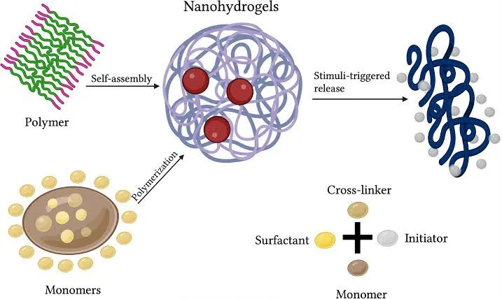
Figure 3.1: 1 Nanohydrogels Based on Polymers
3.2. Liposomes:
Liposomes offer a novel approach to treating dermatophyte infections. Nanotechnology has brought numerous new developments in the treatment of microbial infections. In addition, lipid-based nano systems like liposomes have shown especially attractive physicochemical properties and safety profiles when compared with other nanocarriers.[21] Liposomes are spherical structures made up of one or more phospholipid bilayers that enclose aqueous compartments. The capacity of liposomes for surface modification is a key feature that makes them promising candidates as innovative antifungal treatments, as discussed further below. Surface functionalization with ligands, such as molecules or polymers, allows antibiotics to be delivered in a targeted manner, which is essential for effective drug delivery and therapeutic results.[8]
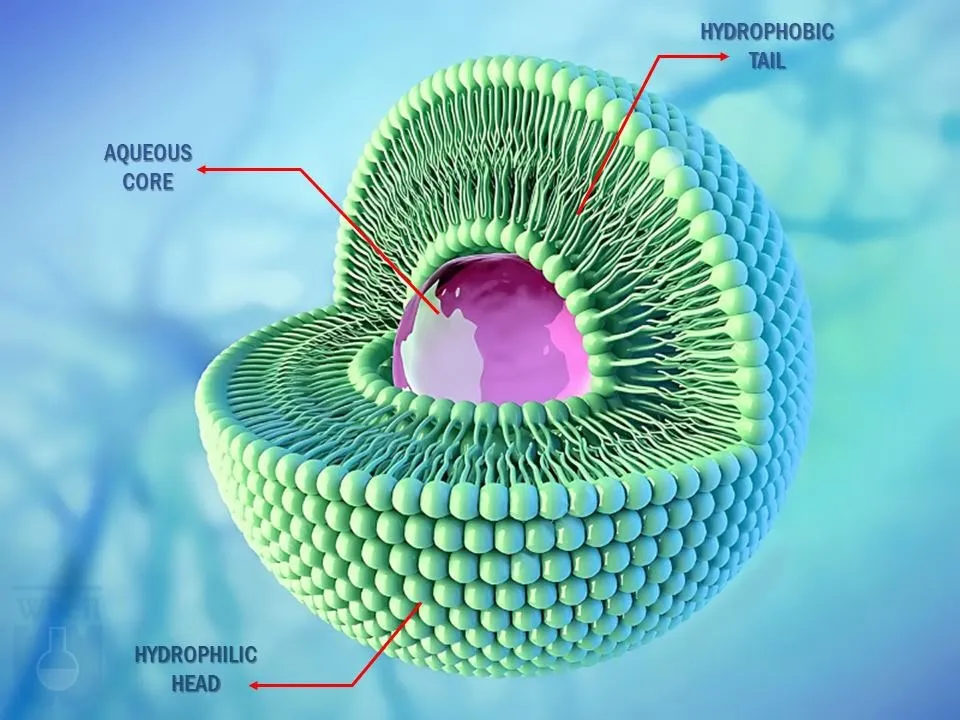
Figure 3.2: 1 Liposomes
3.3. Niosomes:
These are a type of spherical lipid vesicle made using non-ionic surfactants. They interact with the stratum corneum, which leads to a decrease in trans epidermal water loss. Its permeation through the skin depends on the kinds of surfactant, the properties of the drug employed, and the morphological features of niosomal preparations. Ketoconazole was observed to have greater therapeutic activity in niosomal preparations. Niosomes containing itraconazole and miconazole were also shown to be effective, demonstrating that they serve as effective carrier systems for antifungal drugs.[7]
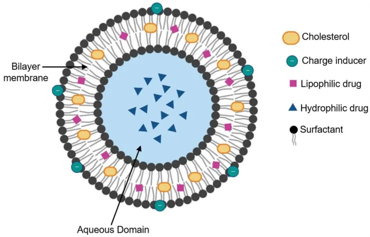
Figure 3.3: 1 Niosomes
3.4 Microemulsion:
These are stable, translucent, and isotropic oil-in-water dispersions, stabilized by surfactants and co-surfactants, used for topical and transdermal drug delivery with a droplet size of 0.1–1.0 μm. Because of their unique capacity to improve drug solubility, they have been described as a highly promising delivery system for anti-fungal agents. The antifungal spectrum of many azole drugs is limited by their poor aqueous solubility. They provide benefits such as enhanced drug solubility, high thermal stability, high permeability, simple manufacturing, optical clarity, and low cost. They exhibit excellent biocompatibility, since microemulsions are a suitable delivery system for topical and transdermal applications. In the microemulsion formulation, the oils and surfactants that are present assist drugs in permeating through the stratum corneum.[7]
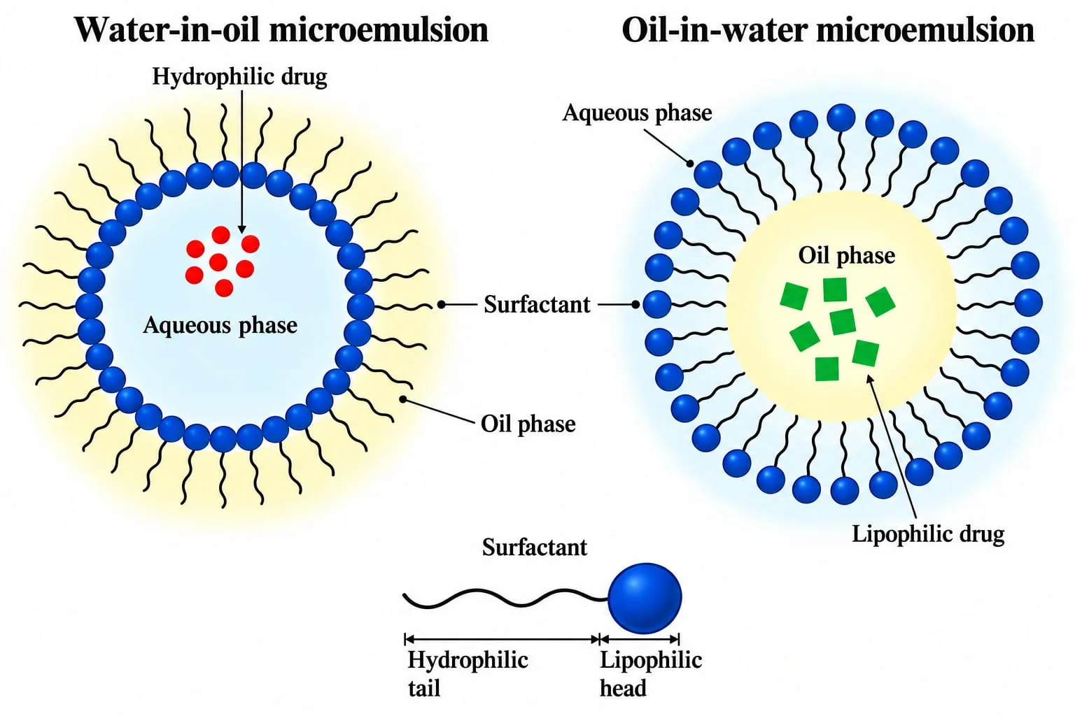
Figure 3.4: 1 Microemulsion
3.5 Nanoemulsion:
It is a single-phase, stable, and isotropic dispersion made up of an emulsified oil phase, water, and amphiphilic molecules, with droplet sizes ranging from 5 to 200 nm. These are both thermodynamically and kinetically stable. Because of their high surfactant concentration, nanoemulsions are regarded as well suited for skin permeation of both hydrophilic and lipophilic drugs.[7]
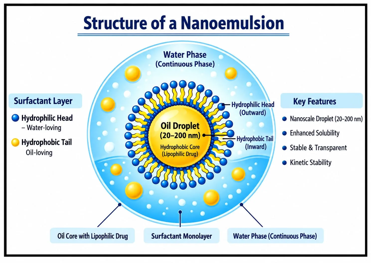
Figure 3.5: 1 Nanoemulsion
4.MECHANISM OF ENHANCED SKIN PENETRATION:
Phospholipid vesicles that aid in enhancing the absorption of active ingredients are most frequently studied in the forms of transferosomes and ethosomes.[9]
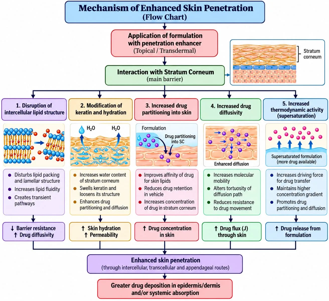
Figure 4.1: Mechanism of Enhanced Skin Penetration
5.FORMULATION STRATEGIES FOR ENHANCED ANTIFUNGAL ACTIVITY:
5.1 Econazole Gel for Topical Use
Carbopol® 940, serving as the gelling agent, was introduced into a sufficient quantity of water at varying concentrations (Table 1) and then stirred at 100 rpm for two hours. Triethanolamine was then used to neutralize the resulting mixture. In addition, econazole nitrate was dissolved in a viscous blend of propylene glycol (a plasticizer), Capmul ® MCM C8 (an emollient and solubilizer), and methyl and propyl paraben (preservatives). Subsequently, this blend was incorporated into the gel base and mixed thoroughly, with distilled water added to bring the gel's final weight to 100 g. After being shaken at 500 rpm for two hours to remove any air bubbles, the resulting combination was left at room temperature for a full day so that its stability and consistency could be assessed.[10] [30]
5.2 Griseofulvin
Batches of solid dispersions (SDs) of griseofulvin were created by combining griseofulvin (melting point 220–223 °C) with PEG-6000 in drug-to-carrier ratios of 1:2, 1:4, and 1:6; the mixture was sieved (80 mesh, ~170 μm) and then microwave-irradiated (IFB 20 L Convection microwave, 20SC2, Metallic Silver) at 900 W for 7 min, melting at 210 degrees Celsius to yield several SD batches; these SDs were subsequently used in the formulation of griseofulvin-loaded lipid nanoparticles (G-LN). Heating continued until the sample reached a constant mass, after which it was left to cool and solidify. For 48 h the solidified mass was stored in a desiccator to eliminate residual moisture; it was then pulverized and subjected to solubility studies and FTIR analysis to evaluate stability. Saturated solutions of the pure drug (griseofulvin) and of griseofulvin SDs with polymer were prepared in various solvents, shaken for 24 h, filtered, diluted, and analysed at 293 nm on a Shimadzu UV spectrophotometer. On the basis of the solubility study, the optimized SD (1:4 ratio) was selected for preparing the G-LNs. A weighed quantity of stearic acid was incorporated into the molten SD, after which the mixture was emulsified with an aqueous Tween 80 solution under mechanical stirring (4,000 rpm, 60 min), ultrasonicated (20 min), cooled in an ice bath (15 min), and then stored at room temperature. By varying the stearic acid, six G-LN batches (B1–B6) were formulated.[11]
5.3 Terbinafine HCL
Development of a Terbinafine HCl Nanoemulsion (TH-NE)
The nano emulsion mentioned previously was produced by means of a self-emulsification approach. First, 0.625 mg of TH was dissolved in 120 µL of olive oil, after which it was combined with a solubilizing agent, S mix (made up of propylene glycol and Span 80 at a 1:2 ratio). Subsequently, this pre-emulsion was subjected to vortex mixing for 10 min, which led to the creation of a nanoscale formulation.[12]
5.4 Luliconazole-loaded nanostructured lipid carrier nanoparticles (LCZ-NLCs NPs)
A modified emulsification-solvent evaporation method was used to prepare the LCZ-NLCs. Briefly, 100 mg of LCZ, 100 mg of Span 80, 150 mg of GMS, and 50 mg of oleic acid were dissolved in 500 µL of chloroform at 60 °C; this solution served as the organic phase. Deionized water (no more than 10 mL) containing 100 mg of Tween 80 was used as the aqueous phase. The aqueous phase was heated until it reached the same temperature as the organic phase. The aqueous phase was then added dropwise to the heated organic phase under a high-shear homogenizer (Heidolph M 036170020, Germany) run at 5000 rpm for 15 min. The resulting homogeneous emulsion was then sonicated at 40% amplitude for 5 min (Bandelin 3100, Germany). Right after sonication, the emulsion was cooled in an ice bath to promote the separation of LCZ-NLCs. The mixture was then stirred overnight at ambient temperature to remove any residual organic solvent. To obtain the LCZ-NLCs, the mixture was centrifuged (19000 rpm, 25 °C, 30 min), washed to remove excess surfactant and free LCZ, and then lyophilized to yield freeze-dried nanoparticles for further studies.[13] [23]
Table 5.1: Comparison of Formulation Strategies for Enhanced Antifungal Activity
| Sr. No. |
Parameter | Econazole Topical Gel | Griseofulvin-Loaded Lipid Nanoparticles (G-LNs) | Terbinafine Hcl Nano emulsion (TH-NE) | Luliconazole Nanostructured Lipid Carriers (LCN-NLCs) |
|---|---|---|---|---|---|
| 1 | Drug used | Econazole nitrate | Griseofulvin | Terbinafine Hydrochloride | Luliconazole |
| 2 | Dosage form | Topical gel | Lipid Nanoparticles | Nanoemulsion | Nanostructured lipid carriers |
| 3 | Gelling agent/polymer | Carbopol 940 | PEG 6000 | Not specified in the described procedure | Not specified |
| 4 | Oil/Lipid phase | Capmul® MCM C8 | Stearic acid | Olive oil | Glyceryl monostearate (GMS) and oleic acid |
| 5 | Surfactants | Not specifically specified | Tween 80 | Span 80 | Tween 80 and Span 80 |
| 6 | Other excipients | Propylene glycol, methyl paraben, propyl paraben, triethanolamine | PEG 6000 | Propylene glycol | Chloroform as organic solvent solvent. |
| 7 | Preparation method | Gel formation & mixing | Microwave-assisted solid dispersion followed by emulsification | Self-emulsification | Emulsification solvent evaporation |
| 8 | Main processing condition | Stirring at 100 rpm for 2 hours; subsequent shaking at 500 rpm for 2 hours | Microwave irradiation at 900 W for 7 minutes; stirring at 4,000 rpm for 60 minutes; ultrasonication for 20 minutes | Vortex mixing for 10 minutes | Homogenization at 5,000 rpm for 15 minutes; sonication for 5 minutes |
| 9 | Cooling/drying | Stored at room temperature for 24 hours | Cooled, dried in a desiccator for 48 hours, then stored at room temperature after formulation | Not specified | Ice-bath cooling followed by overnight stirring, centrifugation, washing, and lyophilization |
| 10 | Optimization parameter | Carbopol concentration | Drug-to-polymer ratio and stearic acid quantity | Oil and surfactant/co-surfactant composition | Lipid and surfactant composition |
| 11 | Evaluation Mentioned | Stability and consistency | Stability and FTIR analysis | Not specified in the supplied method | Further nanoparticle characterization; specific tests not provided |
| 12 | Primary Formulation Objective | Prepare a suitable topical gel | Improve drug solubility and incorporate the drug into lipid nanoparticles | Form a nanoscale emulsion for topical delivery | Develop a lipid-based nanoparticulate drug delivery system |
6.EVALUATION TESTS FOR TOPICAL FORMULATION:
6.1 Determination of pH:
The pH indicates how acidic or alkaline a substance is. This matters greatly in the pharmaceutical field, since some drugs may lose their potency or break down in acidic or alkaline conditions; certain drugs dissolve only partially in acidic or alkaline media, which can influence their bioavailability and therapeutic efficacy, and the pH of biological fluids such as blood and urine can alter the rate and extent of drug release. This highlights how essential pH is in the development, manufacturing, and administration of pharmaceutical products. For topical preparations applied to the skin, a pH range of 4.2 to 6.5 is regarded as suitable. Overly alkaline gels cause the skin to become scaly. On the other hand, skin becomes irritated when the pH is too acidic.[14][27]
6.2 Viscosity Measurement
The rheological behaviour of gels differs, and rather than flowing under low shear stresses, they undergo reversible deformation, much like what occurs in elastic materials. flowing when shear stresses are low, which resembles the behaviour of elastic materials. Once the yield value of shear stress is surpassed, they behave like liquids and flow. Broadly speaking, once the shear stress yield value is surpassed, they flow in the manner of liquids. Broadly speaking, the consistency of gel compositions is reflected in their viscosity. Shear thinning, also referred to as non-Newtonian flow, shows that the consistency of gel compositions is reflected in their viscosity. Shear thinning, also called non-Newtonian flow, shows how gel viscosity drops as the shear rate rises, which is desirable since gels perform well under high shear conditions thanks to their low flow resistance. The rheological property governs how drugs diffuse out of a gel, and this property corresponds to low flow resistance.[14] [29]
6.3 Distribution of Droplet Sizes:
Analysis of the droplet size distribution was performed at room temperature using light scattering on a Malvern Mastersizer 2000 (Malvern Instruments, Malvern, UK) equipped with a Hydro S accessory. Distilled water was used to disperse the sample. Using a stirrer set at 1250 rpm, the diluted sample was added dropwise into the sample chamber, which held approximately 150 mL of water, until a laser obscuration of 10% to 20% was reached. For each sample, the distributions were measured in triplicate. The size data were reported as the relative volume distribution of droplets and expressed as diameter values at the 10%, 50%, and 90% percentiles, along with the span value (mean ±SD). The span value is a statistical parameter that is helpful for describing how wide the droplet size distribution is, as defined by the following equation:
Span = [d (90) - d (10)] / d (50)[15]
6.4 In Vitro Release Testing (IVRT)
The experiments were performed in accordance with the OECD "Guidance document for the conduct of skin absorption studies". Clotrimazole release was measured with static vertical Franz diffusion cells, which had a diffusion area of 1 cm2 and a receptor compartment of 3 mL. The membranes (Tuffryn® membranes, Pall Europe, Hampshire, UK) were rinsed and equilibrated in ethanol/water (1:1), after which they were mounted between the donor and receptor compartments. The receptor medium, an ethanol/water mixture (1:1), was selected after appropriate screening based on Clotrimazole solubility studies so that sink conditions were maintained throughout the experiment. The release medium was stirred continuously with a magnetic bar and kept at (35 ±2)◦C by a thermostatic water pump, which ensured a temperature of 32 ◦C at the membrane surface (mimicking skin conditions).[14]
7.CHALLENGES:
SLNs—no deeper penetration for SLNs; drug expulsion during storage is caused by lipid polymorphism for SLNs, along with the risk of gelation for SLNs. NLCs—no deeper penetration.[25]
Liposomes—require special storage conditions, higher production cost, poor permeation into the viable epidermis and dermis, poor physicochemical characteristics for long-term stability (storage at 4–6 °C, in a refrigerator, and adjustment of the pH of the dispersions to pH values close to neutral).[23][24]
Niosomes, transferosomes, ethosomes—skin irritation and toxicity (erythema and eschar formation value 0∼1) caused by chemical excipients.[16] [26]
Microemulsion formulations require a high concentration of surfactant combined with co-surfactants in order to cover a wider interface, achieve complete emulsification of the ingredients, and ensure long-term stability. However, undesirable residues left on the substrate may lead to local skin toxicity with prolonged use, so local toxicity must be considered, especially when the products are meant to be used over a longer period.[17]
In nanocarrier-based dermatologic formulations, the challenge lies in the biological variability among patients, since differences in skin thickness, lipid composition, microbiota and barrier integrity can considerably affect the penetration of nanocarriers and drug release. Disease-specific changes, such as hyperkeratosis in psoriasis or impaired barrier function in eczema, may modify therapeutic performance and make dose optimization more complicated.[18] [28]
8.FUTURE PROSPECTS:
Antifungal drugs in current use may gain from innovative formulations and delivery approaches, thus helping to enable the creation of customized treatments that enhance efficacy and reduce toxicity, consequently improving patient outcomes. Over the past ten years, repurposing has drawn growing interest as a viable and accelerated route for developing antifungal medications. In contrast to the laborious, lengthy, and expensive process of de novo drug development, repurposing involves investigating new therapeutic applications for already approved medications. Existing antifungal drugs could gain from innovative formulations and methods of delivery, which in turn would help develop customized therapeutics that boost efficacy while reducing toxicity. The concepts behind antifungal pharmacokinetics and pharmacodynamics (PK/PD) are likewise beneficial; better therapeutic monitoring, more accurate clinical antifungal PK/PD predictions, and improved in vitro and in vivo PK/PD models are able to refine antifungal dosing and raise the chances of positive results for patients suffering from fungal infections.[19]
Research is increasingly directed toward the development and assessment of transdermal delivery systems for antifungal drugs, aiming to overcome the drawbacks of traditional dosage forms. An additional perspective may also be taken into account.
1. Studies on long-term safety and efficacy: Thorough clinical trials are needed to establish the long-term safety and efficacy of transdermal antifungal delivery systems under real-world conditions, while also addressing concerns about possible side effects.
2. Investigating combination therapies: Pairing antifungal agents with other therapeutic strategies—immunomodulators or wound healing promoters, for instance—might boost treatment results. As such, researchers ought to explore novel combinations in order to boost how effective treatments are.
3. Investigating natural compounds that show antifungal activity and
incorporating plant extracts or essential oils into topical formulations
might provide alternative treatment options.[20]
CONCLUSION:
This review article examined the transdermal delivery of antifungal agents for the treatment of dermatophytosis, highlighting the considerable advancements and insights in this area. Across these studies, the main objective was to develop a range of transdermal delivery enhancement strategies capable of improving the permeation of antifungal agents through the skin and thereby overcoming the limitations linked to conventional routes of administration. These approaches include nanostructured lipid carriers, ethosomes, liposomes, and nano/microemulsions. In addition, most of these strategies were incorporated into gels to improve adhesion properties and sustain drug release. Furthermore, chemical enhancers, iontophoresis, microneedle arrays, and other physical enhancement techniques were explored to enhance the penetration of antifungal agents. The results from these studies showed promising outcomes, indicating the feasibility and effectiveness of transdermal delivery for antifungal agents. Commonly reported benefits include targeted delivery, enhanced permeation, sustained release, and improved therapeutic outcomes. However, it is important to recognize challenges such as skin irritation, formulation stability, and the scalability of these delivery systems. Future research should focus on tackling these challenges and refining the developed strategies for practical use in clinical settings. In summary, this study emphasized the potential of transdermal delivery systems as a feasible and beneficial method of administering antifungal agents. Continued efforts to improve these systems and resolve the challenges linked to them will help develop innovative and clinically relevant solutions for treating fungal infections.[20]
REFERENCES

Christina Viju, Asmita Sawant, Adrija Pramanick, Manas Pandere, Pruthviraj Jangam, Shrutika Patil, Advances in Topical Drug Delivery for Dermatophytosis Formulation: Strategies, Evaluation Parameter, Therapeutic Perspectives, Int. J. of Pharm. Sci., 2026, Vol 4, Issue 11, 557-571. https://doi.org/10.5281/zenodo.23938264
 10.5281/zenodo.23938264