

We use cookies to ensure our website works properly and to personalise your experience. Cookies policy
B.R.Harne college of Pharmacy.
Visceral leishmaniasis (VL), also known as kala-azar, is a potentially fatal neglected tropical disease caused primarily by Leishmania donovani and Leishmania infantum. Despite advances in antileishmanial therapy, its management remains challenging due to treatment-associated toxicity, drug resistance, high treatment costs, and limitations in drug administration. Amphotericin B (AmB), a polyene macrolide antibiotic with potent antileishmanial activity, has emerged as an important therapeutic agent in the management of VL. However, conventional amphotericin B deoxycholate is associated with dose-limiting nephrotoxicity, infusion-related adverse reactions, and the requirement for intravenous administration. To overcome these limitations, lipid-based formulations, particularly liposomal amphotericin B, have been developed to improve drug distribution, enhance tolerability, and maintain therapeutic efficacy. Formulations such as AmBisome and the indigenous Indian preparation FUNGISOME have demonstrated considerable clinical relevance, although their cost, parenteral administration, and storage requirements remain important challenges. In this context, oral drug delivery has gained attention as a potential alternative to conventional injectable therapy. Self-emulsifying drug delivery systems (SEDDS) and self-nanoemulsifying drug delivery systems (SNEDDS) offer promising approaches for improving the dispersion and delivery of poorly water-soluble drugs such as amphotericin B. Preclinical investigations of oral lipid-based formulations, including iCo-010, have demonstrated encouraging antileishmanial activity and reductions in parasite burden in experimental models. Nevertheless, poor gastrointestinal absorption, complex physicochemical properties, formulation stability, and potential toxicity remain significant barriers to successful oral delivery.
Neglected tropical diseases (NTDs) are a diverse group of infectious diseases caused by viruses, bacteria, parasites, fungi, and toxins According to the WHO, more than 1.5 billion people worldwide are affected by these diseases, with the greatest burden observed in tropical and subtropical areas where poverty, inadequate sanitation, unsafe drinking water, and limited access to healthcare are prevalent. [1]
Global and Indian burden of VL
Leishmaniasis is one of the 20 neglected tropical diseases that collectively affect over one billion people worldwide, predominantly impacting impoverished communities across more than 90 countries in Asia, Africa, the Middle East, and Central and South America. [2/5]
The disease is closely associated with poverty, as it can interfere with employment, education and social activities, further increasing social and economic difficulties.[5]
- Burden in India: India faces a considerable NTD burden, with at least 12 different diseases prevalent in the country.
- Health and social consequences: NTDs can cause pregnancy complications, disability, stigma, and delayed physical and intellectual development in children, potentially affecting school attendance.
- Economic impact: These diseases reduce work productivity, limit employment, and increase household healthcare expenses, contributing to social and economic difficulties for families, communities, and the country.[1]
Leishmaniasis is a vector-borne disease caused by obligate intracellular protozoan parasites of the genus Leishmania, belonging to the family Trypanosomatidae. The parasites are transmitted to humans and other animals through the bites of infected female phlebotomine sandflies and cause cutaneous, mucocutaneous, and visceral forms of the disease in the Old and New Worlds. [2/5]
Clinical forms of leishmaniasis
Old World species: Leishmania tropica, L. major, and L. aethiopica.
New World species: Leishmania mexicana and L. braziliensis.
The disease causes skin ulcers, mainly on exposed areas such as the face and limbs. Multiple lesions may result in permanent scarring, physical disability, and social stigma.
Diffuse cutaneous leishmaniasis (DCL) is a rare form of the disease, even in endemic regions. It is associated with Leishmania mexicana and L. aethiopica.
The disease initially appears as a painless pimple at the site of infection and may progress into widespread, non-ulcerating skin lesions, including macules, nodules, and plaques.These lesions contain large numbers of intracellular amastigotes and commonly affect the face and limbs.
Mucocutaneous leishmaniasis (MCL) is a severe form of leishmaniasis that requires prompt treatment. It is associated with Leishmania (Viannia) braziliensis, L. (V.) amazonensis, L. (V.) panamensis, and L. (V.) guyanensis.
Disease severity depends on host cell-mediated immunity and parasite virulence. It may develop after cutaneous leishmaniasis, initially affecting the nasal mucosa and progressing to destruction of facial cartilage and oronasopharyngeal tissues. Complications include secondary infections, facial disfigurement, and airway obstruction. [16]
Visceral leishmaniasis (VL) is a widespread form of leishmaniasis caused by Leishmania parasites.Unlike cutaneous leishmaniasis (CL), mucocutaneous leishmaniasis (ML), and post-kala-azar dermal leishmaniasis (PKDL),Visceral leishmaniasis (VL) primarily involves internal organs, making early diagnosis challenging and complicating effective treatment [3]
Morphological Forms of Leishmania
Leishmania exists in two principal morphological forms: amastigote and promastigote.
1.Amastigote: An intracellular, non-motile, aflagellate form found within reticuloendothelial cells of mammalian hosts. It is round to ovoid, measures approximately 3–6 × 1–3 µm, and is also known as the Leishman–Donovan (LD) body.
2. Promastigote: An extracellular, motile, flagellated form found in the alimentary tract of phlebotomine sandflies. It is slender and spindle-shaped, measures approximately 15–30 × 1–5 µm, and possesses an anterior flagellum and a kinetoplast near the flagellar pocket. [4/ 16]
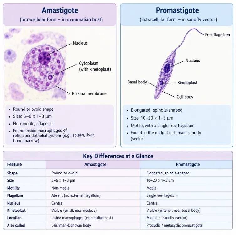
Figure 1: Morphological Forms of Leishmania: Intracellular Amastigotes in Mammalian Hosts and Extracellular, Flagellated Promastigotes in the Sandfly Vector.
Life Cycle of Leishmania
Leishmania has two major asexual forms: amastigotes in mammalian hosts and promastigotes in sandflies.
Life cycle: During feeding, infected sandflies transmit promastigotes to susceptible mammalian hosts. These forms are engulfed by phagocytic cells, transform into amastigotes, and multiply through binary fission within the host cells.[16]
Flowchart:
Infected female sandfly bite ↓ Promastigotes injected into mammalian skin ↓ Phagocytosis by macrophages ↓ Transformation into amastigotes (12–24 h) ↓ Amastigotes multiply by binary fission inside macrophages ↓ Macrophage ruptures → release of amastigotes ↓ Infection of new macrophages ↓ Another sandfly takes a blood meal and ingests amastigotes ↓ Amastigotes transform into flagellated promastigotes (24–48 h) ↓ Promastigotes multiply in the sandfly gut ↓ Migration to pharynx and buccal cavity ↓ Promastigotes transmitted to a new mammalian host during the next blood meal
Key conditions:
[4]
Source of infection and reservoir hosts
Visceral Leishmaniasis
Visceral leishmaniasis (VL) was recognized by the World Health Organization (WHO) as a neglected tropical disease (NTD) in its 2007 report and received renewed attention in 2015 due to limited public awareness, its high mortality burden, and its prevalence in impoverished endemic regions, with more than 20,000 deaths reported in 2015. [3]
VL contributes substantially to the global disease burden, with approximately 2.4 million disability-adjusted life years (DALYs) reported annually.
VL also produces a considerable economic burden because of treatment costs, hospitalization, monitoring, and loss of productivity.[7]
In India, approximately 165.4 million people are estimated to be at risk of visceral leishmaniasis (kala-azar).
The disease is endemic in four states: Bihar (33 districts), Jharkhand (4 districts), West Bengal (11 districts), and Uttar Pradesh (6 districts).[17]
Visceral leishmaniasis (VL), also called kala-azar, is the most severe form of leishmaniasis and can become fatal if appropriate treatment is not provided.
- Causative species: Leishmania donovani and Leishmania infantum are the major species associated with VL. L. donovani is also associated with post-kala-azar dermal leishmaniasis (PKDL).[5]
- Potential complications: Untreated VL may lead to life-threatening complications, including bleeding, secondary infections, and liver dysfunction.
- Economic burden: VL can impose substantial financial pressure on affected families because treatment may require hospitalization and prolonged medication, alongside income loss during illness.
- Impact in India: The economic burden of the disease is especially high in endemic areas, where limited access to low-cost healthcare can make diagnosis and treatment difficult to afford.
Treatment expenses may force vulnerable households to borrow money or use a substantial proportion of their income for medical care.
- Public health significance: Inadequate diagnosis, underreporting, and limited access to treatment can contribute to an underestimation of the overall burden of VL.[5]
Types of visceral leishmaniasis (VL)
Visceral leishmaniasis (VL) is classified into two types:
Treatment of Visceral Leishmaniasis (VL)
The management of visceral leishmaniasis (VL) remains challenging because of the limited availability of effective drugs and the increasing development of drug resistance.
Chemotherapy is the principal treatment approach for VL.
The major drugs used include pentavalent antimonials, paromomycin, miltefosine, and amphotericin B.[15]
Pentavalent antimonial drugs, including sodium stibogluconate and meglumine antimoniate, were historically used as first-line treatments for visceral leishmaniasis. However, the emergence of drug resistance, particularly in India, has limited their therapeutic use.[19]
their use is limited by the prolonged treatment duration, the need for injections, and the possibility of serious adverse effects.
Reported adverse effects include:
Miltefosine is another approved treatment, while paromomycin and pentamidine also remain among the drugs used against leishmaniasis.[19]
Miltefosine (MF) is an alkylphospholipid compound initially developed for its potential anticancer activity, particularly against breast cancer.
It is the oral medication for VL
Miltefosine remains in the body for an extended period due to its long elimination half-life. When combined with inadequate treatment practices, this may increase the risk of resistance development in Leishmania parasites.
It is teratogenic and therefore cannot be used during pregnancy.
Its major adverse effects include diarrhoea, vomiting, and dehydration[18]
Paromomycin (PM) is an aminoglycoside antibiotic that acts by inhibiting protein synthesis in the parasite.
It is administered parenterally, commonly through intramuscular injection.
Its relatively low cost and effectiveness make it a useful therapeutic option for VL.
Its principal limitations are the prolonged injection-based treatment course and the risk of nephrotoxicity and ototoxicity, which can affect kidney function and hearing, respectively.[18]
Amphotericin B
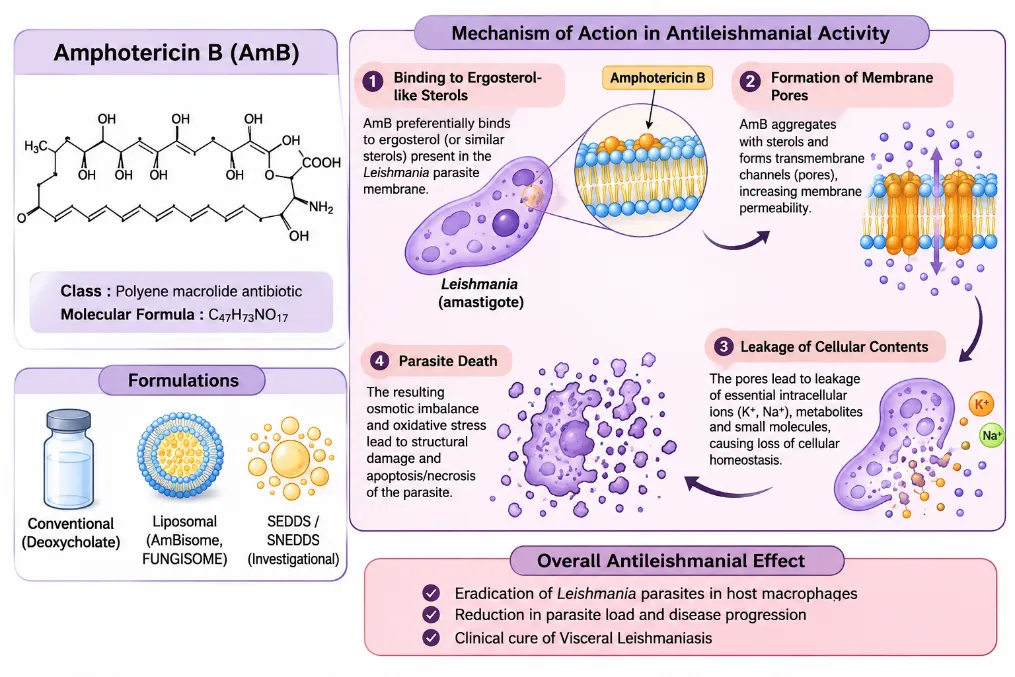
Figure 2: Chemical Structure, Major Pharmaceutical Formulations and Proposed Mechanism of Antileishmanial Action of Amphotericin B Through Interaction with Parasite Membrane Sterols and Disruption of Membrane Integrity.
Conventional Amphotericin B in VL
Intravenous administration requires trained healthcare personnel, appropriate supplies, and controlled storage and transportation.[22]
Nephrotoxicity is a major dose-limiting adverse effect associated with conventional Amphotericin B deoxycholate.[8]
Conventional Amphotericin B is an effective treatment option; however, its administration requires repeated intravenous infusions, and treatment may be associated with tolerability problems.[8/10]
Case Study: Reduced Response to Conventional Amphotericin B
Despite its therapeutic effectiveness, conventional amphotericin B has an important limitation: its toxic adverse effects. These concerns encouraged the development and use of liposomal formulations to improve its safety profile.[12]
. Lipid-based formulations of Amphotericin B have therefore attracted interest because they can provide effective treatment through shorter dosing regimens.[8]
Liposomal Amphotericin B generally produces fewer infusion-related toxic effects than conventional Amphotericin B and some other lipid formulations.[8]
. Development of Lipid-Based Amphotericin B
Liposomal encapsulation represents an important formulation approach for the clinical use of amphotericin B in visceral leishmaniasis. The study describes liposomal amphotericin B as an established treatment option for VL and reports its use in single-dose and short-course treatment regimens. Liposomal formulations allow amphotericin B to be administered intravenously while maintaining high antileishmanial efficacy.[6]
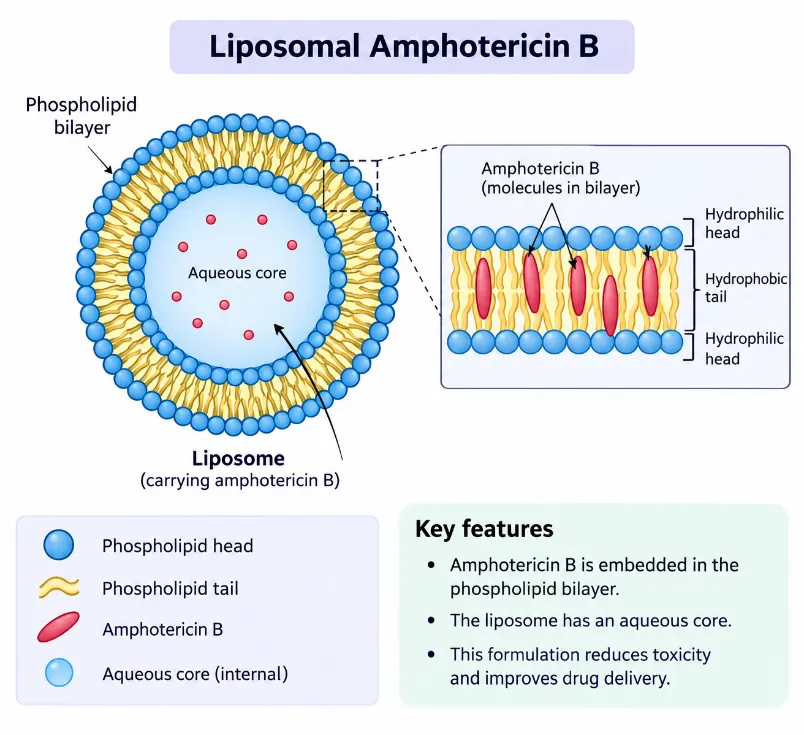
Figure 3: Schematic Representation of Liposomal Amphotericin B Showing the Phospholipid Bilayer, Aqueous Core and Incorporation of Amphotericin B within the Liposomal Structure.
Liposomes are spherical vesicles consisting of a lipid bilayer surrounding an aqueous core.
AmBisome contains hydrogenated soy phosphatidylcholine, distearoylphosphatidylglycerol and cholesterol.
These components help maintain the drug within the liposomal structure and influence its stability and distribution in the body.
The formulation was designed for parenteral administration, particularly intravenous delivery.[21]
It has demonstrated excellent efficacy in the treatment of VL[7]
Liposomal amphotericin B: AmBisome
Liposomal amphotericin B (LAmB) was developed to improve drug tolerability while retaining the activity of amphotericin B.
Encapsulation within liposomes changes the drug’s distribution and pharmacokinetic behaviour.[21]
Oral administration is an attractive alternative because it could reduce dependence on injectable treatment, hospital-based administration and costly storage arrangements[13]
Indian Liposomal Amphotericin B Formulation: Fungisome™
Its development was supported by Indian government research initiatives, including the Department of Biotechnology and the programme supporting technological self-reliance. The formulation was developed as an alternative to internationally marketed liposomal amphotericin B products.[24]
Fungisome™ is an Indian liposomal preparation of amphotericin B that has been used for the treatment of visceral leishmaniasis. According to the study, Fungisome differs chemically from AmBisome in its formulation. Amphotericin B in Fungisome is encapsulated in liposomes composed of phosphatidylcholine and cholesterol and suspended in physiological saline. The formulation is administered by intravenous infusion and requires sonication before administration to convert multilamellar vesicles into smaller unilamellar vesicles.[6]
Safety Profile and Adverse Effects of Amphotericin B Formulations
Table 1: Comparative Safety Profile and Adverse Effects of Conventional Amphotericin B Deoxycholate and Liposomal Amphotericin B.
| Adverse effect / safety parameter | Conventional Amphotericin B (Deoxycholate) | Liposomal Amphotericin B (L-AmB / AmBisome) |
|---|---|---|
| Nephrotoxicity | High risk of nephrotoxicity due to damage to renal cell membranes | Lower nephrotoxicity compared with the conventional formulation because liposomal encapsulation reduces interaction with human cell membranes. |
| Fever and chills | Frequently observed during drug infusion. | May occur as infusion-related reactions. |
| Nausea | Commonly reported during infusion. | May occur during infusion. |
| Vomiting | - | Reported as an infusion-related adverse effect. |
| Hypertension | May occur during infusion. | May occur during infusion |
| Tachycardia | - | Reported as an infusion-related adverse effect. |
| Electrolyte disturbances | Hypokalaemia and hypomagnesemia are identified among the toxic effects associated with Amphotericin B. | Formulation-specific comparative data are not provided in the source. |
| Renal tubular damage | Associated with renal toxicity and injury to tubular cells. | Lower toxicity is attributed to reduced interaction with human cell membranes. |
Future Scope of Antileishmanial Drugs for Visceral Leishmaniasis
The development of new therapeutic approaches for visceral leishmaniasis (VL) remains important because currently available treatments are associated with limitations such as drug toxicity, high treatment costs, prolonged administration and the emergence of drug resistance. Future research should focus on identifying safer, more effective and affordable treatment options, particularly for populations in endemic regions.
Combination therapies: Improve treatment outcomes and reduce drug resistance.
New drug candidates: Discover effective compounds with fewer adverse effects.
Natural products: Explore plant-derived compounds for antileishmanial activity.
Novel drug targets: Investigate parasite-specific targets such as trypanothione metabolism and protein kinases.
Drug resistance: Understand and monitor resistance to existing drugs, including Amphotericin B.
Oral treatments: Develop safer, affordable and better-tolerated oral therapies.
Clinical evaluation: Establish the safety and efficacy of promising treatments through further studies.[25]
Need for oral formulations?
The need for parenteral administration is a major limitation because it makes treatment less convenient and can complicate patient management.
Although lipid-based formulations, including liposomal amphotericin B, have been developed to reduce toxicity, the article identifies the need for further improvements in drug delivery.
These limitations support investigating alternative delivery systems that may improve drug absorption, therapeutic performance and administration convenience.
AmB is difficult to formulate for oral use because of its poor water and lipid solubility and low gastrointestinal absorption.
An effective oral formulation could simplify administration and help overcome practical limitations associated with injectable treatment.[22]
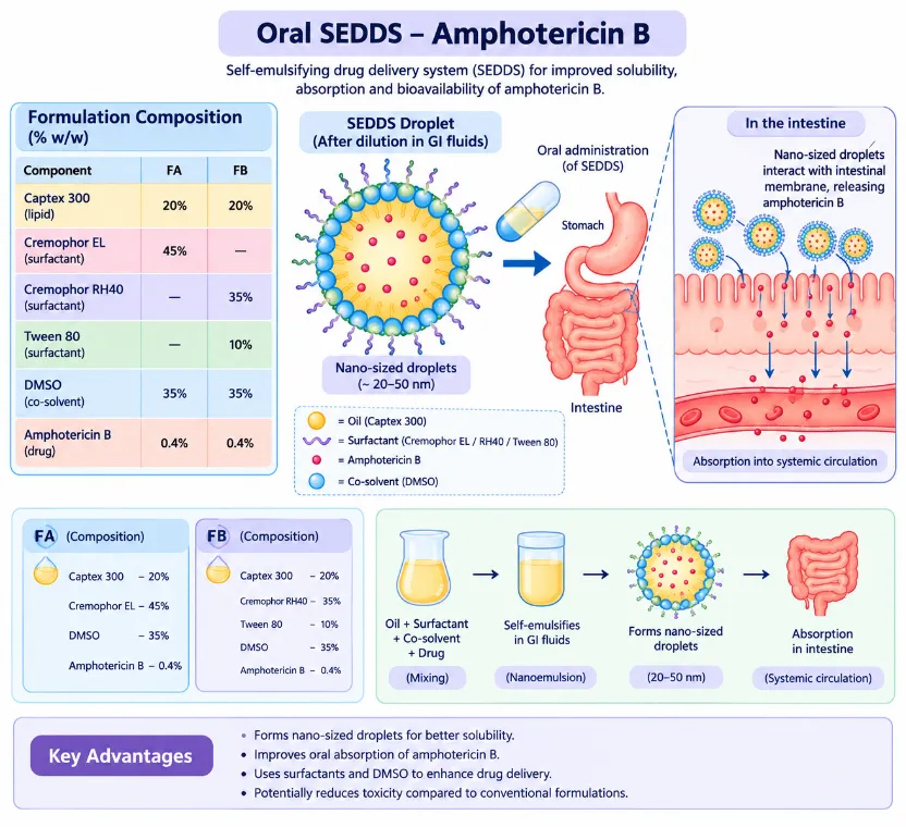
Figure 4: Schematic Representation of an Oral Self-Emulsifying Drug Delivery System (SEDDS) Containing Amphotericin B, Illustrating Formulation Composition, Spontaneous Emulsification, Droplet Formation and the Proposed Intestinal Absorption Pathway.
Why oral Amphotericin B is difficult to develop
Aggregation: AmB can exist in different aggregation states, which influence its behavior and safety profile. The article describes the monomeric state as more selective for ergosterol, while dimeric forms are associated with greater toxicity
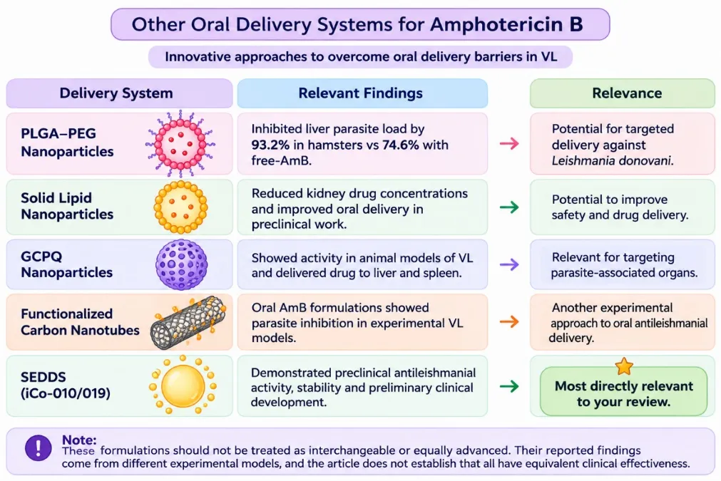
Figure 5: Overview of Emerging Oral Delivery Approaches Investigated for Amphotericin B, Including PLGA–PEG Nanoparticles, Solid Lipid Nanoparticles, GCPQ Nanoparticles, Functionalized Carbon Nanotubes and Self-Emulsifying Drug Delivery Systems.
[15]
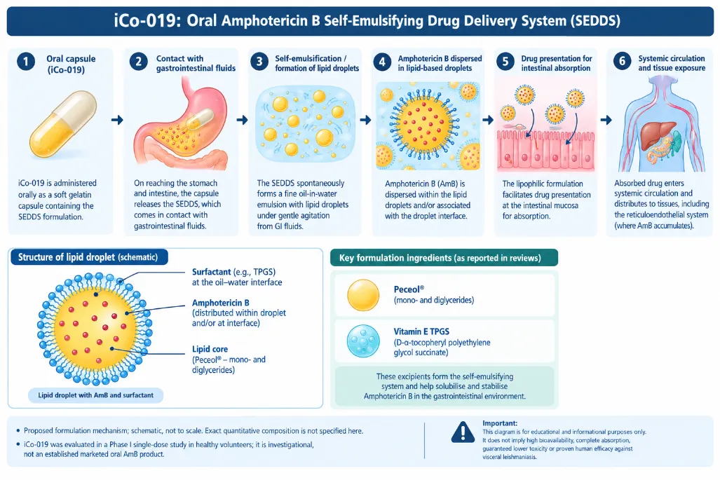
Figure 6: Schematic Representation of the Oral iCo-019 Amphotericin B Formulation, Illustrating Capsule Administration, Self-Emulsification, Gastrointestinal Dispersion and the Proposed Absorption and Systemic Distribution Processes.
Wasan et al. (2010) investigated a tropically stable oral lipid-based formulation of amphotericin B, designated iCo-010, as a potential approach to treating visceral leishmaniasis. The formulation contained mono- and diglycerides with vitamin E TPGS and was evaluated for drug stability, self-emulsification behaviour and antileishmanial activity.
The researchers examined its stability in different temperature conditions, its behaviour in simulated intestinal fluid, and its activity in mice infected with Leishmania donovani.[23]
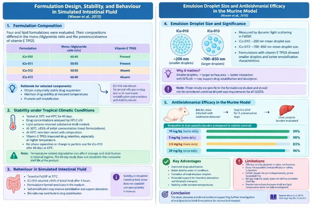
Figure 7: Formulation Characteristics, Stability Evaluation, Self-Emulsification Behaviour and Preclinical Antileishmanial Efficacy of the Oral Amphotericin B Formulation iCo-010 in a Murine Model of Visceral Leishmaniasis.[23]
The study demonstrated that the oral lipid-based formulation iCo-010 showed good temperature stability, self-emulsification properties, and substantial reduction in liver parasite burden in Leishmania donovani-infected mice. These promising preclinical findings support further investigation of oral amphotericin B delivery, but human safety and clinical efficacy remain to be established.
CONCLUSION
Amphotericin B remains an important therapeutic agent in the treatment of visceral leishmaniasis. However, the limitations associated with conventional Amphotericin B, particularly nephrotoxicity, infusion-related adverse effects, and the requirement for intravenous administration, highlight the need to explore alternative drug delivery approaches. Liposomal Amphotericin B has addressed some of these concerns by modifying drug distribution and improving tolerability, although its cost, parenteral administration, and storage requirements continue to present practical challenges.
In this context, an important question arises: Could oral Self-Emulsifying Drug Delivery Systems (SEDDS) offer a feasible alternative for delivering Amphotericin B? The ability of SEDDS to disperse poorly water-soluble drugs provides a rationale for investigating this approach. However, the poor gastrointestinal absorption, complex physicochemical properties, and potential toxicity of Amphotericin B make its oral delivery a challenging area of formulation research.
Further questions remain regarding whether an oral SEDDS formulation can achieve adequate drug absorption, maintain antileishmanial activity, provide a consistent safety profile, and deliver sufficient drug concentrations to the organs affected by visceral leishmaniasis. It is also necessary to determine whether any potential benefits would be maintained during long-term storage and under practical conditions of use. From a pharmacovigilance perspective, the identification, assessment, and monitoring of adverse drug reactions would remain essential. In parallel, the regulatory requirements for quality, safety, efficacy, and clinical evaluation would need to be addressed before such a formulation could be considered for clinical use in India.
Therefore, the key consideration is not whether oral SEDDS is already superior to conventional or liposomal Amphotericin B, but whether it can be developed into a reliable, safe, effective, and practically accessible delivery system. Addressing these questions through systematic formulation development, preclinical studies, and appropriately designed clinical investigations would help establish its potential role in the future management of visceral leishmaniasis.
REFERENCES


Prachi Salunkhe, Dnyaneshwar S. Vyavhare, Mahesh Sherkar, Amphotericin B in Visceral Leishmaniasis: From Conventional and Liposomal Formulations to SEDDS-Based Oral Delivery — Challenges and Future Prospects, Int. J. of Pharm. Sci., 2026, Vol 4, Issue 11, 3109-3124. https://doi.org/10.5281/zenodo.24360236
 10.5281/zenodo.24360236