

We use cookies to ensure our website works properly and to personalise your experience. Cookies policy
Department of Pharmacology, Sarojini Naidu Vanita Pharmacy Maha Vidyalaya (Co-Ed.), Tarnaka, Hyderabad, Telangana, India-500017.
Urolithiasis is a common urinary disorder characterized by the formation of stones in the urinary tract, particularly calcium oxalate stones. It is associated with urinary supersaturation, crystal nucleation, growth, aggregation, oxidative stress, and renal tissue injury. Although several treatment approaches are available, recurrence and adverse effects associated with conventional therapies have increased interest in exploring natural compounds with potential anti-lithiatic properties. Usnic acid, a naturally occurring dibenzofuran derivative obtained from lichens, has demonstrated various pharmacological activities, including antioxidant, anti-inflammatory, and antimicrobial effects. These properties provide a scientific basis for investigating its possible protective role in experimental urolithiasis. The present review focuses on the potential anti-lithiatic activity of usnic acid, its pharmacological properties, possible mechanisms of action, and its relevance in calcium oxalate stone formation. Particular attention is given to the inhibition of crystal formation and deposition, reduction of oxidative stress, and protection against renal epithelial damage. Male Wistar rats with urolithiasis induced by ethylene glycol (0.75% v/v) and ammonium chloride (1% w/v) can be used for experimental evaluation. Urinary, serum biochemical, and histopathological parameters can be assessed to determine its protective effects. However, further investigations are necessary to establish its efficacy, safety, and therapeutic potential
Urolithiasis, also known as urinary stone disease or nephrolithiasis (when stones are located in the kidneys), is a common urological disorder characterized by the formation of solid crystalline deposits within the urinary tract. These stones may develop in the kidneys and subsequently migrate into the ureters, urinary bladder, and urethra, causing varying degrees of discomfort and urinary complications. The clinical manifestations of urolithiasis depend on the size, composition, and location of the stones. Common symptoms include severe renal colic, hematuria (blood in urine), dysuria (painful urination), frequent urination, urinary obstruction, and, in severe cases, deterioration of renal function [1,2]. The prevalence of urolithiasis varies considerably across different geographical regions and is influenced by several factors, including age, sex, dietary habits, climatic conditions, lifestyle, genetic predisposition, and environmental factors [3]. Dehydration, inadequate fluid intake, excessive consumption of salt and animal proteins, and metabolic abnormalities are important risk factors associated with kidney stone formation. Among the different types of urinary stones, calcium oxalate stones are the most commonly occurring type, accounting for approximately 70–80% of all renal stones [4]. These stones are primarily formed due to increased concentrations of calcium and oxalate in urine, resulting in supersaturation and subsequent crystal formation.
The pathogenesis of urolithiasis is a complex process involving urinary supersaturation, crystal nucleation, crystal growth, aggregation, and retention within the renal tubules. Under normal physiological conditions, several urinary inhibitors, such as citrate, magnesium, and certain proteins, prevent excessive crystal formation and aggregation. However, disturbances in these protective mechanisms may promote the development of kidney stones. Various factors, including reduced urine volume, hyperoxaluria, hypercalciuria, urinary tract infections, metabolic disorders, and oxidative stress, contribute to the development of urolithiasis [5]. The deposition and accumulation of calcium oxalate crystals in renal tissues can cause mechanical injury to renal epithelial cells, inflammation, and abnormalities in normal kidney function. Ethylene glycol-induced urolithiasis is one of the most widely used experimental animal models for evaluating the antiurolithiatic activity of potential therapeutic agents. Ethylene glycol is metabolized in the body through several metabolic pathways, ultimately producing oxalic acid. The increased oxalate production causes hyperoxaluria and encourages the kidney to form and deposit calcium oxalate crystals. These crystals accumulate within renal tubules and produce pathological changes similar to those observed in human calcium oxalate stone disease [6]. Therefore, this experimental model is extensively used in pharmacological investigations to evaluate the preventive and therapeutic effects of different natural and synthetic compounds against kidney stone formation.
Oxidative stress and inflammation also play important roles in the development and progression of urolithiasis. Increased oxalate concentrations and calcium oxalate crystal deposition stimulate the production of reactive oxygen species (ROS) in renal epithelial cells. Excessive ROS production can cause lipid peroxidation, cellular membrane damage, and impairment of antioxidant defence mechanisms. Additionally, oxidative stress causes renal tissue damage by triggering inflammatory pathways and encouraging the release of pro-inflammatory mediators. resulting in renal tissue injury. Damaged epithelial cells provide suitable surfaces for crystal adhesion and retention, thereby accelerating calcium oxalate crystal accumulation and stone formation [7,8].
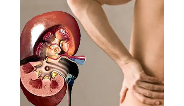
Figure 1: Kidney Stone
1.1 Epidemiology &Risk factors of Urolithiasis
The development of urinary stones is influenced by multiple interacting factors including Diet, dehydration, genetic predisposition, climate, obesity, and metabolic changes are risk factors for kidney stone development. These variables differ depending on socioeconomic status and geographic location, which explains why urolithiasis prevalence varies globally [2,3]. However, epidemiological trends indicate a global rise, particularly in regions with warm climates and notable dietary shifts. Dietary factors also affect the risk of developing stones. Inadequate protective dietary factors combined with high sodium and animal protein intake can change the levels of calcium oxalate, citrate, and uric acid in the urine. Kidney stones are known to be a risk factor for various conditions, including diabetes, heart disease, bone fractures, and kidney disease. [9-10]. However, kidney stones also raise the chance of getting these conditions. Metabolic disorders like hypercalciuria, hyperoxaluria, hypocitraturia, and hyperuricosuria have been linked to urinary calcium stones.[11].
1.2 Types and composition of urinary stones
Urinary calculi (stones) are primarily categorized based on their mineral or chemical composition. The most clinically significant varieties include calcium oxalate, calcium phosphate, uric acid, struvite, and cystine stones. Stones with mixed compositions are also frequently encountered in practice.
1.2.1 Calcium oxalate stones
The most prevalent component of urinary stones is calcium oxalate. It occurs mainly in two crystalline forms
1.Calcium oxalate monohydrate
CaC₂O₄·H₂O
2.Calcium oxalate dihydrate
CaC₂O₄·2H₂O
In particular, hypercalciuria, hyperoxaluria, and hypocitraturia are linked to calcium oxalate stones [12].
1.2.2 Calcium phosphate stones
Calcium phosphate stones represent a significant category of calcium-based urinary calculi. They mainly consist of minerals such as carbonate apatite, hydroxyapatite, and brushite. Their formation is promoted by alkaline urinary conditions and may be associated with hypercalciuria, reduced urinary citrate levels, and conditions such as distal renal tubular acidosis.[13].
1.2.3 Uric acid stones
Uric acid stones mainly consist of uric acid and commonly develop in persistently acidic urine. A low urinary pH reduces uric acid solubility, favoring crystal formation, while increased urinary uric acid excretion may further promote stone development. They can occur either as pure stones or together with other stone components[14].
1.2.4 Struvite stones
Struvite stones, commonly referred to as infection-related stones, mainly consist of magnesium ammonium phosphate hexahydrate (MgNH₄PO₄·6H₂O). They are typically associated with urinary tract infections caused by urease-producing bacteria. Urease breaks down urea, increasing urinary ammonia and raising urine pH, there by promoting the formation of struvite and carbonate apatite crystals. Struvite can occur as a pure stone but is also frequently found together with calcium oxalate and other mineral components in mixed calculi.[14].
1.2.5 Cystine stones
Cystine stones are rare metabolic calculi made up of cystine, a sulfur-containing amino acid with poor solubility. They arise mainly in people with cystinuria, a hereditary condition marked by defective renal tubular reabsorption of cystine and other dibasic amino acids. Because cystine dissolves poorly in urine—especially when the urinary pH is low—elevated cystine levels can lead to crystal precipitation and stone formation. These stones account for only a small fraction of urinary calculi. For instance, a large German stone-analysis study found that cystine stones represented about 0.4% of all s stones examined.[13].
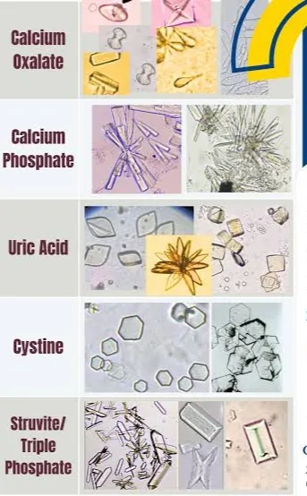
Figure 2: Types of Stones
1.3 Calcium oxalate urolithiasis
Calcium oxalate (CaOx) primarily in its monohydrate (COM) and dihydrate (COD) forms serves as the primary constituent of renal calculi. The pathogenesis of CaOx nephrolithiasis is a multi-step cascade comprising crystal nucleation, growth, aggregation, and attachment to renal tubular cells [14]. Reactive oxygen species (ROS) significantly mediate this stone formation process [15]. Uncontrolled oxidative stress triggers free radical propagation, damaging essential cellular components including proteins, lipids, and nucleic acids . Furthermore, elevated urinary oxalate levels induce oxidative stress within renal epithelial cells, driving an surge in intracellular ROS [16]. Renal tubular epithelial cell damage enhances crystal adhesion, creating a surface that facilitates calcium oxalate retention and accelerates the transition from crystalluria to kidney stone formation [8]. Recent experimental findings further show that inhibiting calcium oxalate crystal attachment to these epithelial cells prevents crystal retention and halts stone progression [9].
1.4 Pathogenesis of urolithiasis
The pathogenesis of kidney stones is a multi-step process driven by urinary supersaturation, crystal nucleation, expansion, clumping, and adherence, alongside damage to the renal epithelium. Calcium oxalate forms the majority of these stones, though calcium phosphate, uric acid, struvite, and cystine variants are also observed [5]. Key drivers of stone development include metabolic dysregulation, dietary habits, urinary pH and volume, genetic risk factors, and shifts in the balance between endogenous crystallization promoters and inhibitors. [9]
1.4.1 Urinary supersaturation
Urinary supersaturation serves as a critical trigger in kidney stone pathogenesis. It arises when levels of stone-forming solutes—including calcium, oxalate, phosphate, and uric acid surpass their solubility threshold in urine. [17].Factors such as low urine volume, elevated excretion of calcium, oxalate, or uric acid, and altered urinary pH elevate supersaturation levels, thereby encouraging crystal formation.[18].Thermodynamics dictates that this supersaturated state drives dissolved minerals to precipitate and evolve into urinary crystals.
1.4.2 Crystal growth
Crystal growth occurs as existing crystals enlarge through the steady accumulation of calcium, phosphate, oxalate, and other dissolved ions found in supersaturated urine. This growth rate is determined by factors such as the level of urinary supersaturation, pH balance, ionic strength, and the influence of substances that promote or inhibit crystallization. [18].Higher levels of calcium and oxalate in the urine accelerate calcium oxalate crystal development, leading to larger structures that are more likely to become trapped in the kidney tubules.[19].
1.4.3 Crystal aggregation
Crystal aggregation is the process in which tiny individual crystals join together to form larger clusters. As these aggregates grow, they become less likely to pass smoothly through the renal tubules. Urinary macromolecules such as proteins and other organic compounds can either encourage or inhibit aggregation depending on their characteristics and the urinary environment. Increased aggregation contributes significantly to the development of urolithiasis and encourages crystal retention in the kidneys. [20].
1.4.4 Crystalline adhesion and retention
The attachment and trapping of crystals within the kidney are critical milestones in kidney stone formation. While small crystals are usually flushed out in urine under normal conditions, factors like kidney cell damage, altered cell surface properties, and reduced urine flow can cause crystals to stick to the tubular lining. Specifically, calcium oxalate crystals can adhere to injured epithelial cells, preventing their normal excretion. When these crystals remain in the tubules over an extended period, they have enough time to grow larger and clump together, driving stone development.
1.4.5 Renal epithelial injury, oxidative stress and inflammation
Renal epithelial injury plays a central role in the development of calcium oxalate urolithiasis. Elevated urinary oxalate levels and calcium oxalate crystal deposits trigger the production of reactive oxygen species (ROS), which drive oxidative stress, lipid peroxidation, mitochondrial dysfunction, and damage to renal tubular epithelial cells. This cellular injury enhances crystal adhesion and retention within the kidney. Crystal deposition and epithelial damage also stimulate inflammatory pathways, notably the NLRP3 inflammasome, leading to the release of pro-inflammatory cytokines such as interleukin‑1β (IL‑1β). Consequently, oxidative stress and inflammation worsen renal injury and further promote stone formation. In experimental models of urolithiasis, increased levels of malondialdehyde (MDA) and reduced concentrations of glutathione (GSH) are widely used as markers of oxidative stress.
1.4.6 Crystal retention and stone development
Crystal retention occurs when crystals persist within the renal tubules or attach to papillary surfaces, enabling continued deposition of stone-forming materials. As these crystals enlarge and cluster, they can develop into sizable calculi that obstruct tubular flow or urinary outflow. Retention is often accompanied by tubular blockage, epithelial damage, inflammation, and alterations in kidney function.The progression of stone development is influenced by several factors: the level of urinary supersaturation, the presence or absence of crystallization inhibitors, the efficiency of crystal clearance, and the extent of renal tissue injury.
1.5 Role of Oxidative stress and Inflammation in urolithiasis
1.5.1 Role of oxidative stress
The pathophysiology of calcium oxalate nephrolithiasis is thought to be significantly influenced by oxidative stress. Experimental animal studies have demonstrated that hyperoxaluria increases the production of reactive oxygen species (ROS), resulting in oxidative stress and injury to renal tubular epithelial cells. This process may involve mitochondrial dysfunction, endoplasmic reticulum stress, and activation of NADPH oxidase, an important source of ROS generation [22–24].
Under normal physiological conditions, ROS participate in intracellular signalling pathways and maintain cellular homeostasis. However, excessive ROS production can overwhelm the antioxidant defence system, resulting in oxidative stress, cellular damage, and apoptosis. NADPH oxidase contributes to ROS generation and may aggravate renal tissue injury associated with kidney stone formation [25,26]. Oxidative stress can also disrupt the integrity of renal tubular epithelial cells, creating favourable conditions for calcium oxalate crystal adhesion and retention.
Nuclear factor erythroid 2-related factor 2 (Nrf2) is an important transcription factor that regulates cellular antioxidant defence mechanisms. Activation of the Nrf2 signalling pathway promotes the expression of antioxidant and cytoprotective genes, helping to maintain redox balance and reduce oxidative damage [28]. By protecting renal epithelial cells from oxidative injury, Nrf2-mediated antioxidant responses may help reduce calcium oxalate crystal adhesion and retention, potentially limiting kidney stone formation [29].
1.5.2 Role of Inflammation
Inflammation plays a significant role in the development and progression of urolithiasis. Both local inflammatory responses and tissue injury contribute to the formation of calcium-containing kidney stones, as demonstrated by studies linking inflammatory processes with calcium stone development [30]. During urolithiasis, calcium oxalate crystals can interact with and adhere to renal tubular epithelial cells, causing cellular injury and promoting crystal aggregation and retention within the kidneys [31, 32].
The adhesion and accumulation of calcium oxalate crystals can stimulate renal epithelial cells to release pro-inflammatory cytokines and other inflammatory mediators [33]. These inflammatory responses may further damage the renal tubular epithelium, alter cellular surface properties, and create an environment that favours crystal retention and the growth of larger crystal aggregates. Consequently, persistent inflammation may contribute to the progression of calcium oxalate nephrolithiasis.
1.5.3 Interrelationship Between Oxidative Stress and Inflammation
Oxidative stress and inflammation are closely interconnected processes in the pathogenesis of urolithiasis. Excessive ROS production can activate inflammatory signalling pathways and promote the release of pro-inflammatory mediators. Conversely, inflammatory responses may increase ROS generation, further intensifying oxidative stress and renal tissue damage. This bidirectional interaction can establish a cycle of oxidative injury, inflammation, crystal adhesion, and crystal retention, ultimately contributing to calcium oxalate kidney stone formation.
Therefore, targeting oxidative stress and inflammation may represent a potential therapeutic strategy for preventing renal epithelial injury and reducing calcium oxalate crystal deposition in experimental urolithiasis
1.6 Experimental models of urolithiasis
1.6.1 Sodium oxalate-induced urolithiasis
Sodium oxalate-induced urolithiasis occurs when there is an excess of oxalate in the body, leading to the formation of urinary stones [34]. Sodium oxalate is a salt form of oxalate, and its ingestion can contribute to the development of kidney stones . The mechanism involves several steps: after ingestion, sodium oxalate is absorbed in the gastrointestinal tract and enters the bloodstream [35]. The kidneys then filter the blood, removing waste products and excess substances, including oxalate, which is excreted in the urine [36].
1.6.2 glycolic acid induced urolithiasis
The development of uroliths, or urinary stones, due to exposure to glycolic acid is known as glycolic acid-induced urolithiasis [37]. This condition is linked to several metabolic disorders, such as primary hyperoxaluria type 1 (PH1), where excessive oxalate production leads to its accumulation and the formation of oxalate-based kidney stones. Glycolic acid contributes to urolithiasis through its metabolism in the liver [38].
1.6.3 Ethylene glycol-induced urolithiasis
Ethylene glycol-induced urolithiasis is associated with the toxic effects of ethylene glycol, a chemical commonly found in antifreeze products. Although ethylene glycol itself does not directly cause urinary stone formation, its metabolic breakdown products contribute to the development of urinary calculi. [39].
1.6.4 Hydroxy-L-proline (HLP)urolithiasis
Hydroxy-L-proline (HLP) is an amino acid derivative that may contribute to the development of urinary stones under certain conditions [40]. It is produced during the metabolism of collagen, a structural protein present in connective tissues, and can be excreted through urine [41]. The potential role of HLP in urolithiasis is associated with its metabolic effects and interactions with urinary calcium ions, which may influence the formation and deposition of urinary crystals.
1.6.5 calcium oxalate-induced urolithiasis.
The most prevalent kind of kidney stones is caused by calcium oxalate-induced urolithiasis. A series of physiological and biochemical processes lead to the formation and growth of calcium oxalate crystals in the urinary tract. When calcium and oxalate levels in the urine are elevated, the urine becomes supersaturated with these ions [42].
1.6.6 Ethylene Glycol and Ammonium Chloride-Induced Urolithiasis
Ethylene glycol-induced urolithiasis is primarily associated with increased oxalate production, which contributes to oxidative damage and urinary stone formation. Ethylene glycol is metabolized in the liver by alcohol dehydrogenase (ADH) and aldehyde dehydrogenase (ALDH), resulting in the formation of oxalate [43,44]. Since oxalate is not significantly metabolized in animals, it is primarily eliminated through the kidneys [45]. Elevated oxalate levels promote the formation and deposition of calcium oxalate crystals in the renal tubules, leading to kidney injury, oxidative stress, and nephrotoxicity. Recent research has also emphasized the important role of oxidative stress in the progression of ethylene glycol-induced renal damage and urolithiasis.
1.6.7 Sodium Oxalate (NaOx)-Induced Urolithiasis in Rats
Sodium oxalate (NaOx) is used to induce experimental urolithiasis in rats by promoting the rapid formation of renal crystals following intraperitoneal administration. These crystals primarily develop within the lumen of the proximal tubules in the renal cortex [46]. The size, number, and distribution of the crystals may vary depending on the administered dose and the interval between injections [47]. For experimental purposes, sodium oxalate can be dissolved in a suitable vehicle, such as saline solution, to prepare the required concentration for inducing urolithiasis in rats [48].
1.6.8 zinc disc-induced urolithiasis
zinc disc-induced urolithiasis develop bladder stones due to the implantation of zinc disks into the bladder or urinary tract. In the zinc disk implantation model, zinc disks are surgically inserted into the bladders of rats, which are known to weigh and measure [49]. The rats were sedated with sodium pentobarbitone (40 mg/kg, i.p.) [49,50]. A suprapubic incision was made to expose the bladder, followed by a small cut on the bladder's upper surface. The urine was then aseptically aspirated into a sterile container for pH testing and bacteriological analysis.
1.7 cystone
Cystone is a proprietary Ayurvedic polyherbal formulation commonly used as a reference standard in experimental studies of urolithiasis. It is used in the management of urinary calculi and is investigated for its antiurolithic and lithotriptic properties. Cystone is reported to possess diuretic, antioxidant, anti-inflammatory, antimicrobial and anti-adherent activities, which may contribute to its protective effects against kidney stone formation[12]. In experimental animal studies, Cystone is commonly administered orally at a dose of 750 mg/kg body weight as a standard reference drug. In ethylene glycol-induced urolithiasis models, it has been reported to reduce urinary calcium and oxalate excretion, inhibit calcium oxalate crystal deposition and improve renal antioxidant status. It may also help maintain urinary volume and protect renal tissues against oxidative damage.
Mechanism of action
The pharmacological actions of Cystone include:
Antiurolithic activity: Helps reduce the formation and deposition of urinary crystals.
Diuretic activity: Promotes urine flow, which may facilitate the elimination of small crystals.
Antioxidant activity: Helps protect renal tissues against oxidative stress and lipid peroxidation.
Anti-inflammatory activity: May reduce inflammation associated with renal crystal deposition.
Anti-adherent activity: May reduce the attachment of crystals to renal tubular epithelial cells.
Lithotriptic activity: Is traditionally described as helping in the disintegration or passage of urinary calculi.
1.8 Usnic Acid
Usnic acid (UA) is one of the most abundant and characteristic secondary metabolites found in lichens. It is a naturally occurring dibenzofuran derivative that has been identified in several lichen species, particularly those belonging to the genus Usnea, including Usnea diffracta and Usnea longissima.
Over the past three decades, numerous studies have investigated the antimicrobial properties of usnic acid, demonstrating its effectiveness against various bacterial strains. As reported in previous studies [51,52], usnic acid has shown considerable antibacterial activity, particularly against Gram-positive bacteria, including different strains of Staphylococcus and Enterococcus. Furthermore, studies have evaluated its activity against both human and bovine strains of Mycobacterium tuberculosis [53]. The antiviral potential of usnic acid has also been investigated, with studies reporting activity against the Epstein–Barr virus [54]. In addition, usnic acid has demonstrated antifungal activity when used in combination with undecylenic acid for the treatment of tinea pedis (athlete’s foot).
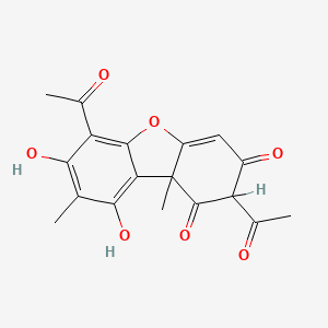
Figure 3: Structure of Usnic Acid
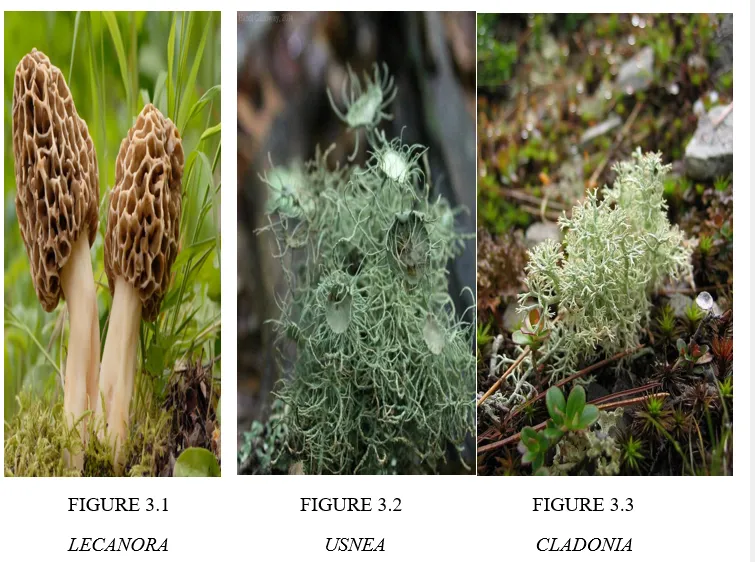
LECANORA USNEA CLADONIA
1.9 Pharmacological properties of usnic acid
1.9.1 Anti-inflammatory
Inflammation is a protective response of the body to foreign substances, infections, or tissue injury. However, excessive or prolonged inflammation may lead to tissue damage and impairment of normal physiological functions. Usnic acid has demonstrated potential anti-inflammatory activity, which may be associated with the inhibition of prostaglandin synthesis. Furthermore, studies have reported that different concentrations of usnic acid and its self-microemulsion formulation can reduce tumor necrosis factor-alpha (TNF-α) production induced by heat-killed Escherichia coli, indicating a concentration-dependent effect [53]. In addition, usnic acid has shown neuroprotective effects in an experimental model of Parkinson’s disease induced by 1-methyl-4-phenyl-1,2,3,6-tetrahydropyridine (MPTP), with improvements in motor dysfunction reported in the experimental [54].These findings suggest that usnic acid may possess anti-inflammatory and neuroprotective properties, although further studies are required to establish its mechanisms of action and therapeutic potential.
1.9.2 Antibacterial and Antimicrobial Activity
Usnic acid is one of the major biologically active compounds found in lichens of the genus Usnea and has been investigated for its antimicrobial properties. It has demonstrated inhibitory activity against several Gram-positive bacteria and certain mycobacterial species, including Mycobacterium tuberculosis. Antimicrobial effects have also been reported against organisms such as Mycobacterium neoaurum, Bacillus cereus, Staphylococcus aureus, Streptococcus pneumoniae, Propionibacterium acnes (currently known as Cutibacterium acnes), and Enterococcus species. Activity against Escherichia coli has also been reported in some experimental studies. The antimicrobial effects of usnic acid may vary depending on the microbial species, concentration, and experimental conditions. These findings indicate that usnic acid has potential antimicrobial properties; however, its clinical applicability requires further investigation.
1.9.3 Antitumor Activity
Usnic acid has been investigated for its potential antitumor activity through mechanisms involving the inhibition of cancer cell proliferation, induction of apoptosis, cell-cycle arrest, and suppression of tumor angiogenesis. Experimental studies suggest that its effects may vary according to the cancer cell type and the molecular signaling pathways involved. Usnic acid has been reported to affect cell-cycle progression in human gastric carcinoma BGC-823 cells, human lung carcinoma A549 cells, human breast cancer MCF-7 cells, and human prostate cancer LNCaP cells [55–57].These effects may be associated with reduced cell viability, altered cell-cycle progression, and the induction of programmed cell death. In particular, cell-cycle arrest at the G1 phase has been reported in certain experimental cancer models. However, usnic acid does not necessarily produce identical effects in all cancer cell lines, as its activity may depend on cell-specific mechanisms and signaling pathways. Therefore, further studies are needed to clarify its antitumor mechanisms, evaluate its safety, and determine its potential therapeutic applications.
1.9.4 Antioxidant
The antioxidant properties of usnic acid are particularly relevant to the present investigation because oxidative stress is implicated in calcium oxalate crystal-associated renal injury [58–59].Experimental investigations have demonstrated antioxidant activity associated with usnic acid or usnic-acid-containing lichen preparations. A 2023 study involving isolation and characterization of usnic acid from Usnea species reported antioxidant activity in experimental assays [60].
1.10 Toxicity and Safety of Usnic Acid
Usnic acid shows promising pharmacological activities, but its safety must be carefully evaluated because oral exposure specially through weight-loss supplements has been linked to liver injury. Reviews have highlighted hepatotoxicity, genotoxic potential, and other adverse effects, with mitochondrial dysfunction and oxidative stress considered major contributors to liver damage[61].
Safety considerations
In experimental studies, the dose should be selected cautiously to avoid exceeding levels associated with toxicity. Usnic acid has produced hepatocellular degeneration, inflammation, and elevated liver enzymes in animal studies, supporting the need for rigorous hepatic safety monitoring.
During the study period, animals should be regularly observed for:
2.Experimental Evaluation of Anti-Lithiatic Activity
The anti-urolithiatic effect of usnic acid may be assessed in male Wistar rats using an experimental model of urolithiasis induced by ethylene glycol and ammonium chloride.
2.1 Experimental Animals
A total of 30 healthy male rats weighing 200-250g were obtained from VAB Labs .Hyderabad and habituated for a week. For the duration of the study, the rats were accommodated in washed propylene (PP) cages with uncontaminated paddy shuck (as bedding) at room temperature (22°c), humidity (50%), and light intensity (325 lux) with a 12h dark-12h light cycle and were given quality feed and RO H2O ad libitum.
2.2 Induction of Urolithiasis
Experimental urolithiasis can be induced by administering ethylene glycol (0.75% v/v) and ammonium chloride (1% w/v) in drinking water. This combination promotes urinary oxalate elevation, calcium oxalate crystal formation, and renal tissue injury[60]
2.3 Administration of Test Drug
Usnic acid can be investigated at different dose levels to evaluate its possible anti-lithiatic activity. Treatment effects can be compared with an appropriate standard drug, such as Cystone
3.Parameters
3.1 Relative Body weight
Every week, the rats' relative body weight (RBW) was determined. Initial weight (IBW) and absolute weight (ABW) are two crucial factors in the computation of RBW. ABW is the rat's actual weight over a given time period, whereas IBW is the rat's weight at the start of the experiment[62]. The formula for calculating RBW is as follows:
RBW (g)=IBW (g)/ABW (g)×100
3.2 Urine analysis
On the 28th day of the study, individual metabolic cages were used to collect 24-hour urine samples from each experimental animal, with water provided throughout the collection period. The harvested samples were then examined under a 10X microscope to evaluate crystal morphology, followed by biochemical analyses to determine total urine volume, calcium, phosphate, magnesium, oxalate, and uric acid levels[63].
3.3 Serum Analysis
Blood samples were collected through cardiac puncture, and the samples were centrifuged at 10,000 rpm for 10 minutes using an REM R8C laboratory centrifuge to separate the serum. The obtained supernatant was then analyzed for serum calcium, serum phosphorus, and blood urea nitrogen (BUN) levels.[64].
3.4 Histopathological examination
Histopathological examination of kidney tissue using haematoxylin and eosin (H&E) staining can help evaluate renal tubular damage, inflammation, and calcium oxalate crystal deposition. Polarized-light microscopy may also be used to identify birefringent calcium oxalate crystals.
4.Need for Investigating Usnic Acid in Experimental Urolithiasis
Urolithiasis is characterized by recurrent renal stone formation and injury to kidney tissue. Despite the availability of various treatment options, there remains a need to identify agents capable of inhibiting crystal development and preserving renal structure and function.
Usnic acid, a naturally derived metabolite of lichens, has been reported to possess antioxidant and anti-inflammatory activities. These properties offer a plausible basis for examining whether it can counteract oxidative stress, inflammatory responses, and renal damage associated with experimentally induced kidney stones.
At present, direct experimental evidence supporting an anti-urolithiatic effect of usnic acid remains limited. Therefore, a systematic investigation using an ethylene glycol–ammonium chloride-induced urolithiasis model in Wistar rats would be valuable for assessing both its potential therapeutic efficacy and safety
CONCLUSION
Usnic acid, a naturally occurring secondary metabolite obtained from lichens, possesses several pharmacological properties, including antioxidant, anti-inflammatory, antibacterial, and antimicrobial activities. Its antioxidant and anti-inflammatory properties provide a scientific rationale for investigating its potential protective effects against calcium oxalate crystal-induced renal injury. By potentially reducing oxidative stress, inflammatory responses, and crystal deposition, usnic acid may offer a promising approach for further investigation in experimental urolithiasis.
The ethylene glycol and ammonium chloride-induced urolithiasis model in Wistar rats provides a suitable experimental approach for evaluating its possible anti-lithiatic activity. Assessment of urinary parameters, serum biochemical markers, and renal histopathological changes can help determine its effects on crystal formation and kidney function. Cystone can be used as a standard reference drug for comparison.However, the direct anti-urolithiatic efficacy of usnic acid has not yet been sufficiently established. Its potential hepatotoxicity and other safety concerns must also be carefully considered during experimental evaluation. Therefore, further mechanistic, pharmacological, dose-dependent, and toxicological studies are required to establish its safety and efficacy. Overall, usnic acid represents a promising lead compound for future anti-urolithiatic research, but additional experimental evidence is essential before considering its therapeutic application.
REFERENCES



Nomula Pavani, Syeda Sumaiya, Madathala Sreekanth, Evaluation of the Anti-Lithiatic Activity of Usnic Acid in Experimental Urolithiasis, Int. J. of Pharm. Sci., 2026, Vol 4, Issue 11, 3051-3065. https://doi.org/10.5281/zenodo.24334639
 10.5281/zenodo.24334639