

We use cookies to ensure our website works properly and to personalise your experience. Cookies policy
1Department Of Pharmaceutical-Microbiology, School Of Pharmacy
2Student Of B.Pharmacy, School Of Pharmacy
3Department Of Pharmaceutics, School Of Pharmacy
4NRI Institute Of Technology University, Pothavarappadu(V), Agiripalli(M), Eluru(Dt), Andhra Pradesh, India – 521212.
Viruses are microscopic infectious agents that require living host cells for replication and survival. They contain either DNA or RNA as genetic material enclosed within a protein coat, and some possess an additional lipid envelope. Viral infections affect humans, animals, and plants, causing diseases that range from mild illnesses to severe epidemics. Transmission occurs through various routes including direct contact, air, blood, body fluids, contaminated materials, and insect vectors. Among important human viral diseases are Herpes Simplex, Ebola Virus Disease, and Zika Fever. Herpes simplex virus causes recurrent oral and genital infections due to its ability to remain latent in nerve cells after primary infection. Ebola virus disease is a highly fatal hemorrhagic illness transmitted through direct contact with infected body fluids and is associated with severe outbreaks and high mortality rates. Zika virus, mainly spread by Aedes mosquitoes, is generally mild in adults but gained global concern because of its link to congenital abnormalities such as microcephaly in newborns. Diagnosis of these viral diseases involves laboratory techniques such as PCR, serological tests, and viral culture. Prevention and control strategies include vaccination, antiviral therapy, sanitation, isolation measures, vector control, and public health awareness. Continuous research in virology has significantly improved understanding of viral pathogenesis, treatment, and prevention, helping reduce the global burden of viral diseases.
Viral disease is one of the most significant human health concerns in modern medicine, manifesting as a broad range of acute infections, chronic diseases and virus-associated malignancies. Viruses such as human immunodeficiency virus (HIV), hepatitis-A,B and C, influenza, rotavirus, chikungunya and dengue affect millions of people each year worldwide. The emergence of novel strains of H1N1 flu[1] and previously unrecognized pathogens such as severe acute respiratory syndrome(SARS) corona virus[2] and SFTS bunya virus[3,4] has highlighted the threats of viral emergence and pandemic with which humanity regularly contends. This, along with the emergence of antiviral resistance among viruses such as influenza, highlights the need to better understand virus replication and the virus-host interaction to identify critical restriction points that can be target for antiviral drug development and vaccine generation.
Common examples for Viral diseases
COVID-19 – Caused by SARS-CoV-2 virus, Influenza (Flu) – caused by influenza virus, AIDS – caused by HIV, Dengue fever, Measles, Herpus virus(HVS), Zika virus, Ebola virus(EVD/EHF) and Hepatitis-A,B and C.
Introduction
Zika virus is a single-stranded RNA virus of the family Flavivirus and the genus Flavivirus and belongs to two phylogenetic types: Asian and African. In the majority of people, infection by the Zika virus is mild and self-limiting[5,6].
In most cases, Zika infection is a mild self-limited illness. Today, zika virus infection is a reportable illness.
Zika virus is a mosquito-borne virus that was first identified in Uganda in 1947 in a Rhesus macaque monkey, followed by evidence of infection and disease in humans in other African countries in the 1950s.
From the 1960s to the 1980s, sporadic human infections were detected across Africa and Asia. However, since 2007, outbreaks of Zika virus disease have been recorded in Africa, the Americas, Asia
During outbreaks over the last decade, Zika virus infection was found to be associated with increased incidence of Guillain-Barré syndrome
Outbreaks of Zika virus disease were identified throughout most of the Americas and in other regions with established Aedes aegypti mosquito populations. Infections were also detected in travellers from active transmission areas and sexual transmission was confirmed as an alternate route of Zika virus infection[7,8].
Etiology
Diseases caused by Zika virus are predominately arboviral and transmitted by the bite of female Aedes aegypti and Aedes albopictus mosquitoes. Person-to-person contact (e.g., sexual contact), blood transfusion, organ transplantation, and perinatally (maternal-fetal vertical transmission) may also transmit infection. Zika virus is related to multiple other arboviral causes of human diseases, including Japanese encephalitis virus, tick-borne encephalitis virus, West Nile virus, dengue virus, and the yellow fever virus[9].
Symptoms and Causes
What causes Zika?
A type of flavivirus (an RNA virus usually spread by mosquitoes) causes Zika infections. The viruses that cause dengue fever and West Nile infections are also types of flavivirus[10].
Epidemiology
The spread of Zika virus (ZIKV) infection around the world caused panic, especially in Latin American and Caribbean nations, with approximately 440 000–1300 000 cases in Brazil during the 2016 outbreak (Carlson and Dougherty, 2016; Gyawali and Bradbury, 2016; Krauer et al., 2017). Also, ZIKV continued to spread wildly, and as of July 21, 2016, 60 nations and territories reported active ZIKV transmission. Reference the aforementioned, the World Health Organization (WHO) pronounced ZIKV as “a public health emergency of global angst” on February 1, 2016 (Imperato, 2016; Oladapo et al., 2016) and consequently, stressed the need of drastic actions to decrease its infection, especially in women of childbearing age and pregnant women (Adebayo and Neumark, 2017; Gyawali and Bradbury, 2016; O'Reilly et al., 2018).
The virus was first identified in the African regions in Kampala, Uganda, in the Zika forest in 1947 in a rhesus monkey (de Paula Freitas et al., 2017; Medin and Rothman, 2017; Imperato, 2016; Duffy et al., 2009; Plourde and Bloch, 2016; Sampathkumar and Sanchez, 2016; Weaver et al., 2016). Five years later, the virus was found spreading wildly in the length of Africa for the first time (DICK & National Institute for Medical Research, 1952; Sakkas et al., 2016; de Oliveira Diasa and Ventura, 2018; Weaver et al., 2016). The virus then migrated to Asia in the 1983s as a different strain from that in Africa (Salehuddin et al., 2017; Weaver et al., 2016; Wikan and Smith, 2016). Besides, the Asian strain has caused isolated outbreaks outside of Asia, resulted in greater outbreaks in French Polynesia during 2007, 2013 and 2014 (Plourde and Bloch, 2016; Salehuddin et al., 2017; Sampathkumar and Sanchez, 2016; Weaver et al., 2016; Wikan and Smith, 2016). The largest ZIKV outbreak in history occurred in May 2015 in Northeastern Brazil and has achieved pandemic proportions (Gyawali and Bradbury, 2016; Krauer et al., 2017).
ZIKV cases were peaked in the Pacific Regions, Region of Americas, and the coast of West Africa territories, with about 1.62 million people estimated to be infected in more than 70 countries around the globe (Basu and Tumban, 2016; Gyawali and Bradbury, 2016).ZIKV has been divided into two main lineages in terms of their phylogenetic: the African and the Asian-based on their geographic origins. The African line of ZIKV is divided into; the East and West African Lineages of ZIKV (Basu and Tumban, 2016; Haddow et al., 2012)[11].
Zika Genome
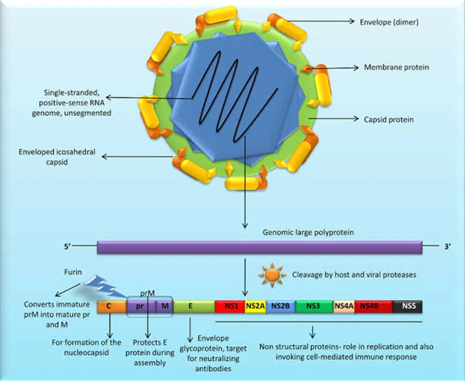
(Fig:1) Set Of Genetic Material Present In The Viral Organism
Pathophysiology
The Zika virus is transmitted by the Aedes mosquito and several other Aedes species. Besides a mosquito bite, the virus can also be transmitted sexually.
Zika virus is primarily transmitted by infected mosquitoes of the Aedes (Stegomyia) genus, mainly Aedes aegypti but also Aedes albopictus; these species also transmit dengue, chikungunya and urban yellow fever. These mosquitoes are most abundant in tropical and subtropical regions, but their geographic range is expanding. Aedes mosquitoes usually bite during the day and mostly outdoors, although they can also feed indoors.
Zika virus is also transmitted from mother to fetus during pregnancy, as well as through sexual contact, laboratory exposure, transfusion of blood and blood products, and possibly through organ transplantation[12].
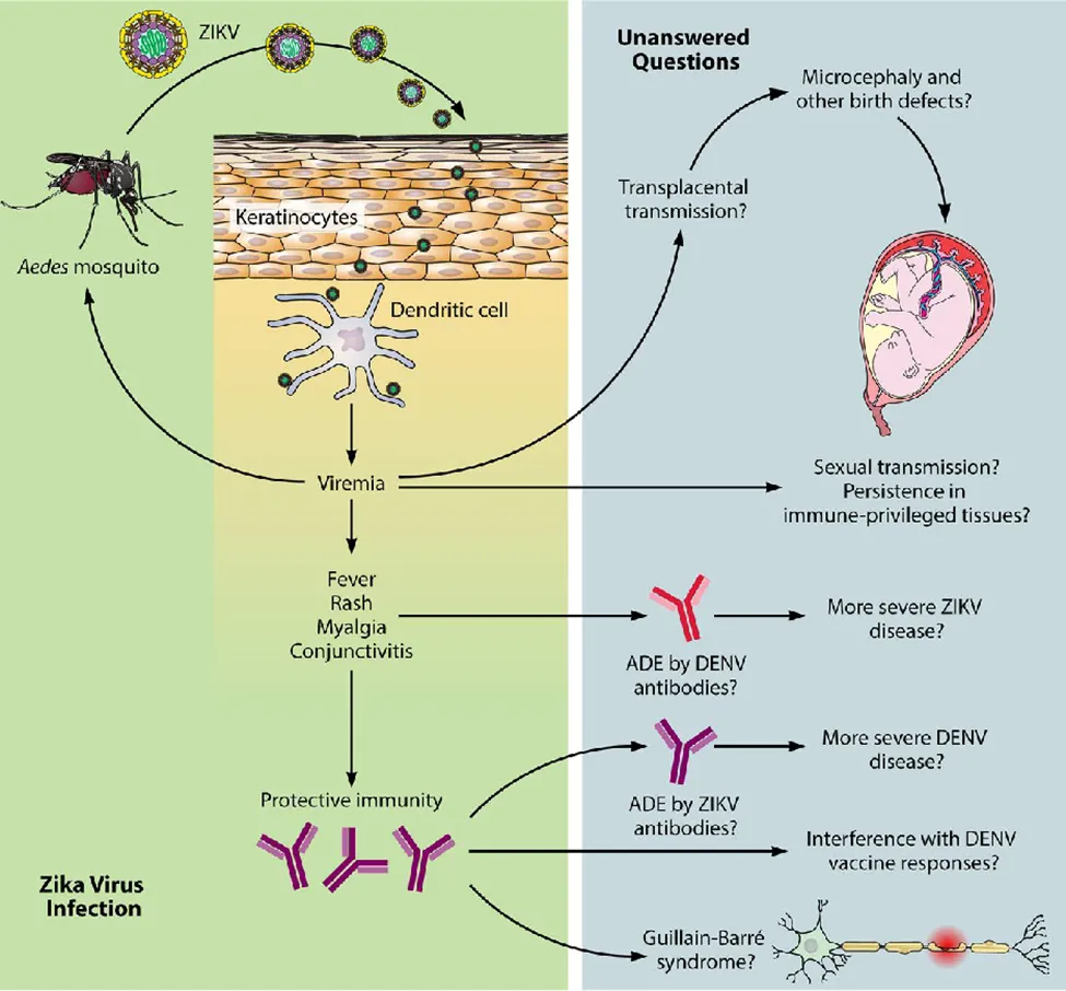
(Fig:2) Transmission of Zika Virus Evaluation
The testing for Zika virus infection is based on the risk of exposure, symptoms, and pregnancy status. Routine laboratory tests are frequently normal, although mild leukopenia, thrombocytopenia, and elevated hepatic transaminases may be seen.
As of April 25, 2017, the United States Centers for Disease Control and Prevention (CDC) testing recommendations are:
Test everyone with Zika exposure (living or traveling in areas with Zika or having sex with someone without a condom who has lived or traveled in a Zika area), and symptoms of Zika.
Test pregnant women with Zika exposure.
Test pregnant women with a fetus whose ultrasound demonstrates findings that might be associated with Zika infection.
Zika testing should be part of the routine obstetrical testing at the first prenatal visit and during the second trimester for pregnant women with Zika exposure.
Test selection for detecting Zika virus infection is guided by the duration of symptoms (less than 14 days, more than 14 days) and pregnancy status.
The virus infection is detected with PCR and antibodies[13][14][15].
ZIKV diagnosis
It manifests as moderate fever, arthralgia, and occasionally arthritis. The second and third days of the fever are characterized by a variable, though potentially high, prevalence of conjunctivitis and a maculopapular rash in patients[16].
This does not last more than three to five days, and the rash will disappear shortly. A moderate amount of progress has been made with protease ZIKV. With a Ki of 361 ± 19 nM, the classic serine protease inhibitor aprotinin (BPTI) is a potent inhibitor of protein fusion [17]. Because of the larger vial volume and extended excretion period, whole blood and urine are also advised for testing in addition to serum[18].
Treatment
There are no antiviral treatments available for Zika virus. Treatment is generally supportive and can include rest, fluids and use of analgesics and antipyretics. Because of similar geographic distribution and systoms, patients with suspected Zika virus infections also should be evaluated and managed for possible dengue or chikungunya virus infection. Aspirin and other non-steroidal anti-inflammatory drugs(NSAIDs) should be avoided until dengue can ruled out to reduce the risk of hemorrhage. People infected with Zika, chikungunya or dengue virus should avoid mosquito exposure during first few days of illness to prevent other mosquitoes from becoming infected and reduce the risk of further transmission[19].
No vaccine or preventive drug is available for Zika virus. All travelers to areas with possible Zika virus transmission should take steps to avoid mosquito bites during the day and night to prevent Zika virus and other vector-borne infections.
Patients with possible exposure to Zika virus should be advised to reduce the risk of sexual transmission through abstinence or using condoms.[19]
Ebola
Introduction
Ebola virus is recognized as an emerging and re-emerging zoonotic disease that causes acute hemorrhagic fever in humans and has a high case fatality rate [20]. The virus infects humans who come into direct contact with sick animals or people, and most Ebola virus disease(EVD) outbreaks are caused by person-to-person transmission. Several thousand people died because of a recent EVD outbreak in West Africa [21]. The main outbreaks have been documented in humans, primarily in central Africa [22].
What Is Ebola?
Ebola is a serious, life-threatening type of viral hemorrhagic fever — a viral infection that damages your blood vessels. Ebola symptoms start off like the flu (influenza). But they can progress to:Severe bleeding (hemorrhage) Neurological disorders (conditions that affect your brain and nerves) Severe vomiting[23]
Etiology
EVD is caused by Ebola virus (EBOV), one of five ebolaviruses in the Filoviridae family. The genome of EBOV consists of an enveloped, single-stranded, negative-sense RNA of about 19 kilobases [24]. Clinically, patients with EBOV infection present with signs or symptoms of fever or history of fever, intense fatigue or weakness, vomiting or nausea, and diarrhea [25]. Previous studies found significant differences in the clinical characteristics between EVD cases and non-cases [26].
Symptoms and causes
The symptoms of Ebola disease can be sudden and include fever, fatigue, malaise, muscle pain, headache and sore throat. These are followed by vomiting, diarrhoea, abdominal pain rash, and symptoms of impaired kidney and liver functions. It is important for health and care workers to be on the lookout for these symptoms.
Despite a perception that bleeding is a common symptom, this is less frequent and can occur later in the disease.[27]
Ebola Genome

(Fig:3) Set of Genetic Material Present In Viral Organism
Transmission
The Ebola virus is transmitted to humans by coming into contact with the blood, organs, or other bodily fluids of infected animals.
The first case of Ebola virus disease during the 2014–2016 West Africa outbreak was traced to exposure to bats [28]. Besides bats, EVD cases have been reported in people who handle infected chimpanzees, gorillas, and forest antelopes, whether alive or dead, in Gabon, the Republic of the Congo, and Cote d’Ivoire [29]. The virus can enter the body through the nose, mouth, eyes, ears, wounds, open wounds, cuts, or mucous membranes [30]. Transmission through sexual contact with a convalescent or survivor of Ebola virus has been documented [31]. Ebola virus is present in all bodily fluids of people with EVD, including blood, vomit, urine, feces, sweat, tears, breast milk, semen, mucus, saliva, and other bodily fluids [32]. Reusing contaminated needles and medical supplies without first sterilizing them can spread the Ebola virus. The virus can survive for weeks on surfaces such as utensils, bedding, clothing, furniture, doorknobs, electrical switches, and other items that can become contaminated by body fluids [33].
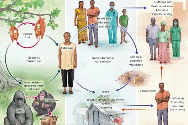
(Fig:4) Transmission Of Ebola Virus
Diagnosis
reverse transcriptase polymerase chain reaction (RT-PCR) assay antibody-capture enzyme-linked immunosorbent assay (ELISA) antigen-capture detection tests
virus isolation by cell culture.
Samples collected from patients are an extreme biohazard risk; laboratory testing on non-inactivated samples should be conducted under maximum biological containment conditions.[34]
Treatment
Over the years, WHO and partners have developed guidance and training that outline how to provide the best possible care for patients and increase their chance of survival, whether or not specific treatments are being used. Called optimized supportive care, this covers the relevant tests to administer, how to manage pain, nutrition and co-infections (such as malaria), and other approaches that put the patient on the best path to recovery.
For Ebola virus disease, WHO made strong recommendations for treatment with mAb114 (ansuvimabTM) or REGN-EB3 (InmazebTM) that are both monoclonal antibodies. For other Ebola diseases, such as SVD or BVD, there are no approved therapeutics, but candidate products are under development and a CORE protocol for clinical trials is available.
Vaccines
For Ebola virus disease:
Two vaccines are approved: Ervebo (Merck & Co.) and Zabdeno and Mvabea (Janssen Pharmaceutica). Ervebo vaccine is recommended as part of outbreak response, see SAGE recommendations of July 2024.
In case of a confirmed Ebola virus disease outbreak, Ervebo vaccines can be accessed through the International Coordinating Group on vaccine provision.
For preventive vaccination of health-care and frontline workers, request of Ervebo vaccines can be made through Gavi Preventive Ebola vaccination.[35]
Prevention and control
Community engagement is key to successfully controlling any outbreak. Outbreak control relies on using a range of interventions, such as clinical care, surveillance and contact tracing, laboratory services, infection prevention and control in health facilities, safe and dignified burials, vaccination (only for Ebola virus disease) and social mobilization.
Raising awareness of risk factors and protective measures that individuals can take is an effective way to reduce human transmission.[36]
Herpus Virus
Introduction
Herpes simplex virus type 1 (HSV-1) is a member of the Alphaherpesviridae subfamily. Its structure is composed of linear dsDNA, an icosahedral capsid that is 100 to 110 nm in diameter, with a spikey envelope. In general, the pathogenesis of HSV-1 infection follows a cycle of primary infection of epithelial cells, latency primarily in neurons, and reactivation. HSV-1 is responsible for establishing primary and recurrent vesicular eruptions, primarily in the orolabial and genital mucosa. HSV-1 infection has a wide variety of presentations, including orolabial herpes, herpetic sycosis (HSV folliculitis), herpes gladiatorum, herpetic whitlow, ocular HSV infection, herpes encephalitis, Kaposi varicelliform eruption (eczema herpeticum), and severe or chronic HSV infection. Antiviral therapy limits the course of HSV infection.[37][38]
Etiology
HSV-1 and HSV-2 contain a large, linear double stranded DNA genome protected by an icosahedral capsid surrounded by a proteinaceous layer termed the tegument and wrapped in an envelope containing viral glycoproteins (Figure 1). Initial attachment to the plasma membrane occurs through binding of glycoprotein B (gB) and gC to glycosaminoglycans (GAG) [Citation12].
5.Heldwein EE, Krummenacher C. Entry of herpesviruses into mammalian cells. Cell Mol Life Sci. 2008;65(11):1653–1668.
The major risk factor for eczema herpeticum is skin barrier dysfunction. This can be seen in atopic dermatitis, Darier disease, Hailey-Hailey disease, mycosis fungoides, and all types of ichthyosis. The increased risk is also associated with mutations in the filaggrin gene, which is seen in atopic dermatitis and ichthyosis vulgaris. Pharmaceutical risk factors for eczema herpeticum include the use of topical calcineurin inhibitors such as pimecrolimus and tacrolimus.
Risk factors for severe or chronic HSV infection include immunocompromised states such as transplant recipients (solid organ or hematopoietic stem cells), HIV infection, or leukemia/lymphoma patients.[39]
Symptoms And Causes
People who develop herpes symptoms may first experience tingling, itching, or burning before sores or blisters form around the mouth or genitals. These blisters break open and leak fluid, which forms a crust before healing
Alongside sores or blisters, herpes may cause:
According to the World Health Organization (WHO)Trusted Source, HSV-1 typically spreads via oral contact, and HSV-2 typically spreads via sexual contact.
A person may contract HSV if they come into contact with:
HSV cannot spread through general contact with objects like toilets, doorknobs, or towels[40]
Epidemiology
It has been hypothesized that approximately one-third of the world’s population has experienced symptomatic HSV-1 at some point throughout his or her lifetime. HSV-1 first establishes primary infection in patients with no existing antibodies to HSV-1 or HSV-2. Non-primary initial infection is defined as infection with one HSV subtype in patients who already have antibodies to the other HSV type (i.e., HSV-1 infection in a patient with HSV-2 antibodies, or vice versa). Reactivation results in recurrent infection and most commonly presents as asymptomatic viral shedding.
Approximately 1 in 1000 newborns in the United States experience a neonatal herpes simplex virus infection, resulting from HSV exposure during vaginal delivery. Women with recurrent genital herpes have a low risk of vertically transmitting HSV to their neonate. However, women who acquire a genital HSV infection during pregnancy have a higher risk.
Epidemiologically, it is important to note that herpes encephalitis is the leading cause of lethal encephalitis in the United States, and ocular HSV infection is a common cause of blindness in the United States.[41][42]
Herpus Genome
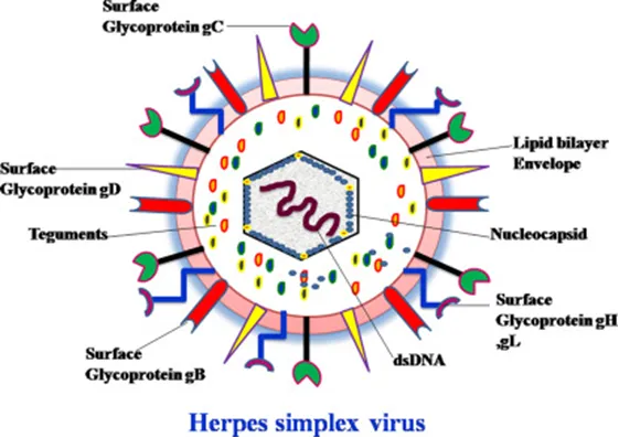
(Fig:5) Set Of Genetic Material Present In The Viral Organism
Etiology
HSV-1 is typically spread through direct contact with contaminated saliva or other infected bodily secretions, as opposed to HSV-2, which is spread primarily by sexual contact. HSV-1 begins to replicate at the site of infection (mucocutaneous) and then proceeds to travel by retrograde flow down an axon to the dorsal root ganglia (DRG). It is in the DRG that latency is established. This latency period allows the virus to remain in a non-infectious state for a variable amount of time before reactivation. HSV-1 is sly in its ability to evade the immune system via several mechanisms. One such mechanism is inducing an intercellular accumulation of CD1d molecules in antigen presenting cells. Normally, these CD1d molecules are transported to the cell surface, where the antigen is presented resulting in the stimulation of natural killer T-cells, thus promoting immune response. When CD1d molecules are sequestered intercellularly, the immune response is inhibited. HSV-1 has several other mechanisms by which it down-regulates various immunologic cells and cytokines.[43]
Transmission
Herpes is passed directly from the affected area of skin (which could be the genitals, face or hands), by direct skin to skin contact, with friction, when the virus is present. (See also asymptomatic shedding, below.) It may come back at or near the place the virus was caught. That means that when you catch it genitally, It does not travel through your body and appear on your face; it won’t be in your saliva.
Herpes simplex gets in easily through mucous membranes. These comprise the moist skin inside the mouth and genital-anal area. Sometimes it gets into ordinary skin, on other parts of the body. This is possible if there is a cut or break in the skin. This can be on the fingers/hands, knees, etc. But this is only if these come into direct contact with the affected area of another person. A finger sore is called a herpetic whitlow.
HSV-1 is mainly transmitted via contact with the virus in sores, saliva or skin surfaces in or around the mouth. Less commonly, HSV-1 can be transmitted to the genital area through oral-genital contact to cause genital herpes. It can be transmitted from oral or skin surfaces that appear normal; however, the greatest risk of transmission is when there are active sores. People who already have HSV-1 are not at risk of reinfection with HSV-1, but they are still at risk of acquiring HSV-2.
HSV-2 is mainly transmitted during sex through contact with genital or anal surfaces, skin, sores or fluids of someone infected with the virus. HSV-2 can be transmitted even if the skin looks normal and is often transmitted in the absence of symptoms.
In rare circumstances, herpes (HSV-1 and HSV-2) can be transmitted from mother to child during delivery, causing neonatal herpes.[44]
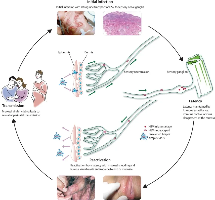
(Fig:6) Transmission of Herpus Virus
Evaluation
The gold standard for diagnosing HSV-1 infection is HSV-1 serology (antibody detection via western blot). The most sensitive and specific mechanism is viral polymerase chain reaction (PCR). However, serology remains the gold standard. Viral culture, direct fluorescent antibody (DFA) assay, and Tzanck smear are alternative methods of diagnosing. It is important to note that the Tzanck smear identifies multinucleated giant cells, so it cannot distinguish between HSV and VZV. The DFA assay, however, can distinguish between the 2 entities.[45]
Diagnosis
Healthcare providers diagnose HSV infections by doing a physical exam and testing. During an exam, your provider will look for signs of infection (like sores). They may take a sample from the sores to send for lab testing. If your provider suspects encephalitis and/or meningitis, they may do a spinal tap.
If you don’t have sores, your provider can use a blood test to check for antibodies against HSV-1 or HSV-2. Antibodies are a sign you’ve been infected with the virus in the past. Test results help your provider plan treatment[46]
Treatment
For the treatment of orolabial herpes, the current recommendation is oral valacyclovir (2 grams twice daily for one day). If the patient has frequent outbreaks, chronic suppression is warranted. For chronic suppression of immunocompetent patients, oral valacyclovir 500 mg daily (for patients with less than ten outbreaks per year) or oral valacyclovir 1 gram by mouth daily (for patients with greater than 10 outbreaks a year) is recommended.
For immunocompromised patients with severe and chronic HSV, treatment is aimed at chronic suppression. For chronic suppression of immunocompromised patients, oral acyclovir 400 to 800 2 to 3 times daily, or oral valacyclovir 500 mg twice daily is recommended.
Recently, two prodrugs have been licensed for the treatment of herpes zoster in the elderly. Valaciclovir, the prodrug of acyclovir, and famciclovir, the prodrug of penciclovir, provide high plasma levels of the parent compounds and offer added efficacy as well as decreased dosing frequency in the management of shingles.[46]
Prevention
The following strategies can reduce the risk of developing or passing on herpes:
Using protection (like condoms or dental dams) for all sexual activity. These will not protect you 100%. But they’ll lower the risk of HSV spreading between partners. Protection doesn’t cover all areas where HSV may shed. But they do cover some areas. Plus, they’ll help protect you against other STI. [47]
Medication
Anti-Viral Medication is suggested to these viruses.
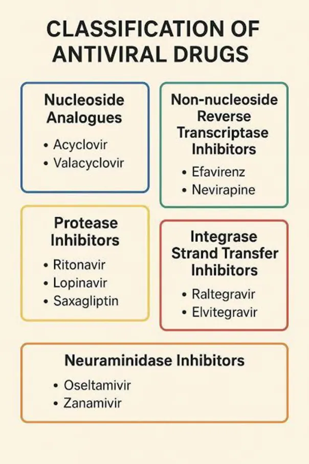
(Fig:7)
Adverse effects
Anti-viral drugs are the medicines used to treat viral infections such as HIV, Dengue Fever,measles, Influenza, Hepatitis-A and B, Zika virus, Ebola virus(EVD/EHF) and Herpus virus (HVS)
Common ADE’s of Anti-viral Drugs :-
Serious ADE’s of Anti-viral Drugs:-
REFERENCES
adilha KP, Resck MEB, Cunha OATD, Teles-de-Freitas R, Campos SS, Sorgine MHF, Lourenço-de-Oliveira R, Farnesi LC, Bruno RV. Zika infection decreases Aedes aegypti locomotor activity but does not influence egg production or viability. Mem Inst Oswaldo Cruz. 2018 Aug 23;113(10):e180290
adilha KP, Resck MEB, Cunha OATD, Teles-de-Freitas R, Campos SS, Sorgine MHF, Lourenço-de-Oliveira R, Farnesi LC, Bruno RV. Zika infection decreases Aedes aegypti locomotor activity but does not influence egg production or viability. Mem Inst Oswaldo Cruz. 2018 Aug 23;113(10):e180290


: A.L.T Kalyani, Gandivalasa Gandiv Sai Charan, Pisipati Aparna, R.V.R., An Outline About Zika, Ebola And Herpes Simplex Viruses, Int. J. of Pharm. Sci., 2026, Vol 4, Issue 7, 4284-4300. https://doi.org/10.5281/zenodo.21479058
 10.5281/zenodo.21479058