

We use cookies to ensure our website works properly and to personalise your experience. Cookies policy
MVM College of Pharmacy, Karnataka -560064, Affiliated to Rajiv Gandhi University of Health Science, Bengaluru, Karnataka -560041
The skin, the body’s largest organ, comprises three primary layers: epidermis, dermis and hypodermis. The stratum corneum, the outermost epidermal layer (~10–20?µm), acts as the principal barrier to transdermal drug delivery. Its brick-and-mortar structure of corneocytes embedded in lipid matrices impedes permeation, particularly for hydrophilic or high-molecular-weight compounds (>500?Da). Conventional approaches patches, chemical enhancers, iontophoresis and electroporation often yield suboptimal flux. This is where microneedles come in as a genuine breakthrough: these are tiny needle arrays, each barely visible to the naked eye, that gently pierce just the outer skin layer to create microscopic channels without ever reaching the nerve endings that cause pain, boosting drug absorption anywhere from 10 to 1000 times compared to passive methods. Microneedles (MNs) represent a transformative strategy, mechanically bypassing the stratum corneum via microchannels without stimulating nociceptors, thus enabling painless, enhanced permeation (10–1000× increase). MN arrays are classified into five types: solid (pre-treatment), coated (surface-bound drug), dissolving (matrix-embedded, biodegradable), hollow (fluid infusion), and hydrogel-forming (swelling-mediated sustained release). Critical quality attributes include drug content uniformity, weight variation, swelling ratio and mechanical integrity (insertion and fracture force). Microscopic techniques (SEM, confocal) assess morphological fidelity, while DSC, FTIR and XRD evaluate drug–polymer interactions and stability. In vitro permeation studies using Franz diffusion cells and ex vivo/in vivo models validate efficacy and skin recovery kinetics. Microneedle systems offer a minimally invasive, patient-compliant platform for transdermal delivery of therapeutics, vaccines, cosmetics and diagnostics.
Topical application is an excellent method of treatment since it does not go through the gastro-intestinal system and first-pass effect, thus causing minimal side effects while targeting the lesions. The skin acts as one of the major biological barriers; the effectiveness of topical application is mainly dependent on the integrity of the stratum corneum since this layer requires permeation before reaching the desired location for bioactivity and disease individualization dosage determination. Therefore, the permeability and personalization of the dosage are critical factors determining the efficacy of this form of therapy.1
Transdermal drug delivery (TDD) represents a globally recognized alternative to oral and parenteral administration, offering distinct clinical advantages. These include the avoidance of hepatic first-pass metabolism, bypass of gastrointestinal degradation, and a significant reduction in systemic side effects.2 TDD systems provide a steady-state delivery profile, which is particularly beneficial for chronic disease management where patient compliance is often a challenge. However, the inherent barrier function of the skin, primarily centered in the stratum corneum, strictly regulates the permeability of exogenous substances. This barrier typically restricts effective permeation to drug molecules with low molecular weight (<500 Da), moderate lipophilicity (log P 1–3) and high potency.3
The limitations of passive diffusion have led to the development of various enhancement strategies, ranging from chemical enhancers and thermal ablation to physical methods such as iontophoresis, electroporation and sonophoresis. While these methods can increase permeability, they are often associated with skin irritation, require complex and expensive equipment, or fail to deliver larger biotherapeutics efficiently.4 Microneedles (MNs) offer a superior solution by mechanically creating micro-conduits that are large enough to allow drug transport but small enough to avoid stimulating dermal nociceptors and blood vessels. This "painless" technology has the potential to transform the delivery of vaccines, hormonal therapies, and dermatological treatments. This review synthesizes the current advancements in microneedle design, fabrication, and characterization, providing a comprehensive roadmap for their successful clinical translation.5
Transdermal drug delivery is a medical approach to treating diseases affecting the skin and other body systems, especially by circumventing first-pass metabolism. In addition, transdermal drug delivery allows for controlled dosage over an extended period of time. Given the complex physiology of the skin with its vast networks of vascular and lymphatic blood vessels, it acts as a perfect point of entry for systemic drug administration.6 Furthermore, the skin serves as a biological reservoir for medications. This is particularly advantageous for drug candidates with short biological half-lives, as it eliminates the need for frequent dosing and maintains stable plasma concentrations. But, the primary evolutionary role of the skin is that of defence, with the stratum corneum (SC) being a strong physical barrier, which impedes the penetration of most drugs into the skin. The main issue in transdermal delivery is, therefore, how to increase the permeability of the stratum corneum barrier.7 Three generations of delivery technologies have emerged to address this problem, with the first generation of drug delivery techniques restricted to only small, lipophilic drugs at relatively low doses. In contrast, the second generation involved the use of chemical enhancers and iontophoresis to enable the delivery of a larger variety of drugs. Recently, the latest generation of technologies involving microneedles, thermal ablation, and electroporation has made it possible to deliver large biological drugs such as insulin and parathyroid hormone.8
2. PHYSIOLOGY AND BARRIER FUNCTION OF THE SKIN
The skin is the largest organ of the human body, accounting for approximately 15% of total body weight. It serves as a crucial interface, providing protection against UV radiation, chemicals, and pathogens, while also performing roles in thermoregulation and immune surveillance.
2.1. Epidermal Layers and the Stratum Corneum
The epidermis is the superficial, non-vascularized layer, ranging in thickness from 50 µm to 1.5 mm. It is composed primarily of keratinocytes in various differentiation stages. The outermost layer, the stratum corneum (SC), is the most significant barrier to drug diffusion. The SC consists of 15 to 20 layers of non-viable, flattened corneocytes densely packed with keratin filaments.9 These cells are surrounded by a continuous lipid matrix composed of ceramides, free fatty acids, and cholesterol. This "brick-and-mortar" structure is remarkably impermeable to most substances, particularly polar and high-molecular-weight molecules.10
2.2. Dermis and the Vascular Network
The dermis, located beneath the epidermis, is a thicker (0.5 to 3 mm) layer composed of collagen and elastin fibres. It contains blood vessels, lymphatic vessels, hair follicles, sweat glands, and nerve endings. The papillary dermis, adjacent to the epidermis, is rich in capillaries, making it the primary target for systemic drug absorption once the stratum corneum barrier is breached. The deep dermis provides structural support and anchors the skin to the underlying hypodermis.11
2.3. Molecular Transport and the rationale for Microneedles
Drug transport through the skin occurs primarily via passive diffusion, governed by Fick’s First Law: $J = -D (dc/dx) $, where $J$ is the flux and $D$ is the diffusion coefficient. The SC’s low diffusion coefficient is the primary bottleneck for systemic delivery. Microneedles circumvent this by physically removing the SC barrier in localized areas, effectively creating "macro-pores" through which drugs can diffuse at rates several orders of magnitude higher than passive permeation. This mechanical disruption is temporary; the skin’s barrier function typically restores itself within 24 to 72 hours through natural repair mechanisms.12
3. MICRONEEDLE DRUG DELIVERY
Transdermal drug delivery has traditionally been based on the duality of topical creams and hypodermic needles. Conventional drug delivery techniques employ metal needles or syringes to administer either subcutaneous or intramuscular injections; however, this method is associated with severe pain and infection caused by blood-borne pathogens. Additionally, most topical creams cannot efficiently deliver medications across the impermeable surface of the skin. To address this problem, microneedle (MN) technology was developed as a combination approach integrating the benefits of physical insertion with those of patches applied directly to the skin.13 Microneedles represent an array of tiny needles attached to a patch-like substrate and designed in a way to penetrate the upper layers of the skin without affecting sensitive pain receptors. With an average thickness of 1500 micrometers, the epidermis is easily pierced by microneedles that have a smaller width. Hence, microneedle technology offers painless delivery of medications across the epidermis with minimal chances of adverse effects. Moreover, microneedles provide precise and rate-controlled delivery of medication in comparison with conventional drug delivery methods. These Microneedles can be prepared using Silicon, Polymer, Glass and Metal.14
The emergence of Microneedle (MN) technology has been recognized as one of the most revolutionary approaches towards transdermal drug delivery, due to their ability to enhance therapeutic potency and efficacy. The absence of any first-pass effect along with the ability to deliver medication without causing stomach irritation, makes Microneedles ideal for the effective delivery of therapeutics. These micron-scale structures, fabricated on a suitable substrate, can deliver therapeutic drugs without causing any irritation and pain since they have the ability to traverse through the outermost layer of the skin without causing any damage. Even though a variety of microneedle types are available in the market, polymers-based systems are the most successful ones in terms of systemic delivery of protein-based medicines.15 Contrary to microneedles made of metals, ceramics, etc., or those that are coated with therapeutic molecules, polymer-based arrays are more efficient for delivering high doses of therapeutics since solid and coated microneedles cannot carry sufficient amounts of drugs.16 Microneedles have been revolutionizing the world of medical treatment through their innovative approach of delivering medications to patients. This is achieved through creating temporary, microscopic channels through the stratum corneum in order to overcome the major barrier presented by the skin, thereby facilitating the systemic absorption of various macromolecules as well as pharmacological agents. The pores that are created by microneedles are small enough not to be detected by pain receptors, thus preventing any form of discomfort to patients. At present, there are five main types of microneedle systems identified based on their structure.17
3.1. Classification of Microneedle Systems
Microneedles are categorized based on their structural design and the method by which they facilitate drug delivery, each offering specific advantages for different therapeutic needs.
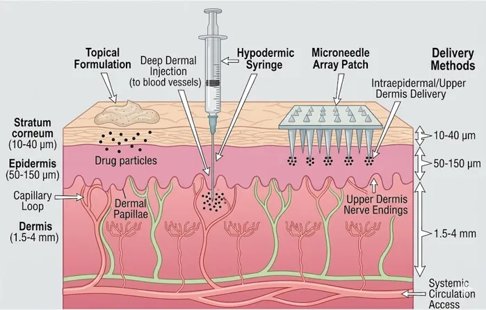
Fig.1: Microneedle delivery methods
3.2. Solid Microneedles
Solid microneedles stand out as the most basic form of microneedle design, developed mainly for the purpose of forming temporary microchannels through the skin barrier layer. This device comprises micro tapered needles made of durable material, which do not contain any drug compounds or inactive carriers. The presence of these micropores facilitates the formation of channels that help bypass the major rate-limiting step in skin, thereby facilitating drug diffusion into the tissue through passive diffusion. As a result, the drug diffused through the pores is delivered directly to the blood capillaries present in the upper dermis layer, which improves the bioavailability of the drug. Solid microneedles are characterized by their superior mechanical strength.18 Solid Microneedles are the simplest form of the technology, typically fabricated from silicon, stainless steel, or titanium. They are utilized in a "poke-and-patch" method: the array is applied to the skin to create micro-pores and then removed, followed by the application of a drug-loaded patch or topical formulation. This pre-treatment significantly enhances the flux of large or hydrophilic molecules.19
Advantages: High mechanical strength, robust penetration, and compatibility with various drug formulations.
Disadvantages: Two-step application process, risk of contamination during the interval between poking and patching, and the potential for patient error.
3.3. Coated Microneedles
Coated microneedles rely on the use of a structurally sound core surrounded by a matrix that dissolves easily when introduced into the skin to promote quick absorption of the pharmacological substance. The success of this type of application heavily relies on the rheology of the substance, where the viscosity levels are optimized to maintain stability.20 Additionally, an appropriate distribution of the drug on the needles requires adjustment of the surface tension of the drug to optimize its behavior. Rapid release of the drugs is the principal advantage of coated microneedles. However, despite an increased dosage level through the repeated coatings of the needles, this technique is limited by volume and dosage size. Thus, coated microneedles are most suited to deliver high-potency drugs and cannot be used with those that require large volumes of administration. Coated Microneedles utilize a "coat-and-poke" approach, where the therapeutic agent is applied as a dry film to the surface of solid microneedle shafts. Techniques such as dip-coating or spray-coating are used to ensure precise drug loading. Upon insertion into the skin, the coating dissolves in the interstitial fluid, rapidly releasing the drug.21
Advantages: Rapid drug release, suitable for low-dose therapeutics like vaccines and potent biologics.
Disadvantages: Limited drug loading capacity determined by surface area, and potential for coating instability during storage.
3.4. Dissolving Microneedles
The dissolving microneedle is an innovation in the transdermal administration method, constructed from a network of biodegradable and water-soluble polymers, where the drug is held. Once inserted into the skin, the water in the skin acts on the needle, leading to the dissolving of both the needle and the drug at the same time. This process eliminates any waste material, improving the bio-compatibility of the drug administration process.22 The ability of the dissolving microneedles to deliver drugs gradually through the degradation process makes them an ideal option for prolonged therapy programs. Dissolving Microneedles are fabricated from water-soluble or biodegradable polymers (e.g., PVP, HA, sugars) that encapsulate the drug within the needle matrix. Upon insertion, the needles dissolve completely, releasing the drug cargo and leaving no sharp waste behind.23
Advantages: Self-disposable (no sharp waste), ideal for home use, and the ability to load drugs specifically in the needle tips ("tip-loading") for maximum efficiency.
Disadvantages: Lower mechanical strength compared to metals, sensitive to environmental humidity, and loading limits for very high-dose drugs.
3.5. Hollow Microneedles
A hollow microneedle can be described as the next level of a syringe, only that it is small in size and has a hollow lumen within it where the drug dispersion or solution will be stored. These microneedles have very tiny openings on their tips, and when inserted into the skin, they release the drugs directly into the epidermis or even the superficial dermis.24 One of the biggest benefits of a hollow microneedle is that it is capable of storing large volumes of medicine, unlike other types of microneedles such as those that are coated or solid. The hollow microneedles also allow for the automatic delivery of potent molecules like the vaccines into the skin due to their ability to inject in large volumes. Hollow Microneedles resemble miniaturized hypodermic needles, featuring an internal lumen for the active infusion of liquid drug formulations. This allows for the delivery of larger volumes (up to 200 µl) and provides control over the delivery rate using pumps or pressurized reservoirs.25
Advantages: Delivery of large volumes and high-dose biologics (e.g., insulin), and precise control over infusion rates.
Disadvantages: Prone to clogging by skin tissue (coring), requires specialized insertion devices, and higher complexity in fabrication.
3.6. Hydrogel-Forming Microneedles
Microneedles, which form hydrogels upon insertion, are fabricated using a cross-linked polymeric structure which does not break down on contact with the skin. Rather than dissolving and releasing an encapsulated drug cargo, these devices serve as a permeable channel, allowing for a constant flow through the microneedles from a reservoir to the skin tissue. Since the drug can be evenly distributed throughout the whole surface area of the patch, this drug delivery method works extremely effectively with medications which cannot be delivered using other kinds of microneedles due to their high dose. The main benefit of this method in pharmacology is the ability of transporting the large amount of the drug through the diffusive flux. Nonetheless, it is worth noting that such a process takes a lot of time, making the wear time for the patch longer than the wear time for other delivery methods. Hydrogel Microneedles are made of cross-linked polymers that do not dissolve but instead swell upon contact with interstitial fluid. This swelling creates a continuous aqueous conduit for the drug to diffuse from an external reservoir into the skin.26
Advantages: Delivery of high doses over extended periods, can be removed intact, and potential for biosensing applications.
Disadvantages: Slower onset of action compared to dissolving MNs, and the need for a separate drug reservoir.27
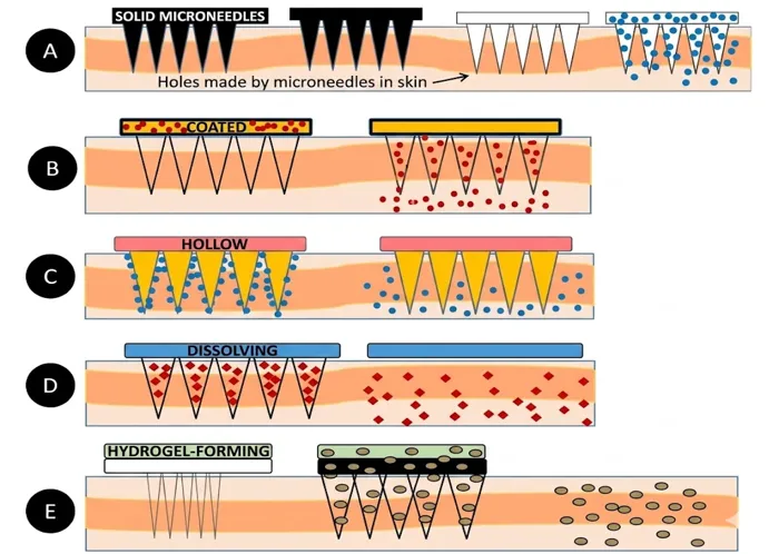
Fig.2: Types of Microneedle Mechanism
4. FABRICATION STRATEGIES FOR MICRONEEDLE ARRAYS
The fabrication of microneedles requires sub-micron precision to ensure needle sharpness, structural integrity, and consistency across the array. The choice of technique is determined by the material properties and the intended application.
4.1. Micro Moulding and Casting
Micro moulding is the most prevalent technique for producing polymeric and dissolving microneedles. The process begins with the creation of a "master mould" using high-precision techniques like laser machining or photolithography. The polymer solution, containing the therapeutic agent and stabilizers, is dispensed into the PDMS mould. To ensure the viscous solution fills the sharp needle tips, vacuum or centrifugal forces (e.g., 4000 RPM for 10 minutes) are applied. The solvent is evaporated under controlled conditions (e.g., 25°C, 40% RH) for approximately 24 hours. A backing membrane is then added to provide mechanical support. This method is highly scalable and cost-effective for mass production.28,29
4.2. Photolithography and Etching
Photolithography is the gold standard for fabricating silicon and metal microneedles. The process involves coating a silicon wafer with a light-sensitive photoresist and exposing it to UV light through a mask to define the needle patterns. Deep Reactive Ion Etching (DRIE) is then used to remove material, creating high-aspect-ratio shafts with vertical walls. Tapered needles can be produced using wet chemical etching, which takes advantage of the crystal orientation of the silicon. While these methods provide exceptional dimensional accuracy, the high cost of equipment and the fragility of silicon have led researchers to explore more robust alternatives like stainless steel and polymers.30
4.3. 3D PRINTING AND ADDITIVE MANUFACTURING
Additive manufacturing has emerged as a disruptive force in microneedle fabrication, offering the ability to create complex and customized geometries that are impossible to achieve with traditional moulding.
Stereolithography (SLA): Uses a UV laser to cure photosensitive resin layer-by-layer. SLA can produce MNs with diverse shapes and sizes with a resolution of ~25 µm.
Two-Photon Polymerization (2PP): This technique uses a highly focused femtosecond laser to trigger polymerization only at the focal point. With a resolution of less than 100 nm, 2PP can produce needles with incredibly sharp tips and complex internal architectures, such as porous or barbed shafts.30
Digital Light Processing (DLP): Offers faster printing speeds than SLA by projecting an entire layer of light at once. This makes it more suitable for small-scale production of customized clinical devices.31
4.4. Thermal Processing and Laser Ablation
Micro-injection moulding is used for high-volume production of thermoplastic MNs, such as those made from polycarbonate. Molten polymer is injected into a cooled micro-mould under high pressure, allowing for rapid cycle times. Laser ablation, using high-power femtosecond or nanosecond lasers, is employed to cut microneedle shapes directly from thin metal or polymer sheets. This method is highly versatile and does not require expensive masks or molds.32
5. Mechanism of Microneedle transdermal drug delivery
Phase-1- Physical Penetration - Application of microneedle patch on the skin
Phase-2 – Microchannel formation- Microneedle penetrate into stratum corneum by fracturing the lipid bilayer in the skin
Phase-3 – Transdermal transport - Drug enters into stratum corneum and diffuses through viable epidermis. This increases the drug permeability
Phase-4– Systemic Pharmacokinetics- Drug enters into the blood stream at the target site with no hepatic first pass effect
Phase-5 – Skin recovery - Microchannels naturally close within some time and stratum corneum restores within 72 hours with no permanent damage to the skin.32
6. ADVANCED CHARACTERIZATION OF MICRONEEDLE SYSTEMS
Rigorous characterization is mandatory to ensure the safety and performance of Microneedle systems. Evaluation focuses on mechanical strength, morphology, and physicochemical stability.
6.1. Mechanical Characterization
The mechanical performance of Microneedles is the primary determinant of their ability to successfully penetrate the skin barrier without breaking.
Axial Compression Testing: A single needle or array is compressed against a hard surface using a texture analyser. The force-displacement curve is monitored to identify the failure point (buckling or fracture). For a 600 µm needle, a fracture force exceeding 0.5 N per needle is typically required to ensure penetration.33,34
Insertion Force: Measured by pressing the Microneedle array against a skin mimic or excised skin. The force at which the first "pop" or drop in resistance occurs represents the insertion threshold. This value should be low (0.1 to 3 N for an array) to allow for manual application.35
Safety Factor: Defined as the ratio of the fracture force to the insertion force. A safety factor of at least 5 is recommended for clinical applications to prevent needle breakage during handling or misapplication.36
6.2. Morphological and Dimensional Evaluation
The geometry of Microneedles including height, base width, and tip radius critically affects the drug delivery rate and the level of pain.
Scanning Electron Microscopy (SEM): SEM provides detailed images of the needle surface, allowing researchers to verify tip sharpness and the fidelity of the fabrication process. It is used to identify defects such as rounded tips, shaft cracks, or surface roughness.37
Confocal Laser Scanning Microscopy (CLSM): CLSM allows for the non-invasive visualization of the Microneedles in the skin. By labelling the drug with a fluorescent dye, researchers can map the three-dimensional distribution and penetration depth (typically 100 to 350 µm) of the drug within the epidermis.38
Optical Microscopy: Research microscopes (e.g., Olympus SZX16) equipped with digital cameras allow for rapid screening and quantitative measurements of array dimensions.39
6.3. PHYSICOCHEMICAL STABILITY AND DRUG LOADING
Drug Content Uniformity: Assessed by dissolving Microneedle patches in a suitable solvent and analysing the solution via HPLC or UV-Vis spectrophotometry. High uniformity (±5%) is essential for consistent clinical dosing.40
Weight Variation and Swelling: Patches are individually weighed to ensure batch-to-batch consistency. Swelling index testing for hydrogel Microneedles measures the capacity to absorb interstitial fluid, which directly correlates with the diffusion-based drug release rate.41
FTIR Spectroscopy: Used to identify potential chemical interactions (e.g., hydrogen bonding) between the drug and the polymer matrix by analysing shifts in characteristic absorption peaks.42
Differential Scanning Calorimetry (DSC): Evaluates the thermal properties and physical state of the drug within the MNs. The disappearance of a drug’s crystalline melting peak suggests the formation of a stable amorphous dispersion, which can enhance solubility and release.43
7. BIOLOGICAL EVALUATION AND PERMEATION METRICS
The transition from benchtop characterization to clinical validation requires rigorous testing in both in vitro and in vivo models to assess efficacy and safety.
7.1. IN VITRO DRUG RELEASE AND PERMEATION STUDIES
Franz diffusion cells are utilized to model the release of drugs from MNs into the body. This setup consists of a donor and a receptor compartment separated by a semi-permeable membrane. The MN patch is applied to a pre-conditioned dialysis membrane or excised skin (porcine or human). The receptor compartment is filled with phosphate-buffered saline (PBS) at pH 6.8 or 7.4, maintained at a physiological temperature of 32°C to 37°C. The receiver phase is stirred using a magnetic bead to maintain sink conditions. Samples are withdrawn at regular intervals (e.g., 0.5, 1, 2, 4, 8, 12, and 24 hours) and analysed for drug concentration via validated HPLC or UV-Vis methods. This allows researchers to calculate the cumulative drug release and determine the release kinetics (e.g., zero-order, first-order, or Higuchi model).44,45
7.2. IN VIVO ANIMAL MODEL STUDIES: SAFETY AND EFFICACY
To validate the pharmacokinetic performance of microneedles, in vivo studies are conducted using animal models, most commonly hairless rats or pigs. These animals are chosen for their skin’s anatomical and physiological similarity to human skin.
Transepidermal Water Loss (TEWL): TEWL is monitored using a vapometer before and after MN application. An increase in TEWL indicates successful disruption of the stratum corneum barrier. Monitoring the return of TEWL to baseline (typically within 24–72 hours) allows for the assessment of skin recovery kinetics.46
Histological Evaluation: Skin biopsies are taken post-treatment, stained with Hematoxylin and Eosin (H&E), and examined under a microscope. This confirms the depth of needle penetration and ensures that no permanent tissue damage or significant inflammatory response has occurred.
Pharmacokinetic (PK) Analysis: Serial blood samples are collected and analysed via HPLC or LC-MS/MS. Key parameters include the time to reach maximum concentration (T_max), the peak concentration (C_max), and the area under the curve (AUC), which reflects the total systemic exposure. These PK profiles are compared with traditional injections to evaluate the relative efficiency of MN delivery.47
8. THERAPEUTIC APPLICATIONS AND FUTURE PERSPECTIVES
Microneedles have moved beyond conceptual frameworks to significant clinical applications in numerous therapeutic areas.
8.1. Vaccination and Global Health
The skin is rich in antigen-presenting cells (APCs), such as Langerhans cells. Delivering vaccines via MNs targets these cells directly, often resulting in a more robust immune response compared to traditional intramuscular injections. This "dose-sparing" effect allows for the use of smaller amounts of antigen to achieve protective immunity. Furthermore, the stability of vaccines in solid-state MN patches eliminates the need for a cold chain, significantly reducing the cost and logistical challenges of mass vaccination campaigns in developing countries.48
8.2. Chronic Disease Management and Hormonal Therapy
For patients with chronic conditions requiring daily injections, such as diabetes (insulin) or growth hormone deficiencies, Microneedles offer a painless and patient-friendly alternative. Sustained-release Hydrogel Microneedles are particularly promising for maintaining steady hormone levels over several days or weeks. Integration with biosensors could lead to closed-loop systems that monitor biomarkers and deliver drugs accordingly.49
8.3. Oncology and Targeted Therapy
Microneedles are being investigated for the local delivery of chemotherapeutic agents directly into tumor sites, particularly for skin cancers like melanoma. This approach maximizes the local drug concentration while minimizing systemic toxicity. Furthermore, MNs can deliver immunotherapeutic agents to modulate the tumor microenvironment.50,51
8.4. Dermatology and Cosmetics
In the cosmetic industry, MN patches are used to deliver anti-aging compounds (e.g., hyaluronic acid, peptides) and treatments for skin disorders like alopecia and hyperpigmentation. MNs also show potential in scar management by delivering drugs directly into the fibrotic tissue. 52,53,54
8.5. Challenges in Clinical Translation and Regulatory Hurdles
Despite their potential, several hurdles remain for the widespread adoption of microneedles. Moving from laboratory-scale micro moulding to high-throughput industrial manufacturing requires standardized processes and robust quality control. As combination products (device + drug), microneedles must satisfy both medical device and pharmaceutical regulations, requiring extensive safety and stability data. Sterilization methods must be validated to ensure they do not degrade the drug or the polymer matrix.
9. CONCLUSION
Microneedle technology stands at the forefront of the next generation of drug delivery systems. By merging the benefits of needle-based and needle-free delivery, it provides a minimally invasive, efficient, and patient-compliant platform for a vast range of therapeutics. Continued advancements in microfabrication, polymer science, and biosensing will likely address current limitations in drug loading and scalability, paving the way for microneedles to become a standard of care in global vaccination campaigns and chronic disease management. The transition from purely physical penetration to intelligent, responsive delivery systems marks the beginning of a new era in personalized medicine.
REFERENCES


Misba Anjum, Ashly Merin George, Agilandeswari Devarajan, An Overview of Microneedle Mediated Transdermal Drug Delivery: Painless Passage through the Skin Barrier, Int. J. of Pharm. Sci., 2026, Vol 4, Issue 9, 176-189. https://doi.org/10.5281/zenodo.22233299
 10.5281/zenodo.22233299