

We use cookies to ensure our website works properly and to personalise your experience. Cookies policy
Department of Pharmacology, Sarojini Naidu Vanita Pharmacy Maha Vidyalaya (Co-Ed.), Taranaka, Hyderabad, Telangana, India, 500017.
The kidneys are exceptionally susceptible to drug-induced injury due to their high blood flow and the concentration of drugs during filtration and excretion. This condition, known as drug-induced renal injury (DIRI), is a significant contributor to acute kidney injury and can lead to chronic kidney disease. A range of common therapeutic agents, including aminoglycosides, cisplatin, non-steroidal anti-inflammatory drugs (NSAIDs), and calcineurin inhibitors, are recognized for their nephrotoxic potential. The resulting damage can affect various renal structures, such as the tubules, interstitium, glomeruli, or vasculature, with severity ranging from subtle, subclinical changes to severe dysfunction. Preclinical research utilizes models like rodents, zebrafish, and rabbits to explore DIRI mechanisms and screen for protective compounds. Key pathogenic pathways involve oxidative stress, inflammation, apoptosis, and mitochondrial damage. Despite these efforts, translating findings to clinical practice is challenging due to interspecies differences and inconsistent experimental designs. A major hurdle in managing DIRI is the delayed detection with traditional biomarkers like serum creatinine and BUN, which only rise after significant injury has occurred. Consequently, more sensitive and early indicators are being investigated. These novel biomarkers, such as Kidney Injury Molecule-1 (KIM-1), Neutrophil Gelatinase-Associated Lipocalin (NGAL), and inflammatory cytokines like IL-18 and TNF-alpha, offer the potential for earlier diagnosis. To improve clinical translation, future research must focus on standardizing study protocols, validating these new biomarkers, and employing advanced models like kidney organoids and microfluidic chips, which better replicate human kidney physiology.
Critical to homeostasis, the kidneys are multitaskers: they produce and release key hormones like renin and erythropoietin, clear metabolic waste from the bloodstream, and keep a tight rein on electrolytes, pH levels, and overall fluid balance. To pull this off, they filter roughly a quarter of the heart's resting output every single minute. Yet, this very efficiency comes with a catch-the sheer volume of substances they process and reabsorb leaves them highly exposed to harmful compounds, making them particularly prone to toxic damage. Given this vulnerability, it's no surprise that a number of medications can lead to Drug-Induced Renal Injury (DIRI). In fact, DIRI is a factor in about 20% of all nephrotoxicity cases and most often shows up as impaired function in the kidney's tubules1.
Epidemiology
CKD prevalence has climbed sharply worldwide, more than doubling from 378 million adults in 1990 to 788 million in 2023 - a shift only partly explained by population growth, since the age-standardised rate also rose to 14.2%. Regional burden is uneven: North Africa and the Middle East carry the highest load, at 18.0% prevalence, while globally most cases remain confined to the earlier stages (1 through 3) rather than advanced disease.
The downstream toll is substantial too CKD was the ninth-leading cause of death and twelfth-leading cause of disability-adjusted life years in 2023, and it contributed to roughly 11.5% of cardiovascular deaths. Three metabolic drivers keep surfacing across analyses of this trend: elevated fasting blood glucose, high BMI, and raised systolic blood pressure2.
According to a 2024 cohort study, drug-induced AKI represents a significant portion of cases at 19.3%, reinforcing the serious threat posed by nephrotoxic drugs. Across a group of 1,398 hospitalized patients, 35.8% received acute kidney support and the mortality rate reached 33.6%. Although patients with DI-AKI had a lower death rate than those with other AKI types, their demand for renal replacement therapy was equally high. Notably, vancomycin emerged as the most nephrotoxic, correlating with the greatest likelihood of both death and the need for advanced support 3..
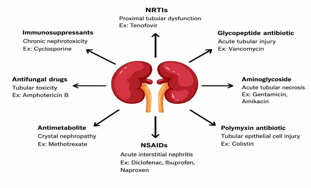
Figure 1: Representative of Drugs Associated with Each Mechanism
Experimental Animal models on Drug induced Renal Injury
Gentamicin induced Renal injury
When mice received daily intraperitoneal injections of gentamicin at 100 mg/kg for a period of 7 to 10 days, this treatment ultimately triggered acute tubular necrosis12,In Rabbits a dose of 150 mg per kg per day was given single injection13, In Hartley guinea pigs species are given with 80mg &100mg/ kg/ day via Subcutaneous route14.
Advantages & Limitations15
From a practical standpoint, this model has a great deal going for it. It is technically uncomplicated, causes minimal distress to the animal, and delivers exceptionally consistent results across repeated trials. The tubular injury it produces follows a clear dose-response relationship, and the core mechanisms-oxidative damage coupled with impaired mitochondrial function-have been thoroughly dissected over the years.
The dosages commonly used in experimental settings are often vastly higher than anything encountered in routine clinical practice. There is also the thorny issue of biological variability: mice appear remarkably resistant, which complicates cross-study comparisons and generalization. More fundamentally, the model operates in a vacuum that bears little resemblance to the complex reality of human patients-it overlooks coexisting illnesses, the confounding effects of multiple drug regimens, and the systemic upheaval caused by sepsis. On top of all that, the renal injury it induces is acute and short-lived, with spontaneous recovery occurring fairly quickly. This makes it ill-suited for exploring the protracted, progressive processes that define chronic kidney disease, limiting its utility to questions about early or reversible damage rather than long-term pathology.
Vancomycin induced Renal injury
Male Wistar rats were given vancomycin intraperitoneally over a period of seven days. In separate experiments, male Sprague-Dawley rats received either a single intravenous dose of vancomycin at 400 mg/kg or daily doses of 150 mg/kg for four days (96 hours) to produce kidney injury16.
In rabbits, vancomycin-induced nephrotoxicity was induced by administering the drug intraperitoneally at 200 mg/kg twice daily for seven days17
Advantages & Limitations16
This model provides several important advantages: it demonstrates the relationship between dosing frequency and nephrotoxicity, enables direct comparisons of once-daily versus twice-daily vancomycin administration, and maintains tightly controlled experimental conditions with few confounding factors. It also allows for the assessment of both biochemical and histological indicators of renal injury, offering a consistent and reproducible preclinical platform for studying vancomycin-induced kidney damage.
The primary limitations include the possibility that results from rats may not translate directly to humans, given differences in kidney function and how vancomycin is processed in the body. The drug doses used in the experiment might not reflect typical clinical levels, and the model does not fully account for real-world conditions such as sepsis, fluid loss, or the use of other kidney-damaging medications. Moreover, the exact mechanism behind kidney damage may not be fully clarified, and standard markers of kidney injury could indicate problems only after significant damage has occurred. Lastly, the small number of subjects involved restricts how broadly the results can be applied.
Cisplatin induced Renal injury
In a study Sprague-Dawley rats were administered with cisplatin single intraperitoneal (IP) dose of of 8 mg/kg for 3 consecutive days to induce nephrotoxicity 18. In this experiment, the animals were selected to be adult male New Zealand White rabbits. Single intraperitoneal (IP) injection of cisplatin at a dose of 4.0 and 6.5 mg/kg body weight was used. Animals were observed after receiving the medicine and on the third day, the degree of renal damage was determined to evaluate acute kidney damage19
Acute renal damage model induced by cisplatin was established on adult guinea pigs. Animals received single intraperitoneal (IP) injection of cisplatin at a dose of 5 mg/kg body weight to induce nephrotoxicity for 7 consecutive days20. Adult zebrafish (Danio rerio) were used to generate an experimental model of cisplatin-induced acute kidney injury. Specifically, nephrotoxicity was induced by intraperitoneal administration of the chemical at a dose of 20 µg per fish. In order to assess the extent of acute kidney injury, cisplatin-damaged kidneys were analyzed at 24-48 hours following treatment21
Advantages & Limitations18
In a study the . employed a cisplatin-induced kidney injury model in Sprague-Dawley rats, offering a consistent and reproducible system for studying acute nephrotoxicity. This model enables the evaluation of renal damage through a range of indicators, including biochemical parameters (creatinine, urea, NGAL), markers of oxidative stress (MDA, CAT, TAS/TOS), and molecular factors (nephrin, synaptopodin, RAC1). Additionally, it facilitated exploration of TRPC5 signaling pathways and the potential protective effects of clemizole in mitigating cisplatin-related kidney injury.
The primary limitations include the use of a rat model, which may restrict how well the results can be applied to humans. The cisplatin dose of 8 mg/kg and the experimental setup might not closely resemble actual clinical treatment conditions. Additionally, the model does not account for various patient-specific factors such as pre-existing illnesses, dehydration, concurrent medications, or the presence of cancer. Since the study primarily examines acute kidney injury, its findings may not extend to long-term or chronic kidney damage.
Diclofenac induced Renal injury
The animals received diclofenac by oral gavage 100 mg/kg once a day for 3days to induce nephrotoxicity in male CD1 mice with subclinical acute kidney injury triggered by renal ischemia-reperfusion22 .Adult New Zealand White rabbits were used to induce nephrotoxicity by administering diclofenac 10 mg/kg body weight by the intramuscular route once a day for 5 days 23.
Transgenic zebrafish embryos (Danio rerio , Tg (wt1b: EGFP) were used to evaluate nephrotoxicity at the developmental stage. At 24 hours post fertilization, embryos were incubated in embryo medium containing 25 µM diclofenac for 24 hours 24.
Advantages & Limitations23
The study indicates that diclofenac may worsen existing subclinical acute kidney injury (AKI) and contribute to its advancement toward chronic kidney disease (CKD). Functional and histological evaluations of the kidneys support this finding, revealing greater fibrosis and tubular damage following exposure to the drug.
The study involved a limited number of male mice, which limits the direct applicability of the results to humans. The doses of diclofenac and the ischemia-reperfusion model used may not accurately represent typical clinical scenarios. Additionally, the research did not include direct measurements of diclofenac concentrations in the blood or levels of renal prostaglandins.
Colistin induced Renal injury
Male Wistar rats were used to establish an experimental model of colistin-induced acute kidney injury. Renal toxicity by IP administration of colistin at a dose of 300,000 IU/kg/day once a day for 7 consecutive days 25
New Zealand White rabbits was induced by a single intravenous loading dose of colistin sulfate followed by intramuscular injections for 7 consecutive days 26. The nephrotoxicity of colistin was studied in zebrafish (Danio rerio) embryos. A water immersion method was used to expose 6 hour post-fertilization (hpf) embryos to 0, 0.01, 0.1, 1, 2, 3, and 10 µM colistin for 96 hours. The survival, embryonic morphology, lateral-line hair cell and epidermal keratinocyte were analyzed to evaluate the toxicity of colistin 27.
Advantages & Limitations25
This experimental model offers a reliable and standardized platform for investigating nephrotoxicity induced by colistin. It facilitates the assessment of renal functional, biochemical, oxidative stress, inflammatory, and morphological alterations. Importantly, the model enables the exploration of underlying molecular mechanisms, specifically regarding autophagy and necroptosis via the AMPK/mTOR and RIPK1/RIPK3/MLKL cascades. Consequently, it serves as a valuable tool for elucidating the pathophysiology of colistin-related renal damage and for evaluating potential nephroprotective interventions, such as omega-3 nanoemulsion
However, several constraints must be acknowledged. Being an animal-based study, the direct applicability of the results to human clinical scenarios is limited. The experimental dosing regimens and treatment parameters are not fully representative of real-world patient exposures. Furthermore, the model fails to account for confounding clinical variables, including severe systemic infection, critical illness, pre-existing renal dysfunction, or polypharmacy. Lastly, the design primarily addresses acute injury, thereby restricting its relevance to chronic or progressive kidney disease
Amphotericin B induced Renal injury
A single intra-peritoneal injection of amphotericin B deoxycholate at a dose of 15mg/kg was administered to male Wistar Albino rats to induce the condition. The animals were kept for a period of seven days[28]. Male C57BL/6 mice were injected with amphotericin B deoxycholate at a dose of 4mg/kg to induce the disease via an intra-venous route29
Advantages & Limitations28
The amphotericin B-induced nephrotoxicity model in Wistar rats provides a well-standardized and repeatable platform for investigating acute kidney injury. It consistently elicits distinct renal dysfunction, reflected by elevated blood urea nitrogen and creatinine levels, alongside overt structural damage such as tubular dilation and necrosis. Beyond these functional and morphological endpoints, this system enables comprehensive evaluation of oxidative stress, antioxidant enzyme responses, apoptotic signaling, and renal tissue integrity. These attributes render it particularly valuable for screening the nephroprotective potential of therapeutic candidates, as exemplified by dapagliflozin
Several significant constraints limit the interpretation of these findings. The study's reliance on a rat model raises questions about direct human applicability, while the use of a single 50 mg/kg amphotericin B dose across just seven days does not necessarily reflect real-world therapeutic exposure. Furthermore, dapagliflozin failed to produce meaningful changes in systemic oxidative-stress or apoptotic markers, undermining the evidence for the suggested protective pathways.
Tenofovir induced Renal injury
Male Wistar rats were induced by adding tenofovir disoproxil fumarate (TDF) to the standard diet strength of 300 mg/kg of food (approximately 30 mg/kg of body weight per day) for 45 consecutive days30. Male C57BL/6 mice were induced by oral gavage of TDF at a dose of 200, 500, or 800 mg/kg/day for 4 weeks 31 .
Advantages & Limitations30
It provides a consistent method for studying kidney damage resulting from long-term drug use. It allows researchers to assess various aspects of renal health, including function, blood flow, oxidative stress, inflammation, cell death, and structural injury to tubules. In addition, examining key signaling pathways-such as SIRT1/Nrf2/HO-1, TLR4/NF-κB, and those involved in mitochondrial apoptosis-helps identify the mechanisms driving kidney injury and aids in evaluating dapagliflozin’s potential protective effects on the kidneys.
The major limitations of the study are that the research was conducted among male Wistar rats, which cannot represent the human population. The experiment was carried out using a single dose of 45-day tenofovir exposure and experimental doses; thus, the results do not consider the clinical setting. Moreover, there were only six to eight animals per group, which is a relatively small sample size. Even though dapagliflozin was proved to be beneficial in terms of organ functions and molecular processes, the study is preclinical, and there is a need for clinical trials to approve the drug for the treatment of tenofovir nephrotoxicity.
Cyclosporine A induced Renal injury
In male Wistar rats, cyclosporine A was delivered intraperitoneally at 20 mg/kg/day over seven consecutive days, leading to nephrotoxicity32. In New Zealand White rabbits, cyclosporine A (CsA) was given subcutaneously at 25 mg/kg/day, divided into two doses per day, for five consecutive days33.
Advantages & Limitation32
The cyclosporine A-induced nephrotoxicity model offers a standardized approach for investigating kidney injury in male Wistar rats. It permits assessment of renal function, oxidative imbalance, inflammatory responses, apoptosis, and histopathological alterations through various biochemical and molecular markers. This model is therefore valuable for understanding the mechanisms underlying cyclosporine-related renal damage and for testing potential protective treatments such as sitagliptin and hesperidin.
The major limitations of the study include the use of male rats as subjects which limits the applicability of the results to both genders. Another limitation is the high dosage of cyclosporine used which may not be applicable in clinical practice. The study also used a small sample size to conduct the experiment which limits the accuracy of the results. Lastly, the study focused on the acute effects of cyclosporine on the kidney which limits the applicability of the results in understanding the effects of long-term use of the drug.
Methotrexate induced Renal injury
Male Wistar albino rats were produced by a IP of methotrexate (20 mg/kg) on the 5th day of a 10-day experiment 34 . Male mice were used Nephrotoxicity was induced by methotrexate in combination with omeprazole for 48, resulting in renal tubular damage, oxidative stress, and apoptosis 35 . Adult rabbits were induced by IP of methotrexate at a dose of 20 mg/kg body weight for 14 days 36
Advantages & Limitations34
The methotrexate-induced nephrotoxicity model may be employed to study the acute nephrotoxicity of methotreaxate. The model enables to assess kidney function and structure, and the influence of the drug on the tubules, endothelium, podocyte morphology, and the ratio between pro-apoptotic BAX and anti-apoptotic BCL2 proteins. Moreover, using this model it is possible to evaluate the nephroprotective effect of L-carnitine against methotrexate-induced toxicity through the analysis of oxidative stress and apoptosis.The significant limitations of the present study include the use of female Wistar rats as a model which may limit applicability to humans and males.
Tabular form of experimental animals models is shown in Table 1
| Drug | Animals | Dose (mg/kg/day) | Route | Duration |
|---|---|---|---|---|
| Gentamicin | Male Mice12 | 100mg/kg | I.P | 7-10 days. |
| Male Adult Rabbits13 | 150mg/kg | Single injection | 4 hours | |
| Male Hartley Guineapig14 | 80mg &100mg/kg | Subcutaneous S.c. | 14 days | |
| Vancomycin | Male Sprague-Dawley rats15 | 150 mg/kg, | Intravenous I.V | 4 Days |
| Male& Female Rabbits17 | 200 mg/kg twice daily | I.P | 7 Days | |
| Cisplatin | Male Sprague-Dawley rats18 | 8mg/kg | I.P | 3 days |
| Male New Zealand White rabbits19 | 4.0 mg/kg & 6.5 mg/kg, | I.P | 3 days | |
| Adult Guinea pigs20 | 5 mg/kg, | I.P | 7 Days | |
| Adult zebrafish21 (Danio rerio) |
120 µg/ml | I.P | 24-48 hours | |
| Diclofenac | Male Sprague-Dawley rats22 | 10 mg/kg | I.P | 5 days |
| Adult New Zealand White rabbits23 | 10 mg/kg | Intramuscular | 5 days | |
| Transgenic zebrafish (Danio rerio)24 | 25 µM diclofenac | Immersion | 24hours | |
| Colistin | Male Wistar rats25 | 300,000 IU/kg | I.P | 7 days |
| Adult NewZealand White rabbits26 | 150,000&300,000 IU/kg | IM | 7 days | |
| Zebrafish (Danio rerio)27 |
10 µM | I.P | 4 days | |
| Amphotericin B | Male Wistar albino rats28 | 15 mg/kg | I.P, | 7 days |
| Male C57BL/6 mice29 | 4mg/kg | I.V | 7 days | |
| Tenofovir | Male Wistar rats30 | 600mg/kg | Oral | 45 days |
| Male C57BL/6 mice31 | 800 mg/kg | Oral | 28 days | |
| Cyclosporine A | Male Wistar rats32 | 20 mg/kg | I.P. | 7 days |
| Male mice33 | 30mg/kg | S.C. | 28 days | |
| Methotrexate | Male Wistar albino rats34 | 20 mg/kg, | I.P | 10 days |
| Male mice35 | 300 mg/kg | I.P | 48 hours | |
| Male Adult rabbits36 | 20 mg/kg | I.P | 14 days |
Biomarkers of Renal injury
Classification of Biomarkers37 shown in Table 3
Serum Creatinine
The most commonly used functional biomarker currently is serum creatinine (SCr). Creatinine, a by product of creatine in skeletal muscle, is typically excreted through glomerular filtration in the kidneys. A decrease in creatinine clearance results in increased serum levels of creatinine. Serum creatinine is a standard clinical laboratory marker for acute kidney injury (AKI). There are several limitations to using SCr for determining AKI development. First, SCr usually rises more than 24-48 hours after an acute decline in kidney function, making it inadequate for the early detection of AKI. It may altered by a number of factors, including age, gender, muscularity, diet, and fluid balance38.
Blood Urea Nitrogen
One of the biomarkers that can be used to indicate the function of the kidneys is blood urea nitrogen (BUN). It determines the amount of nitrogen that is obtained from urea. The liver is responsible for producing urea as a by-product when breaking down protein; approximately 30% is reabsorbed in the renal tubules following glomerulus filtration. However, BUN levels are often unreliable since it can be easily altered by fluid intake and output, liver function, bleeding, and increased production of protein in the body. Thus, blood urea nitrogen is not used exclusively to evaluate kidney injury due to its inconsistency. Creatinine and newer markers for kidney injury are used alongside BUN to determine renal dysfunction because BUN levels increase much later after kidney injury and are easily influenced by other factors39
KIM-1 -Kidney Injury Molecule-1
One of the sensitive structural indicators KIM-1.The gene product is significantly up-regulated in the proximal tubular epithelial cells during damage but is barely detectable in normal kidneys. Like NGAL.It is a good indicator for the early recognition and assessment of AKI because it rises early after injury before the increase in serum creatinine40.
NGAL- Neutrophil Gelatinase-Associated Lipocalin
The 25-kDa glycoprotein is called (NGAL) and is minimally expressed in healthy kidneys, but it is strongly increased in renal tubular epithelial subsequent to kidney injury. The protein is released into the circulation and urine within 2-6 hours after nephrotoxic or ischemia injury. It is an early indicator of tubular injury, enabling timely treatment, unlike the serum creatinine, which usually rises . The severity of kidney damage is directly correlated with the amount of urinary NGAL and plasma NGAL. Moreover, the levels can help predict the recovery of kidney function and the clinical outcome41
TNF-α -Tumor Necrosis Factor-alpha
TNF-α was evaluated as pro-inflammatory biomarker for its association with acute kidney injury (AKI). The study established that AKI patients had distinctly higher levels relative to healthy individuals, suggesting that increased TNF-α levels are linked to more severe renal inflammation. Moreover, higher TNF-α levels were associated with poor clinical outcomes and disease severity. Therefore, TNF-α is a potential prognostic and diagnostic biomarker for AKI and plays an essential role in disease pathology42
IL-6 - Interleukin-6
(IL-6) is rapidly produced during the occurence of (AKI). Notably, the elevation of circulating IL-6 levels is strongly correlated with the degree of renal damage, severity of inflammation, and overall poor outcome, including mortality. Therefore, IL-6 can be considered as a reliable prognostic biomarker, particularly during the first 24-72 hours after AKI onset. Indeed, in a murine model of nephrotoxic kidney damage, IL-6 was prominently induced, whereas the expression of klotho protein was reduced, which was sufficient to ameliorate IL-6-triggered manifestations and protect against kidney injury43
MCP-1 - Monocyte Chemoattractant Protein-1
It is an inflammatory chemokine, which is significantly increased during the early stages of AKI. Early on following the an injury, it might be seen in the urine., which makes it a promising biomarker for the diagnosis of disease. Compared to some traditional biomarkers of kidney injury, urinary MCP-1 is more specific since its levels are rarely elevated due to uremia44.
MDA - Malondialdehyde
A major marker of oxidative stress is malondialdehyde (MDA), which reflects the degree of lipid peroxidation. The augmented production of MDA is initiated by excessive formation of ROS that attack cellular membranes, especially those of the kidney, causing injury. More prominent manifestations of AKI or CKD are related to increased level of MDA in serum or kidney tissue, which is considered to be a reliable oxidative stress biomarker45.
GSH -Reduced Glutathione
As one of the most potent intracellular antioxidants that plays a critical role in protecting renal cells from oxidative damage is glutathione (GSH). Decreased levels of this substance are associated with the progression of kidney disease since oxidative stress provokes increased lipid peroxidation leading to cell damage. its use led to a decrease in blood urea nitrogen and malondialdehyde in serum while reducing the damage to renal tissues and suppressing ferroptosis 46
SOD-Superoxide Dismutase
It neutralizes superoxide radicals by transforming them into oxygen and hydrogen peroxide. Elevated oxidative stress reduces SOD activity, thereby worsening kidney damage. This study found a strong link between the advancement of AKI and levels of urinary SOD1 as well as overall SOD activity. As a result, SOD1 shows potential as a biomarker for identifying severe AKI47.
Table 3: Classification of Biomarkers37
| Functional Biomarkers | Serum Creatinine (SCr) |
|---|---|
| BUN - Blood Urea Nitrogen | |
| Serum Cystatin C (Cysc) | |
| Structural biomarkers | KIM-1 Kidney Injury Molecule-1 |
| NGAL-Neutrophil Gelatinase-Associated Lipocalin | |
| NAG - N-Acetyl-β-D-Glucosaminidase | |
| L-FABP - Liver-Type Fatty Acid-Binding Protein | |
| Clusterin | |
| Inflammatory biomarkers | IL-18 -Interleukin-18 |
| TNF-α -Tumor Necrosis Factor-alpha | |
| IL-6 - Interleukin-6 | |
| MCP-1 - Monocyte Chemoattractant Protein-1 | |
| Oxidative stress biomarkers | MDA - Malondialdehyde |
| GSH -Reduced Glutathione | |
| SOD -Superoxide Dismutase | |
| CAT - Catalase | |
| GPx - Glutathione Peroxidase. |
Future prospects
Looking ahead, progress in drug-induced renal injury research depends on creating preclinical systems that more faithfully mirror human kidney biology, thereby addressing the frequent disconnect between animal study outcomes and clinical reality. Innovative platforms-including kidney organoids derived from human stem cells and microphysiological organ-on-a-chip devices-hold significant promise by more accurately replicating renal architecture and function. These systems allow researchers to examine how different cell types respond to toxic insults, explore genetic influences on susceptibility, and investigate drug metabolism within a tightly controlled setting. When coupled with artificial intelligence and machine learning tools, these models are improving the prediction of nephrotoxic risk during early-stage drug discovery, leveraging chemical structure data and gene expression profiles to flag potential hazards and elucidate underlying mechanisms with growing precision. At the same time, the push to clinically validate and regulatorily endorse newer urinary biomarkers-such as KIM-1, NGAL, and emerging candidates like vanin-1 and microRNAs-is enabling identification of renal damage at much earlier stages, frequently before conventional indicators like serum creatinine register any abnormality. Future efforts will also prioritize the convergence of multi-omics profiling, advanced imaging modalities, and personalized medicine frameworks to account for the complex interplay of genetic variants, biological sex, and concurrent drug therapies that shape individual vulnerability to nephrotoxic effects. Additionally, ongoing initiatives to harmonize experimental protocols and reduce dependence on animal models through New Approach Methodologies are expected to bolster both the reproducibility and ethical integrity of preclinical investigations. Together, these converging innovations are poised to accelerate the translation of laboratory discoveries into clinical practice, ultimately strengthening patient protection and diminishing the overall impact of medication-related kidney damage
REFERENCES

Kompelli Bhanudeepika, Bathula Jayasri, Dr. Venu Talla, Animals Models on Drug Induced Renal Injury, Int. J. of Pharm. Sci., 2026, Vol 4, Issue 11, 3693-3704. https://doi.org/10.5281/zenodo.24413374
 10.5281/zenodo.24413374