

We use cookies to ensure our website works properly and to personalise your experience. Cookies policy
Department of Pharmacology, Sarojini Naidu Vanita Pharmacy Maha Vidyalaya (Co-Ed.), Tarnaka, Hyderabad, Telangana, India-500017.
Metabolic dysfunction-associated steatotic liver disease (MASLD) is a major worldwide health concern, covering a wide range of conditions from basic hepatic steatosis to metabolic dysfunction-associated steatohepatitis (MASH), advancing fibrosis, cirrhosis, and hepatocellular carcinoma. Growing disease burden alongside the scarcity of effective treatment options underscores the urgent need for dependable preclinical models are faithfully mimic the intricate metabolic and pathological aspects of a human condition. This review offers a thorough examination of animal models employed to study MASLD and MASH, focusing especially on their disease induction methods, pathological traits, metabolic characteristics, time required for disease development, and relevance to human translation. Dietary models such as High-fat diet, High-fat high-fructose diet, High-fat high-cholesterol diet, methionine and choline-deficient diet, choline-deficient L-amino acid defined diet, GAN diet, and ALIOS models recapitulate various stages of metabolic dysfunction and liver damage. Chemical models including streptozotocin, carbon tetrachloride, diethylnitrosamine, and thioacetamide prove valuable for hastening fibrosis and hepatocarcinogenesis. Genetically modified models like ob/ob, db/db, foz/foz, B6-Alms1-del, Ldlr?/?, and Apoe?/? mice shed light on the genetic and metabolic factors driving disease progression. Combination models merge dietary, chemical, and genetic strategies to more accurately reproduce advanced MASH and fibrosis. Emerging infection- and immunity-induced models further broaden opportunities to explore specific disease mechanisms.
Metabolic dysfunction-associated steatotic liver disease (MASLD), formerly called non-alcoholic fatty liver disease (NAFLD), covers many different liver conditions marked by excess fat buildup in the liver among individuals who have one or more key cardiometabolic risks, including excess body weight, type 2 diabetes, abnormal blood lipids, and high blood pressure. Updated terminology excludes liver steatosis caused by excessive alcohol intake or other specific conditions and emphasizes that metabolic dysfunction is the primary factor driving the disease (Figure_1) [1].
Growing knowledge about fatty liver disease has led to important changes in how the condition is named and diagnosed. First metabolic dysfunction-associated fatty liver disease (MAFLD), highlighting that problems are central to the development of liver fat accumulation, rather than defining the disease mainly by ruling out excessive alcohol use. Later, an international journal derived the term metabolic dysfunction-associated steatotic liver disease (MASLD) and placed it within a broader group of steatotic liver diseases (SLD). This revised classification better represents the underlying biological processes, provides clearer diagnostic criteria, and promotes greater consistency in clinical care and scientific research [2].
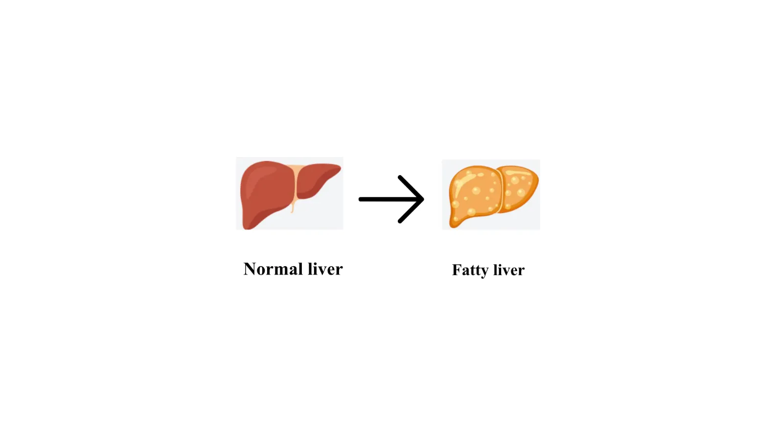
Figure 1: Comparison of Normal Liver and Fatty Liver
EPIDEMIOLOGY
MASLD is now recognized as one of the most common chronic liver diseases worldwide, with around 1.3 billion adults affected. It is present in roughly 25-30% of the adult population, but its prevalence is considerably higher among people with obesity, reaching 80-90%, and among individuals with type 2 diabetes, where it affects nearly 70%. MASLD also rises with age, estimates ranging from 7-14% in children to about 38% in adults [3][4].
The increasing occurrence of MASLD in children is particularly concerning. Global estimates indicate that its prevalence rose from 4.62% in 2000 to 8.77% in 2024, while rates as high as 52.5% have been reported among children with obesity. These trends highlight the need for effective preventive measures and early intervention during childhood [5][6].
The highest disease burden has been reported in Asia and Africa, while Western Europe has experienced a fastest proportional rise in cases since 1990. And also seen a significant increase in MASLD prevalence. This growing trend is largely linked to rapid urbanization, higher consumption of calorie-dense foods, and lower levels of physical activity linked to shifting lifestyle patterns. Current epidemiological projections suggest that the global burden of MASLD will continue to rise, with more than 55% of adults potentially affected by 2040. Among these individuals, approximately 20-30% may develop MASH, further increasing burden of a liver disease [7] [8].
ETIOLOGY
MASLD develops and progresses through the combined effects of genetic predisposition and metabolic disturbances. Cardiometabolic conditions are among major factors linked to increased disease severity and progression. Insulin resistance promotes hepatic fat buildup by boosting the synthesis of new fatty acids and impairing the body’s capacity to utilize and clear lipids efficiently. At the same time, dyslipidemia promotes further lipid deposition in the liver, worsening hepatic steatosis. Hypertension may also accelerate disease progression by causing endothelial dysfunction, vascular damage, and persistent inflammation, all of which can contribute to ongoing liver injury [9] [10].
Beyond genetic and metabolic drivers, lifestyle and environmental factors significantly shape MASLD onset and progression. Diets rich in sugar and saturated fat, limited access to healthy foods, sedentary behavior, inadequate sleep, disturbed circadian rhythms, and chronic psychological stress are all associated with greater disease risk and worse outcomes. Collectively, these elements foster metabolic dysregulation by elevating oxidative stress, sustaining low-grade inflammation, and worsening IR, thereby rendering the liver more prone to fat deposition and fibrosis [11] [12].
PATHOPHYSIOLOGY OF MASLD
Hepatic steatosis causes more fat buildup in hepatitis it is hallmark of MASLD. It arises when triglycerides-rich lipid droplets accumulate within hepatocytes and is typically diagnosed when fat is detected in over 5% of liver cells. [13]. Most of the triglycerides stored in the MASLD liver are formed when glycerol combines with free fatty acids (FFAs). Fat accumulates when the delivery and production of FFAs exceed the liver’s ability to oxidize or export them, leading to progressive storage of triglycerides within hepatocytes [14].
Obesity, particularly when accompanied by IR. Obesity-related metabolic dysfunction, increased tumour necrosis factor-α activates IκB kinase β, which disrupts insulin signalling by decreasing insulin receptor phosphorylation thereby substrates 1 and 2, contributing to IR [15]. IR drives hepatic steatosis via several linked mechanisms. First, diminished insulin-mediated suppression of hormone-sensitive lipase in adipose tissue accelerates lipolysis, elevating circulating FFAs and their hepatic uptake. Second, compensatory hyperinsulinaemia and raised glucose levels. Third, elevated insulin suppresses mitochondrial beta-oxidation of FFAs, limiting the liver’s capacity to clear excess lipids. Collectively, these alterations boost FFA availability and triglyceride synthesis, thereby fostering the onset and progression of hepatic steatosis in MASLD (Figure_2) [16].
PATHOPHYSIOLOGY OF MASH
Advancement from simple steatosis to metabolic steatohepatitis (MASH) affects approximately one-third of individuals with MASLD. On histology, MASH is defined by the simultaneous occurrence of hepatic steatosis, hepatocellular damage, and hepatic inflammation, fibrosis and cirrhosis emerging to differing extents [17].
Beyond steatosis and inflammation, MASH shows a set of typical histopathological changes: ballooned hepatocytes, fibrosis, apoptotic cell death, sinusoidal collagen deposition, megamitochondria, hepatic iron overload. The processes driving the shift from isolated steatosis to MASH were initially described by the “two-hit” hypothesis, in which insulin resistance (IR) and metabolic dysfunction act as the primary insult causing hepatic lipid accumulation, followed by secondary hits that trigger inflammation and hepatocellular damage [18]. However, growing evidence now supports a more complex “multiple‑hit” model involving several concurrent and interacting pathways. In this model, the initial metabolic disturbance driven by IR is followed by additional pathogenic processes, such as oxidative stress, inflammatory cytokine–mediated hepatocyte injury, dysregulated lipid metabolism, free fatty acid (FFA) induced toxicity, excessive intrahepatic cholesterol accumulation, hyperinsulinaemia, hyperleptinaemia, and reduced adiponectin levels. Genetic susceptibility may further contribute to inter‑individual differences in disease progression and severity. Among the many mechanisms implicated in MASH, oxidative stress and inflammatory cytokine signalling are regarded as central drivers of hepatocellular injury and fibrotic progression (Figure_2) [19].
Oxidative stress
Growing evidence points to oxidative stress as a key driver of the severity and progression of MASH. Studies in both patients and experimental models consistently show elevated markers of oxidative stress in MASH. A major contributor to this imbalance is the excess of free fatty acids (FFAs). When FFA influx outstrips the capacity of these pathways, mitochondrial fatty acid oxidation becomes overload, leading to heightened production of ROS, chronic oxidative stress tips the balance between ROS generation and the liver’s antioxidant defenses, causing oxidative damage, together with ROS, disrupt organelle function, trigger hepatocyte injury and death, and activate hepatic stellate cells, ultimately driving the onset and progression of hepatic fibrosis [20].
Proinflammatory cytokines
MASH is closely linked to ongoing liver inflammation and disturbed cytokine signaling. People with MASH often show elevated levels of proinflammatory mediators, especially TNF-alpha and IL-6. These cytokines also alter adipokine balance by lowering adiponectin and while raising leptin expression (Figure 2) [21].
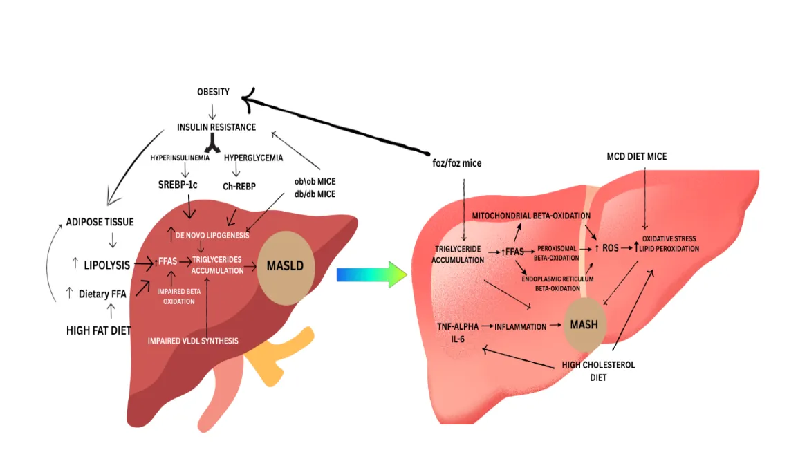
Figure 2: Key Mechanisms Involved in MASLD and MASH Progression
CLASSIFICATION OF MASLD PRECLINICAL ANIMAL MODELS
This review examines available MASLD animal models, focusing on their ability to replicate key human pathological features and support therapeutic development.
1. Animal models for Diet-Induced MASLD & MASH
1.1 High-Fat-Diet
In these models, animals receive diets in which fat provides approximately 60% of total calories, typically ranging from 45% to 75%, compared with less than 20% in standard laboratory diets [22].
Short-term HFD exposure, lasting approximately 6 to 12 weeks, increases fat accumulation, modelling the early stage of human MASLD, or simple steatosis. Longer dietary exposure of about 16 weeks produces more pronounced metabolic disturbances.
In rodents, MASLD is commonly induced by long-term HFD feeding for approximately 8–36 weeks. These models effectively reproduce key features of early MASLD, particularly hepatic lipid accumulation and simple steatosis. However, they often produce limited fibrosis and hepatic injury, reducing their ability to replicate the progression from uncomplicated MASLD to advanced MASH (Table 1) [23].
1.2 High-Fat, High-Fructose Diet (HFHF)
High intake fructose-rich, calorie-dense foods is strongly associated with MASLD onset in humans and increased hepatic fibrosis severity.
Male mice fed HFHF diet for 16 weeks consisting of high-fat diet (58kcal% from fat) and drinking water with 42g/L carbohydrates (55% fructose and 45% sucrose by weight) developed more oxidative stress, macrophage infiltration, and fibrosis than mice given the high-fat diet alone. Both groups, however, showed gains in body weight and fat mass, along with hepatic steatosis, elevated fasting glucose, and insulin resistance, suggesting that fructose drives MASLD progression beyond lipid accumulation [24].
HFHF feeding produces time-dependent liver pathology in mice, with steatosis appearing after approximately 4 weeks, steatohepatitis by 8 weeks, and fibrosis after 16 weeks. This progression resembles key stages of human MASLD and provides a useful model for studying advanced complications, including cirrhosis and HCC (Table 1) [25].
1.3 HIGH-FAT, HIGH-CHOLESTEROL DIET (HFHC)
Excessive cholesterol leads to metabolic disturbances, including dyslipidemia and IR. Evidence from animal and human studies indicates that increased cholesterol exposure promotes hepatic inflammation and accelerates MASLD progression [26].
Male C57BL/6 mice fed an HFHC diet for 30 weeks developed marked obesity, severe steatosis, increased inflammation, and perisinusoidal fibrosis. In contrast, mice fed HFD alone or a cholesterol-enriched diet showed lipid accumulation but minimal inflammation and no detectable fibrosis. HFHC-fed mice also had higher circulating cholesterol and lower serum adiponectin, reflecting broader metabolic disruption during disease progression (Table 1) [27].
1.4 METHIONINE AND CHOLINE-DEFICIENT (MCD) DIET
It is widely used to induce experimental MASH. It is typically rich in sucrose (approximately 40%), contains moderate fat (around 10%), and is deficient in methionine and choline key nutrients required for hepatic beta oxidation and VLDL production. Their absence disrupts hepatic lipid metabolism, leading to steatosis, inflammation, hepatocellular ballooning, ROS-associated injury, and fibrosis [28].
Steatosis develops within 1–2 weeks of MCD feeding, followed by marked necroinflammation at approximately 2 weeks and “chicken-wire” fibrosis by 6 weeks. MCD-fed mice also show increased serum ALT and hepatocellular ballooning, indicating substantial liver injury [29]. Compared with HFD and Western-style diets, the MCD model reproduces several features of advanced human MASH more rapidly, including steatosis, inflammation, and fibrosis (Table 1) [30].
1.5 Choline-Deficient L-Amino Acid (CDAA) Diet
It is modified MCD model where choline deficiency is retained, but the protein source is replaced with a defined L-amino acid mixture. Unlike MCD-fed mice, CDAA-fed animals generally avoid severe weight loss and do not develop IR, offering metabolic advantages over the conventional MCD model. After approximately 3 weeks, mice show increased circulating TNF-α, enhanced TLR4 and CD14 expression, hepatic lipid accumulation [31].
Adding high fat to the CDAA diet accelerates disease progression, producing severe steatosis and inflammation within 7 weeks and fibrosis. This phenotype resembles early-stage fibrotic human MASH (Table 1) [32].
1. 6 Gubra-Amylin NASH (GAN) Diet-Induced Obese (Dio) Model
It is designed to mimic the main metabolic and liver disease features seen in humans with MASH, including gradual buildup of fibrosis. In this model, mice receive diet for more than 39 weeks. This causes mice to develop obesity, high cholesterol, high insulin levels, and problems with blood sugar regulation. Alongside these metabolic issues, the mice also show liver problems such as fat buildup (steatosis), inflammation, and damage to liver cells. Recent studies have shown that using older mice speeds up disease development. In 56-week-old mice, just 12 weeks on the GAN diet can cause liver fat accumulation, inflammation, and damaged liver cells. If the diet continues for 21 weeks, the mice develop significant liver fibrosis and more severe disease features similar to advanced MASH, more so than younger mice.
Because this model closely reflects the key metabolic, tissue, and molecular changes seen in human MASH, it is a valuable tool for studying how the disease works, finding new treatment targets, and testing potential drugs (Table 1) [33].
1.7 American Lifestyle-Induced Obesity Syndrome (ALIOS) MODEL
It is useful tool for studying how MASLD increasese because it mimics many of the metabolic and liver changes seen in people who eat a Western-style diet.
In this model, mice are placed on HFD, provided with drinking water sweetened with high-fructose corn syrup (55% fructose, 45% sucrose). This regimen induces obesity, IR, and hepatic fat accumulation, accompanied by inflammatory liver injury and hepatocellular death. If the mice stay on this diet for up to 12 months, they develop more advanced liver disease similar to MASH and become more susceptible to liver cancer (HCC).
Studies analyzing the liver metabolites in these mice show major changes in fat composition, including lower levels of beneficial polyunsaturated fats and accumulation of other types of fats. There are also decreases in antioxidants and metabolites produced by gut bacteria. These changes together help drive liver injury and the progression of MASLD in this model (Table 1) [34].
Table 1: Dietary Models of MASLD and MASH
| Model | Diet composition | Obesity | Insulin resistance | Steatosis | MASH | Duration | Ref |
|---|---|---|---|---|---|---|---|
| HFD | 45-75% of the animal’s total calorie intake is derived from fat. HFD model comprised 71% fat, 11% carbohydrates and 18% protein | Yes | Yes | Yes | Yes | MASLD: 6-12 weeks MASH: 16-24 weeks |
[22] |
| HFHF | HFD is supplemented with fructose, (usually 23g/L in drinking water) | Yes | Yes | Yes | Yes | MASLD: 8-12 weeks MASH: 12-16 weeks |
[24] |
| HFHC | Approximately 1% cholesterol is added to the diet and fed alongside with HFD, which typically contains 15% to 40% fat. | Yes | Yes | Yes | Yes | MASLD: 4-8 weeks MASH: 8-12 weeks |
[27] |
| MCD | Diet typically provides 40% of its energy from sucrose and 10% from fat, but it lacks methionine and choline | No | No | Yes | Yes | MASLD:3-4 weeks MASH: 4-9 weeks |
[28] |
| CDAA | 0.1% methionine and 45% fat 60% fat and 0.1% methionine |
No | No | Yes | Yes | MASLD:3-4 weeks MASH: 3-6 weeks |
[32] |
| GAN diet | 40% saturated fat, 22% fructose, and 25% cholesterol | Yes | Yes | Yes | Yes | MASLD:12-16 weeks MASH: 38 weeks |
[33] |
| ALIOS | The regimen includes a HFD containing 30% trans fatty acids along with high-fructose corn syrup (55% fructose and 45% sucrose) provided in drinking water | Yes | Yes | Yes | Yes | MASLD:8-16 weeks MASH: 6-12 months |
[34] |
1. CHEMICAL MODELS
2.1 Stelic animal model (STAM)
The STAM mouse model is used to study how diabetes-related metabolic problems leads to the development of MASH related liver cancer. In STAM model, liver fibrosis develops quickly, usually within 8 to 12 weeks. The mice are created by giving them streptozotocin and then feeding them a high-fat diet, which speeds up metabolic issues and liver disease. According to Iida et al., STAM mice are a good model for MASLD research because they not only develop liver cancer but also show metabolic changes seen in humans, such as higher liver enzymes (AST, ALT) and disturbances in amino acid metabolism. This makes them useful for studying MASLD progression and complications (Table 2) [35].
2.2 Carbon Tetrachloride (CCL4)
Carbon tetrachloride (CCL4) is commonly employed in research to investigate liver fibrosis. However, using CCl₄ by itself mainly causes fibrosis and doesn’t mimic important metabolic issues seen in MASLD, like obesity and insulin resistance. To better replicate human disease, CCl₄ is often combined HFD. Kubota & colleagues created a model where mice were fed an HFD and then given repeated CCl₄ injections. They found that the high-fat diet alone mostly caused fat buildup in the liver (steatosis) but didn’t lead to clear signs of liver inflammation or damage (steatohepatitis) (Table 2) [36].
2.3 Diethylnitrosamine (DEN)
DEN is a strong chemical that causes cancer and is often used in liver cancer research because it disrupts cell growth and helps drive the progression from chronic liver damage and fibrosis to liver cancer (HCC). Thompson and colleagues developed a mouse model combining a high-fat diet with DEN exposure. They found that 60% of these mice developed liver cancer, along with increased oxidative stress in the liver, highlighting how metabolic problems can contribute to DEN-driven cancer development (Table 2) [37].
2.4 Thioacetamide model (TAA)
TAA is a liver-toxic chemical often used in research to cause liver injury and cirrhosis. It becomes harmful after being processed by the enzyme CYP2E1, producing reactive substances that damage the liver. In mice, TAA causes liver fibrosis in about 6-7 weeks. Hyun Seo and colleagues created a MASLD model by combining TAA injections with fasting-refeeding cycles and diets like high-fat or CDAHFD. These mice developed fat buildup and other typical MASLD liver changes, making this model useful for testing new treatments (Table 2) [38].
Table 2: Chemical Models MASLD and MASH
| Chemical | Dose | Route | Duration | Reference |
|---|---|---|---|---|
| STAM model | 0.2-1.0 mL/kg | IP | 4-12 Weeks | [35] |
| Carbon Tetrachloride (CCL4) | 35-65 mg/kg | IP or SC | 8-20 Weeks | [36] |
| Diethylnitrosamine (DEN) | 100-300 mg/kg | IP | 8-16 Weeks | [37] |
| Thioacetamide model (TAA) | 25-100 mg/kg | IP | eeks | [38] |
2. Genetic induced models
3.1 ob/ob Mice Model
The ob/ob mouse has a natural mutation causing leptin deficiency, which leads to severe insulin resistance, overeating, low activity, and obesity. This causes fat to build up abnormally in the liver and other tissues, making these mice a good model for studying early-stage MASLD, especially simple fat accumulation. However, they are less prone to liver fibrosis, limiting their use for advanced disease studies.
Administering an MCD diet to ob/ob mice for 4 weeks pushes their condition from simple fat buildup to MASH, causing liver inflammation and fibrosis. Similarly, the GAN diet causes more severe fibrotic MASH in ob/ob mice than in normal mice, showing gene activity patterns similar to human MASH, making it useful for testing treatments (Table 3) [39].
3.2 db/db mouse model
It has mutation in leptin receptor that disrupts signaling of leptin, causing obesity, overeating, insulin resistance, liver fat buildup, and diabetes. This makes it useful for studying type 2 diabetes-related MASLD. It causes greater inflammation in adipose tissue but less in the liver, along with distinct patterns of fat distribution.
Their metabolic traits resemble early human MASLD, like IR, but they rarely develop advanced fibrosis on their own. To cause severe MASH with inflammation and fibrosis, additional factors like special diets or chemicals are needed. Iron overload speeds up disease by increasing oxidative stress and harming insulin function.
While db/db mice model early MASLD well and some early fibrosis, they don’t fully mimic late-stage disease or liver cancer (Table 3) [40]. `
3.3 foz/foz Mouse Model
Mouse harbors changes in Alms1 and causes increase in weight, IR, fat accumulaton, and it is useful model for MASLD research. When fed a Western diet, foz/foz mice advance through the major stages of human MASLD: they develop steatosis in 1 to 2 weeks, exhibit features such as inflammation and ballooning of liver by 4 weeks, fibrosis by 12 weeks. Disease severity is influenced by genetic background, with C57BL/6J mice exhibiting greater hepatic fibrosis than BALB/c mice. Transcriptomic studies further support the translational relevance of this model (Table 3) [41].
3.4 B6-Alms1-del Mice model
To better understand how Alms1 deficiency drives MASLD progression, researchers created B6-Alms1-del model on C57BL/6J background using CRISPR/Cas9 to delete 11 base pairs in exon 8. Unlike the foz/foz model, which develops disease rapidly. Metabolic disturbances including obesity, elevated blood sugar, IR, and abnormal lipid levels appear by 6 weeks of age, with clear steatohepatitis developing around 21 weeks. Mild liver fibrosis develops gradually between 5 and 8 months, while the mice maintain overall metabolic stability over the long term. Feeding a HFHS increases both metabolic dysfunction and hepatic injury. Because these mice survive longer and maintain a stable metabolic profile, they overcome key limitations of other genetic models (Table 3) [42].
3.5 Low-Density Lipoprotein Receptor-Deficient (Ldlr-/-) Mice And Apolipoprotein E-Deficient (Apoe-/-) Mice
Both are well-established models. When fed Western-style diets, they also develop high blood lipid levels, IR, liver fat accumulation, followed by inflammation. The LDL receptor (LDLR) family mediates uptake of lipoproteins such as ApoE and is critical for lipid homeostasis. In Ldlr−/− mice, liver disease progresses in a diet-dependent manner: high-fat, high-cholesterol feeding triggers steatosis and inflammation within a week, while longer exposure boosts oxidized LDL uptake and worsens hepatic injury. Diet composition plays a crucial role. Together, these dietary patterns capture key features of MASH with fibrosis. Similarly, Apoe-/- mice naturally develop high cholesterol and lipid levels due to impaired lipoprotein clearance (Table 3) [43].
Table 3: Genetic Models of MASLD and MASH
| Models | Obesity | Insulin Resistance | Steatosis | Steatohepatitis | Duration | Ref |
|---|---|---|---|---|---|---|
| ob/ob mice | Yes | Yes | Yes | No | MASLD: 6-12 weeks MASH:12-16 weeks |
[39] |
| db/db mice | Yes | Yes | Yes | No | MASLD:6-12 weeks MASH:12-16 weeks |
[40] |
| foz/foz mice | Yes | Yes | Yes | No | MASLD:1-2 weeks MASH: 4 weeks | [41] |
| B6-Alms1^del Mice | Yes | Yes | Yes | No | MASLD: 6-21 weeks MASH: 21-25 weeks |
[42] |
| Ldlr-/- mice and Apoe-/- mice | Yes | Yes | Yes | Yes | MASLD: 6-21 weeks MASH: 7-10 weeks |
[43] |
4. COMBINATION MODES
4.1 High-Fat Diet + CCL4
In the starting face of combined CCL4 and high-fat diet (HFD) exposure, CCl₄ induces hepatocellular injury and initiates fibrogenesis by activating hepatic stellate cells and promoting collagen buildup. With continued treatment (8–12 weeks), the synergy between diet-driven and combination of metabolic stress and repeated chemical injury results in advanced MASH, characterized by liver cells ballooning, inflammation. By 16 to 24 weeks, it exhibits accelerated fibrosis and an increased tendency to develop HCC. Overall, HFD+CCl₄ combination yields stronger fibrotic responses and faster hepatocarcinogenesis than CCl₄ alone (Table 4) [44].
4.2 Streptozotocin + High-Fat-Diet
The classic STAM model involves giving 2-day-old C57BL/6J mice a single subcutaneous injection of 200mg streptozotocin followed by feeding them a HFD starting at 4 weeks of age to accelerate MASH-associated hepatocellular carcinoma (HCC). Early STZ exposure causes irreversible β-cell damage and persistent hyperglycaemia. On HFD, these mice rapidly develop MASH in 6 to 8 weeks, with fibrosis and HCC emerging around 20 weeks. However, the model has major drawbacks. A refined alternative uses repeated low-dose intraperitoneal injection of streptozotocin (STZ) at 40mg/kg for 5 days 7 weeks old mice, and HFD for 8 weeks. This approach produces slower, more human-like disease progression: steatosis by around 14 weeks, MASH by 20 weeks (Table 4) [44].
4.3 CDAA DIET + CCL4
Mice fed a CDAA diet and given i.p injection of CCl₄ develop more severe NASH and HCC-like pathology than those on CDAA alone. The combination amplifies hepatic steatosis, lobular inflammation, and fibrosis. While only about 35% of CDAA-fed mice develop HCC, adding CCl₄ induces tumors in all animals, with larger average tumor sizes, highlighting the enhanced carcinogenic potency of the combined model (Table 4) [45].
4.4 WD + Foz/Foz mice
Combining metabolically stressful diets with genetically susceptible backgrounds can accelerate disease progression. It is associated with human Alstrom syndrome a condition characterized by obesity, IR, and cardiometabolic complications. When fed a western diet it rapidly causes increased weight, high insulin levels, abnormal lipid profiles, and severe steatohepatitis within just 12 weeks much faster than models relying on diet alone. A key advantage of the Foz/foz model produces complications commonly seen in MASLD. However, since pathogenic ALMS1 mutations are rare in the general population, this model reflects an accelerated disease course rather than the common genetic drivers of MASLD, such as PNPLA3 and TM6SF2 variants (Table 4) [45].
Table 4: Combination Models of MASLD and MASH
| Models | Obesity | Insulin Resistance |
Steatosis | Inflammation | Duration | Ref |
|---|---|---|---|---|---|---|
| HFD + CCl4 | Yes | Yes | Yes | Yes | MASLD: 2-4 weeks MASH: 8-12 weeks |
[44] |
| HFD + STZ | No | No | Yes | Yes | MASLD: 6-8 weeks MASH: 6-8 weeks |
[44] |
| CDAA + CCl4 | No | No | Yes | Yes | MASLD: 4-5 weeks MASH: 4-8 weeks |
[45] |
| WD + Foz/Foz mice | Yes | Yes | Yes | Yes | MASLD: 1-2 weeks MASH: 4-5 weeks |
[45] |
5. MISCELLANEOUS
5.1 Infection induced models
Microbial and infectious agents can accelerate MASLD progression by intensifying liver inflammation and fibrosis. For instance, intravenous infection with Cnm- and PA-positive Streptococcus mutants, a major bacterium linked to tooth decay, has been shown to aggravate MASH-like liver damage. These findings under-scores how specific bacterial virulence factors can contribute to liver injury. Similarly, liver fluke infections in hamsters provoke robust inflammatory and fibrogenic responses, promoting periductal fibrosis and facilitating the shift from simple steatosis to MASH, mirroring clinical patterns seen in patients. [46].
5.2 IMMUNITY INDUCED MODELS
5.2.1 HIL Mice
Chronic activation of the immune system especially by CD4 T cells is a major driver of MASH progression. Mice fed HFHC diet for 20 weeks, researchers observed classic signs of MASH, including liver fat accumulation, inflammation, and fibrosis. These changes coincided with greater infiltration of human cd4 memory T cells into the liver and higher levels of proinflammatory cytokines such as IL-17A, highlighting the important role of adaptive immunity in advancing the disease [46].
5.2.2 Pfp/Rag2−/− mice
Pelz and colleagues developed a diet-induced model of MASH using immunodeficient mice to evaluate human mesenchymal stem cell derived hepatocyte like cells as a therapy. By feeding these mice MCD diet, they reproduced key MASH features in an immunocompromised setting, enabling detailed cellular and molecular analysis of disease progression. This approach offers the first platform to directly assess how transplanted human stem cell–derived hepatocytes influence MASH, providing a valuable tool for evaluating regenerative therapies [46].
FUTURE PROSPECTS
Future progress in preclinical animal models for MASLD and MASH hinges on making them more translatable, reproducible, and reflective of the human condition. Since no single model captures all metabolic, inflammatory, fibrotic, and histological aspects of the disease, researchers should use complementary models tailored to specific questions. Diets should better mimic real-world intake by combining fats, fructose, refined carbs, and cholesterol to drive realistic metabolic dysfunction and liver injury. Combination approaches can be refined to ensure steady progression from simple steatosis to steatohepatitis and fibrosis, while avoiding harsh chemical triggers. Models must also represent diverse patient types lean MASLD, obesity-linked disease, diabetes, and genetically prone individuals and account for sex, age, strain, and genetic background, which shape disease and drug responses. Genetically engineered and humanized models can illuminate human-specific pathways, and incorporating gut–liver axis elements (microbiota shifts, barrier function, bile acids, and microbial metabolites) will deepen mechanistic insight. Multi-omics and microbiome profiling will help match animal phenotypes to human disease at the molecular level. Standardizing diet recipes, induction timelines, biochemical tests, histology scoring, and molecular endpoints will boost reproducibility across labs. The field should prioritize systematic validation against human disease features and test promising therapies across multiple models before moving to clinical trials, accelerating the discovery of safe, effective treatments.
REFERENCES

Bathula Jayasri, Kompelli Bhanudeepika, Dr. Venu Talla, Preclinical Animal Models of MASLD and MASH: A Review of Disease Induction Strategies, Int. J. of Pharm. Sci., 2026, Vol 4, Issue 11, 3705-3717. https://doi.org/10.5281/zenodo.24413495
 10.5281/zenodo.24413495