

We use cookies to ensure our website works properly and to personalise your experience. Cookies policy
Dayanand Institute of Pharmacy, latur.
Multidrug-resistant (MDR) Klebsiella pneumoniae has become a major public health concern because of its ability to cause severe infections and resist multiple classes of antimicrobial agents. The emergence of extended-spectrum beta-lactamase-producing and carbapenem-resistant strains has significantly reduced the effectiveness of conventional antibiotic treatment. Bacteriophage therapy has gained attention as an alternative and complementary antimicrobial strategy because bacteriophages are viruses that specifically infect and destroy bacterial cells. Lytic bacteriophages can recognize bacterial surface receptors, introduce their genetic material, replicate inside susceptible bacteria, and cause bacterial cell lysis. In addition, certain phages produce depolymerase enzymes that degrade bacterial capsules and may help disrupt biofilms. Phage cocktails, personalized phage therapy, phage-derived enzymes, and phage–antibiotic combination therapy are important areas of current research. Preclinical investigations and selected clinical case reports have demonstrated promising results; however, therapeutic outcomes depend on bacterial susceptibility, phage characteristics, infection site, and treatment conditions. Important challenges include the narrow host range of individual phages, the development of phage resistance, immune responses, formulation difficulties, and limited controlled clinical evidence. This review discusses the biology, mechanisms, therapeutic applications, advantages, limitations, and future perspectives of bacteriophage therapy against MDR K. pneumoniae. Further clinical trials, standardized manufacturing procedures, and regulatory guidance are necessary to establish its safety and effectiveness in routine clinical practice.
Klebsiella pneumoniae is a Gram-negative, encapsulated, non-motile, rod-shaped bacterium belonging to the family Enterobacteriaceae. It is commonly found in the human gastrointestinal tract and in environmental sources. Although it may exist as part of the normal microbial community without causing disease, it can become an opportunistic pathogen under suitable conditions. It is particularly important in hospitalized patients, intensive care unit patients, newborns, elderly individuals, and people with weakened immune systems.K. pneumoniae can cause several serious infections, including pneumonia, bloodstream infections, urinary tract infections, wound infections, surgical-site infections, liver abscesses, and other healthcare-associated infections. Its ability to acquire antimicrobial resistance and virulence-associated characteristics makes it a significant clinical concern. The World Health Organization has highlighted the global spread of carbapenem-resistant and hypervirulent K. pneumoniae as an important public health challenge.Antibiotics are the principal treatment for bacterial infections. However, the increasing and inappropriate use of antimicrobial agents has contributed to the emergence and spread of resistant bacteria. Multidrug-resistant K.
pneumoniae may resist several antibiotic classes, including beta-lactams, fluoroquinolones, and aminoglycosides. Some isolates produce extended-spectrum beta-lactamases (ESBLs), while others produce carbapenemases such as KPC, NDM, or OXA-48-like enzymes. These mechanisms can substantially limit treatment options.Bacteriophages, commonly called phages, are viruses that infect bacteria. They were discovered in the early twentieth century and have been investigated as antibacterial agents because of their ability to recognize specific bacterial hosts. Unlike conventional antibiotics, lytic bacteriophages multiply within susceptible bacteria and may cause the destruction of bacterial cells. Their specificity and potential activity against antibiotic-resistant bacteria have renewed scientific interest in phage therapy .Bacteriophage therapy is not a universal replacement for antibiotics. Instead, it is being investigated as an additional treatment strategy for selected infections, especially when conventional antimicrobial options are limited. Recent research has explored phage cocktails, personalized phage selection, phage-derived enzymes, and combinations of phages with antibiotics. These approaches may help improve antibacterial activity and address the challenges associated with phage resistance .
Multidrug-Resistant Klebsiella pneumoniae
Definition and clinical importance
Multidrug resistance refers to acquired non-susceptibility to at least one agent in three or more antimicrobial categories under commonly used international definitions. MDR K. pneumoniae presents a particular challenge because it can acquire multiple resistance mechanisms and spread within healthcare settings.
Carbapenem-resistant K. pneumoniae is especially concerning because carbapenems are important agents for treating many serious infections caused by resistant Gram-negative bacteria. Infections caused by these organisms may require specialized antimicrobial selection and close clinical monitoring.
Mechanisms of antibiotic resistance
Several mechanisms contribute to antibiotic resistance in K. pneumoniae.
Beta-lactamase production: ESBLs can inactivate many penicillins and cephalosporins. Carbapenemases, including KPC, NDM, and OXA-48-like enzymes, can compromise carbapenem activity.
Reduced membrane permeability: Changes or loss of outer-membrane porins may reduce the entry of some antibiotics into bacterial cells.
Efflux pumps: These systems transport certain antimicrobial compounds out of bacterial cells and may contribute to reduced antibiotic susceptibility.
Target modification: Genetic changes in antibiotic targets can reduce the binding and effectiveness of particular drugs.
Biofilm formation: Bacteria within biofilms may show increased tolerance to antimicrobial treatment because of the protective matrix, altered physiological activity, and other community-associated mechanisms.
These mechanisms can occur together, resulting in strains with limited treatment options. The appropriate antibiotic regimen must therefore be selected according to clinical circumstances, microbiological results, and current treatment guidance.
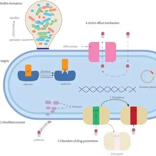
Fig.An-overview-of-antimicrobial-resistance-mechanisms
Role of virulence factors
The polysaccharide capsule is an important virulence factor of K. pneumoniae. It can protect bacteria from host immune defenses and influence their interactions with the environment. Other factors, including adhesins, siderophores, lipopolysaccharide, and biofilm-associated properties, may contribute to infection and persistence. Some hypervirulent strains cause invasive disease, while certain strains also carry antimicrobial-resistance determinants. The combination of resistance and virulence can create particularly difficult clinical situations. Bacteriophage research therefore considers not only bacterial killing but also interactions between phages, capsules, resistance mechanisms, and virulence traits.
Overview and Classification of Bacteriophages
Definition of bacteriophages
Bacteriophages are viruses that infect bacterial cells and use bacterial cellular machinery to produce new phage particles. They are widely distributed in soil, water, sewage, food, and the human body. Their natural abundance and diversity provide a large source of potential antibacterial agents. A typical bacteriophage contains genetic material, either DNA or RNA, enclosed within a protein capsid. Many phages that infect Gram-negative bacteria possess tail structures that help them attach to receptors on the bacterial surface and deliver their genetic material into the host cell. The interaction between a phage and its bacterial host depends on the bacterial surface structure, phage receptor-binding proteins, and other molecular factors. In K. pneumoniae, the capsule, lipopolysaccharide, and outer-membrane proteins may influence phage attachment and infection.
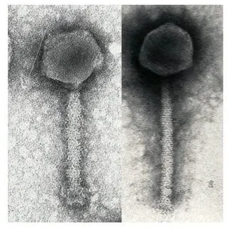
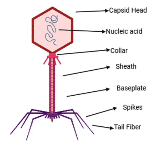
Fig.Electron microscope picture of a bacteriophage (left) and phage diagram (right).
Classification according to life cycle
Bacteriophages can be broadly classified according to their replication strategies.
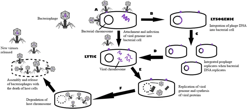
Lytic bacteriophages: These phages infect susceptible bacteria, replicate within the cells, and cause bacterial lysis. They are generally preferred for therapeutic development because their life cycle directly results in bacterial destruction.
Temperate bacteriophages: These phages can establish a lysogenic relationship with their host. Their genetic material may integrate into the bacterial genome or persist without immediately destroying the cell. Such phages require particular caution in therapeutic development because lysogeny and genetic exchange may introduce undesirable traits.
For therapeutic applications, phages should undergo detailed characterization to confirm their identity, host range, genome characteristics, lytic activity, purity, and safety. The selection of appropriate phages is an essential step in the development of a therapeutic preparation [6].
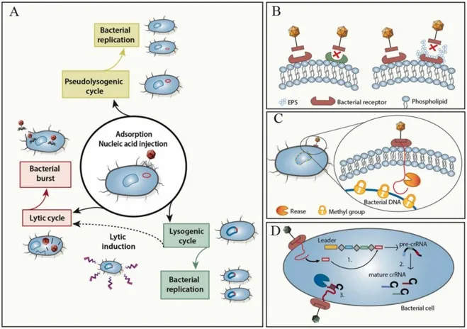
Fig. Phage invasion of bacteria and bacterial evasion mechanisms: A. After phage adsorption to a bacterial host and injection of the genome into the host cell, the phage can undergo a lysis cycle leading to bacterial lysis or enter a lysogenic or pseudolysogenic cycle that does not lyse the host bacteria; B. Innate bacterial phage evasion mechanisms by mutation of extracellular phage receptors or receptor masking; C. Innate bacterial phage genomes cleaved after entry into the bacterial cell bacterial phage evasion mechanism; D. Adaptive bacterial anti-phage mechanism exemplified by the CRISPR-Cas system that includes three phases:acquisition, expression, and interference .
Mechanism of Action of Bacteriophage Therapy
The antibacterial action of a lytic bacteriophage occurs through a sequence of biological events.
1. Attachment to the bacterial surface
The first step involves recognition and attachment of the phage to specific receptors on the bacterial surface. In K. pneumoniae, these receptors may include capsular polysaccharides, lipopolysaccharides, or outer-membrane proteins. Receptor recognition determines whether a particular bacterial strain is susceptible to the phage.
2. Penetration of genetic material
After attachment, the phage introduces its genetic material into the bacterial cell. The genetic material contains instructions required for producing new phage components and controlling the subsequent stages of infection.
3.Replication and synthesis
The phage uses the host cell's machinery to replicate its genome and produce viral proteins. The number of new phage particles produced depends on the particular phage, bacterial strain, environmental conditions, and availability of cellular resources.
4. Assembly of new phage particles
Newly synthesized genetic material and structural proteins are assembled into complete phage particles. These particles accumulate inside the infected bacterial cell before release.
5.Bacterial cell lysis
Lytic phages commonly produce proteins, including endolysins and holins, that contribute to disruption of the bacterial cell envelope. The infected cell breaks open and releases newly formed phages. These particles may then infect other susceptible bacteria.
This process can result in repeated cycles of bacterial infection and destruction while susceptible host bacteria remain available. However, the degree of bacterial reduction depends on phage susceptibility, phage concentration, bacterial density, host conditions, and other factors.
6. Capsule degradation
Certain K. pneumoniae phages produce depolymerase enzymes that degrade capsular polysaccharides. Because the capsule can contribute to immune evasion, its degradation may expose bacterial cells to host immune defenses and improve access to other antibacterial treatments. Capsule degradation does not always result in complete bacterial killing. Some depolymerases primarily modify the capsule, while the phage's lytic activity is responsible for cell destruction. The activity and specificity of these enzymes must be assessed for each candidate phage.
Diagnosis And Treatment
Diagnosis of Antibiotic Resistance in Klebsiella pneumoniae
1. Sample Collection
Common samples include urine, blood, sputum, and wound swabs. Samples must be collected using aseptic techniques and transported promptly to the microbiology laboratory. Proper sample collection helps prevent contamination and ensures reliable results. Accurate collection is the first step in identifying Klebsiella pneumoniae and determining its antibiotic resistance pattern.
2. Bacterial Culture
The collected sample is cultured on suitable laboratory media to isolate Klebsiella pneumoniae. The organism is identified based on colony characteristics and laboratory identification tests. K. pneumoniae typically produces lactose-fermenting colonies on MacConkey agar. Further identification may be performed using biochemical tests or automated identification systems. Bacterial culture confirms the presence of the organism and provides an isolate for antibiotic susceptibility testing.
3. Antibiotic Susceptibility Testing (AST)
Antibiotic susceptibility testing determines whether Klebsiella pneumoniae is susceptible or resistant to specific antibiotics. Common methods include the Kirby–Bauer disk diffusion method and broth microdilution. In the disk diffusion method, antibiotic disks are placed on inoculated agar, and the inhibition zones are measured. Broth microdilution determines the minimum inhibitory concentration (MIC). The results help identify effective antibiotics and detect resistance to important drugs, including carbapenems and cephalosporins.
4. Molecular Methods
Molecular methods detect specific genes responsible for antibiotic resistance in Klebsiella pneumoniae. Molecular testing can provide rapid information about resistance mechanisms. However, detecting a resistance gene does not replace susceptibility testing because the complete resistance pattern must also be determined.
5. Detection of Specific Resistance Mechanisms
Specific laboratory tests help identify important resistance mechanisms in Klebsiella pneumoniae. The modified carbapenem inactivation method (mCIM) can detect carbapenemase production. The EDTA-modified carbapenem inactivation method (eCIM) can help differentiate metallo-beta-lactamases from certain other carbapenemases. Phenotypic tests for extended-spectrum beta-lactamases (ESBLs) can also identify resistance to extended-spectrum cephalosporins. These methods help laboratories investigate multidrug-resistant strains and support appropriate infection-control measures.
6. Interpretation of Results
Laboratory results are interpreted according to established guidelines, such as those published by the Clinical and Laboratory Standards Institute (CLSI) or the European Committee on Antimicrobial Susceptibility Testing (EUCAST). Antibiotic susceptibility results are classified using the categories defined by the applicable standard. The minimum inhibitory concentration (MIC), inhibition-zone diameter, and detected resistance mechanisms are considered. Resistant K. pneumoniae isolates may require alternative treatment and additional infection-control precautions. The final treatment decision is made by the treating clinician based on susceptibility results, infection site, and patient condition [.
Treatment for Antibiotic Resistance in Klebsiella pneumoniae
Ceftazidime–avibactam contains two active components.
Ceftazidime: It is a cephalosporin antibiotic that binds to penicillin-binding proteins and inhibits peptidoglycan cross-linking during bacterial cell-wall synthesis. This weakens the cell wall and can lead to bacterial cell death.
Avibactam: It is a non-β-lactam β-lactamase inhibitor. It inhibits several class A, class C, and certain class D β-lactamases, protecting ceftazidime from enzymatic degradation.
Together, these components restore ceftazidime activity against certain bacteria that produce β-lactamases. However, avibactam does not inhibit class B metallo-β-lactamases, including NDM, VIM, and IMP.
Role in Multidrug-Resistant Infections
Ceftazidime–avibactam is used for selected infections caused by susceptible Gram-negative bacteria. Its clinical importance includes infections caused by KPC-producing and OXA-48-like-producing Enterobacterales. It may also be used for susceptible strains of Pseudomonas aeruginosa.
It should not be considered effective against every carbapenem-resistant organism.
Ceftazidime–Avibactam Combination Therapy
Ceftazidime–Avibactam Ceftazidime–avibactam (CZA) is a combination of ceftazidime, a third-generation cephalosporin antibiotic, and avibactam, a non-β-lactam β-lactamase inhibitor. It is used to treat difficult-to-treat infections caused by resistant Gram-negative bacteria, including carbapenem-resistant Enterobacterales (CRE) and Pseudomonas aeruginosa. CRE infections are associated with high mortality rates, reaching up to 50% in some studies. In 2015, the US FDA approved CZA for complicated intra-abdominal infections (with metronidazole) and complicated urinary tract infections, including pyelonephritis, under specified conditions. In 2016, the European Medicines Agency approved its use for complicated urinary tract infections, complicated intra-abdominal infections, and hospital-acquired pneumonia, including ventilator-associated pneumonia. CZA has also been investigated for CRE bloodstream infections, either alone or combined with other antibiotics, because effective treatment options are limited. Systematic reviews and network meta-analyses help compare the effectiveness of CZA monotherapy and combination therapy in reducing mortality among patients with CRE infections.
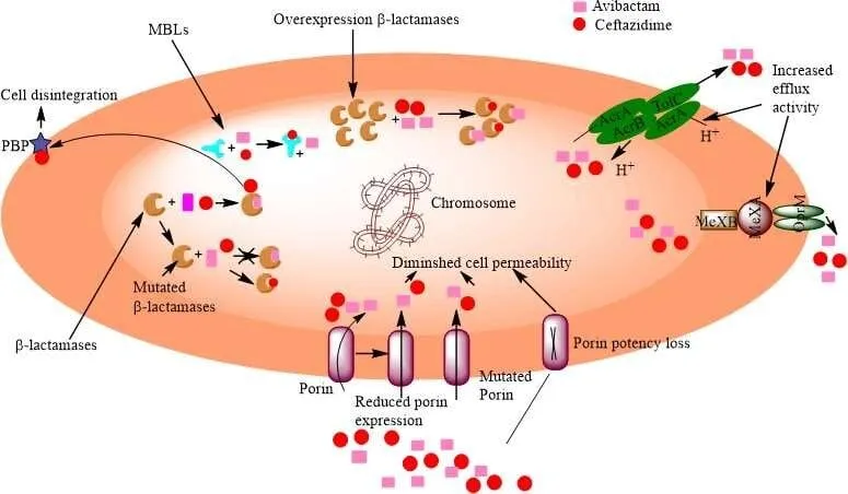
Fig.Mode of action of avibactam and ceftazidime
Clinical Applications
Ceftazidime–avibactam may be used for selected complicated urinary tract infections, complicated intra-abdominal infections in combination with metronidazole, hospital-acquired pneumonia, ventilator-associated pneumonia, and other susceptible Gram-negative infections according to approved indications and clinical guidelines.It is also an important treatment option for selected carbapenem-resistant Enterobacterales infections. The combination with aztreonam is particularly relevant to metallo-β-lactamase-producing Enterobacterales. Clinical evidence supports its use in appropriate cases, although outcomes vary with the organism, infection site, and severity.
Mechanism of Action of Ceftazidime–Avibactam
Ceftazidime–avibactam is a combination antibiotic used to treat certain bacterial infections caused by multidrug-resistant (MDR) Gram-negative bacteria. It consists of two components: ceftazidime, a third-generation cephalosporin antibiotic, and avibactam, a β-lactamase inhibitor. These components work together to overcome certain bacterial resistance mechanisms and inhibit bacterial growth.
1. Binding to Penicillin-Binding Proteins (PBPs)
Ceftazidime binds to penicillin-binding proteins (PBPs), which are enzymes involved in the final stages of bacterial cell-wall synthesis. These proteins are essential for maintaining the structure and integrity of the bacterial cell wall.
2. Inhibition of Cell-Wall Synthesis
After binding to PBPs, ceftazidime inhibits the cross-linking of peptidoglycan, an important component of the bacterial cell wall. This weakens the cell wall and interferes with the bacterium's ability to maintain its normal shape and structural stability.
3. Inhibition of β-Lactamase Enzymes
Many resistant bacteria produce β-lactamase enzymes that break down β-lactam antibiotics, including ceftazidime. Avibactam inhibits several important β-lactamases, including many class A enzymes (such as KPC), class C enzymes (AmpC), and certain class D enzymes (such as OXA-48-like enzymes).
4. Protection of Ceftazidime from Degradation
By inhibiting susceptible β-lactamase enzymes, avibactam prevents or reduces the enzymatic breakdown of ceftazidime. This allows ceftazidime to retain its antibacterial activity against bacteria that produce these enzymes. However, avibactam does not inhibit every type of β-lactamase.
5. Inhibition of Bacterial Growth
Once protected from susceptible β-lactamases, ceftazidime can continue to bind to PBPs and inhibit bacterial cell-wall synthesis. The resulting structural weakness interferes with bacterial survival and growth.
6. Bacterial Cell Death
Continued inhibition of cell-wall synthesis can lead to bacterial cell death, particularly in actively growing, susceptible bacteria. The effectiveness of treatment depends on factors such as the bacterial species, resistance mechanism, antibiotic concentration, and susceptibility of the bacterial isolate.
Limitations and Challenges
1 Narrow host range
Many bacteriophages infect only particular bacterial strains or closely related groups. Consequently, a phage effective against one K. pneumoniae isolate may not work against another. This limits the use of fixed phage preparations and creates a need for susceptibility testing.
2 Development of phage resistance
Bacteria can develop resistance to phages through changes in surface receptors, capsule structure, restriction systems, or other defense mechanisms. Resistance may reduce treatment effectiveness or allow bacterial regrowth.
Phage cocktails and combination strategies may help address this problem, but they do not eliminate the possibility of resistance.
3 Immune responses
The human immune system can recognize and remove phage particles. Antibody production and other immune mechanisms may affect the activity of phages, particularly during repeated administration. The extent of this effect depends on the phage, route of administration, treatment duration, and individual patient.
4 Pharmacokinetic and delivery challenges
Phage distribution and clearance differ from those of conventional antibiotics. Reaching the infection site, maintaining an effective concentration, and preserving phage activity in biological fluids can be challenging. Appropriate delivery methods must be evaluated for each infection type.
5 Manufacturing and quality control
Therapeutic phage preparations require careful control of identity, potency, purity, stability, and microbiological quality. Bacterial contaminants, residual bacterial products, and undesirable genetic elements must be addressed through validated manufacturing and testing procedures.
6 Limited clinical evidence
Although laboratory studies and selected clinical cases are encouraging, evidence remains limited for many phage–pathogen–infection combinations. Differences in phage preparations, doses, administration routes, concurrent antibiotics, and outcome measurements make comparisons difficult.
7 Regulatory and ethical challenges
Regulatory requirements for phage products may differ across jurisdictions, particularly for personalized or rapidly modified preparations. Additional concerns include informed consent, access to treatment, product traceability, environmental release, and long-term monitoring.
Addressing these challenges will require coordinated work involving microbiology, pharmaceutical sciences, infectious disease medicine, clinical trial design, and regulatory authorities[6,10] .
Recent Advances and Future Perspectives
1 Improved phage screening
Advances in bacterial identification, genome sequencing, and laboratory susceptibility testing may help researchers select suitable phages more efficiently. Rapid screening could reduce delays between pathogen identification and therapeutic assessment.
2 Expanded phage libraries
Phage banks containing well-characterized bacteriophages may improve access to candidates with activity against a wider range of resistant bacterial isolates. Reliable databases linking phage characteristics to bacterial susceptibility could support future clinical development.
3 Artificial intelligence and computational tools
Computational methods and machine learning are being investigated to predict phage–host interactions, analyze phage genomes, identify candidate receptors, and guide engineering strategies. Such methods may improve candidate selection, but predictions require experimental validation.
4 Combination treatment development
Future studies should investigate phage cocktails, phage–antibiotic combinations, and phage-derived enzymes under standardized experimental conditions. Controlled studies are needed to determine which combinations offer meaningful benefits and whether they reduce the emergence of resistance.
5 Clinical trials and personalized medicine
Well-designed clinical trials should evaluate safety, effectiveness, pharmacokinetics, appropriate treatment duration, and patient outcomes. Research should also examine which patient groups and infection types are most likely to benefit from phage therapy.
6 Standardization and regulation
Standardized protocols for phage characterization, susceptibility testing, manufacturing, formulation, and clinical monitoring will be essential for wider use. Regulatory frameworks should balance product safety and quality with the need to respond to rapidly evolving resistant bacterial infections.Recent reviews identify phage engineering, computational prediction, combination treatment, and better clinical development pathways as important areas for future progress .
CONCLUSION
Bacteriophage therapy represents a promising emerging approach for the management of multidrug-resistant Klebsiella pneumoniae. Its ability to selectively infect susceptible bacteria, potentially disrupt biofilms, and complement selected antibiotics has created renewed interest in its therapeutic application. Phage cocktails, personalized phage selection, and phage-derived enzymes offer additional opportunities to improve antibacterial strategies.However, bacteriophage therapy should not currently be regarded as a universal replacement for conventional antibiotics. Its clinical effectiveness depends on the susceptibility of the bacterial isolate, the properties of the phage preparation, the site of infection, and the patient's condition. Challenges related to phage resistance, immune responses, pharmacokinetics, manufacturing, and regulatory approval must also be addressed.Future progress will require well-designed clinical trials, rapid phage-susceptibility testing, reliable manufacturing systems, and standardized treatment protocols. With continued research and appropriate clinical oversight, bacteriophage therapy may become a valuable complementary option for selected patients with difficult-to-treat MDR K. pneumoniae infections.
REFERENCES



Todkar Ganesh, Dnyaneshwari Marlapalle, Amarja Bhoge, Nikita Kale, Anvirappa Katte, Ramprasad Khandare, Shredatta Shetkar, Bacteriophages Therapy as an Emerging Alternative for Multidrug-resistance Klebsiella Pneumonia, Int. J. of Pharm. Sci., 2026, Vol 4, Issue 11, 722-732. https://doi.org/10.5281/zenodo.23939724
 10.5281/zenodo.23939724