

We use cookies to ensure our website works properly and to personalise your experience. Cookies policy
Lokmanya Tilak Institute of Pharmacy, Navi Mumbai, Maharashtra, India.
Sleep disorders, particularly insomnia and circadian rhythm disturbances, are common health concerns affecting the overall well-being of individuals. Melatonin, an endogenous hormone secreted by the pineal gland, plays an important role in regulating the sleep–wake cycle and maintaining circadian rhythm. However, conventional oral melatonin preparations may have variable bioavailability due to extensive hepatic first-pass metabolism and gastrointestinal absorption. Buccal drug delivery has emerged as a promising alternative to overcome these limitations. This review focuses on the therapeutic role of melatonin in sleep disorders and its potential delivery through mucoadhesive buccal films and other transmucosal formulations. It discusses melatonin synthesis, pharmacological properties, mechanism of action, buccal mucosal absorption and the advantages of buccal administration. Various formulation approaches, including mucoadhesive polymers, permeation enhancers, liposomes and niosomes, along with important evaluation parameters, are also highlighted. Overall, buccal delivery of melatonin may offer improved drug absorption and patient compliance. However, further formulation development, pharmacokinetic investigations and clinical studies are required to establish its safety and therapeutic effectiveness compared with conventional oral preparations.
1.1 Importance of sleep:
Sleep plays an essential role in the maintenance of physical and mental health. High-quality sleep is critically important for cognitive function and memory, the immune system, reproductive function, and hormone regulation. By contrast, insufficient sleep has been linked to cardiovascular disease, stroke and hypertension, obesity and type 2 diabetes, and increased susceptibility to infections and inflammatory disorders, contributing to higher overall mortality rates. Sleep abnormalities are common in a variety of medical conditions and can occur at any age, ranging from childhood and adolescence through adulthood and older age.
Insomnia can be treated by non-pharmacological and pharmacological approaches. The former includes behavioural and environmental interventions such as sleep hygiene advice, sleep–wake scheduling, timed physical activity, light exposure therapy, somatic interventions, and cognitive–behavioural therapy.[1]
1.2 Need for melatonin therapy:
Melatonin (N-acetyl-5-methoxytryptamine) is a hormone that is produced naturally and primarily by the pineal gland as a function of the circadian timing system. Its secretion typically increases during darkness, thereby triggering biological processes that make the body aware that it is nighttime. Through its action on MT1 and MT2 receptors, melatonin plays a key role in circadian rhythms and timing of sleep. As a consequence, its exogenous administration can have a therapeutic effect on certain sleep disorders, particularly those related to a circadian misalignment, delayed sleep timing and difficulty falling asleep. Its role must be differentiated from that of typical sedative–hypnotic drugs, since its main therapeutic effect is to regulate the sleep timing and circadian physiology, and not to depress non-specifically the central nervous system.[2]
2. CIRCARDIAN RHYTHM:
2.1 Overview:
Circadian rhythms are present in almost all organisms, and most physiological processes have inherent circadian rhythmicity. These rhythms are endogenously driven and persist in the absence of external time-giving stimuli. The main circadian pacemaker is the suprachiasmatic nucleus (SCN), a paired structure, located in the anterior hypothalamus. In humans, the SCN contains about 20,000 neurons, whose firing follows self-sustained oscillations with a period of approximately 24 hours. The SCN is a central command centre for circadian rhythms; it receives and relays information about timing to other brain regions and virtually all peripheral tissues.
Thus, most physiological processes in humans follow circadian oscillations. The circadian rhythm of wakefulness and sleep is the best-known example of a circadian oscillator. Circadian rhythm disorders may arise as a consequence of disruption of endogenous circadian oscillation, or desynchronization between endogenous and exogenous circadian rhythms. Such disorders may adversely affect the sleep-wake cycle and many other physiological functions including metabolism, cognition, cardiovascular, gastrointestinal, and genitourinary functions.[3]
2.2 Characteristics of circadian rhythm:
Circadian rhythms possess several distinctive properties that differentiate them from other biological rhythms. They are endogenous and self-sustained, continuing to oscillate under constant environmental conditions even in the absence of external time cues, a phenomenon known as free-running. Their intrinsic period is close to, but not exactly, 24 hours, distinguishing them from rhythms directly driven by environmental cycles. Circadian rhythms can be synchronized with the external environment through a process known as entrainment, primarily by the light–dark cycle, which acts as the principal Zeitgeber (time-giving cue]. Other factors, such as temperature, physical activity, and social interactions, may also influence their timing. The response of the circadian clock to environmental stimuli depends on the phase at which the stimulus is received, resulting in either a phase advance or a phase delay. This phase-dependent response enables organisms to adjust their internal rhythms to changes in environmental conditions. Another important characteristic is temperature compensation, whereby the circadian period remains relatively stable despite variations in ambient temperature, ensuring reliable biological timekeeping.[4]
2.3 Suprachiasmatic Nucleus (SCN):
The suprachiasmatic nucleus or SCN is the body’s master clock. It is located in the hypothalamus and responsible for synchronizing circadian rhythms. The SCN receives signals from the retina via the retino hypothalamic tract and conveys information about the light-dark cycle to different parts of the body. The SCN coordinates physiological processes and behaviours such as sleep and the sleep cycle, feeding and metabolism, blood pressure, kidney function, and many others. The peripheral tissues possess their own circadian clocks that are synchronized by the central clock or other core clocks. At the molecular level, circadian clocks are controlled by transcription/translation feedback loops or TTFLs. These feedback loops are formed by CLOCK-BMAL1 proteins that turn on the expression of the Period (PER) and Cryptochrome (CRY) genes. The proteins PER and CRY suppress the activity of CLOCK-BMAL1 and inhibit their expression. The degradation of the proteins allows the cycle to begin again. Other genes are involved in the feedback loops through the interaction of REV-ERB and ROR proteins, which regulate the production of BMAL1. Therefore, circadian clocks regulate how much certain genes are expressed. In particular, they control the expression of clock-controlled genes on a regular basis, allowing the synchronization of physiological functions of all tissues and cells in the body.[5]
2.4 Factors affecting circadian rhythm:
Circadian rhythms are endogenous processes that cycle approximately every 24 hours and regulate physiological functions, such as the sleep–wake cycle, hormone secretion, body temperature, and metabolism.[6] Several factors can influence circadian rhythms, including light exposure, sleep–wake cycle, eating, exercise, aging, genetics, shift-work, jet lag, and caffeine. Exposure to light, particularly at night, can suppress the amount of melatonin secreted and delay the timing of sleep, whereas exposure to light at the right time and the administration of melatonin may help reset circadian rhythms.[7]
3. MELATONIN PHARMACOLOGY:
3.1 Synthesis of melatonin:
Melatonin (N-acetyl-5-methoxytryptamine) is an indole compound initially believed to be produced exclusively by the pineal gland. However, it is now known to be synthesized in various other tissues, including the retina, skin, bone marrow, gastrointestinal tract and immune system. It plays an important role in regulating the suprachiasmatic nucleus and peripheral biological clocks, making it an important marker of circadian rhythm. Melatonin synthesis begins with the conversion of tryptophan into serotonin through hydroxylation and decarboxylation. Serotonin is then converted into N-acetyl serotonin by N-acetyltransferase, followed by methylation by hydroxyl indole-O-methyltransferase to form melatonin. Melatonin is also present in saliva, urine, semen, amniotic fluid and breast milk. It is rapidly metabolized, mainly in the liver, and its metabolites are excreted through urine. The major urinary metabolite is 6-sulfatoxymelatonin, while brain metabolism produces N1-acetyl-N2-formyl-5-methoxykynuramine and N1-acetyl-5-methoxykynuramine.[8]
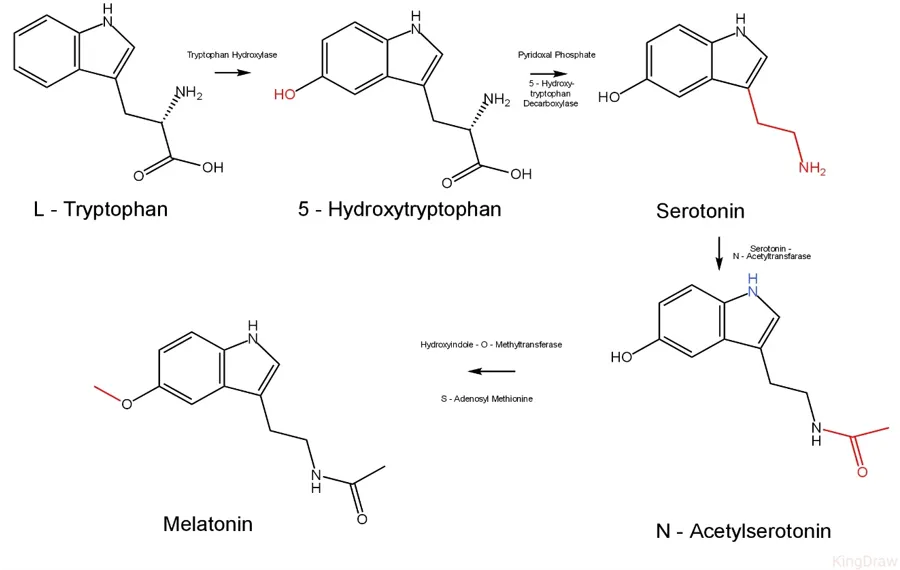
Figure 1: Melatonin Synthesis from Tryptophan
3.2 Melatonin receptor:
The first evidence of melatonin receptors came from studies on amphibian skin cells (melanophores), where melatonin was found to cause pigment aggregation. Later studies confirmed the presence of specific melatonin-binding sites in mammalian tissues, including the brain and retina.
Melatonin receptors belong mainly to the G-protein-coupled receptor (GPCR) family and were initially classified into two subtypes:
1. MT1 (Mel1a): Involved in regulating circadian rhythm and sleep.
2. MT2 (Mel1b): Plays a role in circadian rhythm regulation and phase shifting.
Another receptor subtype, MT3, was identified, but its classification as a true GPCR remains uncertain. The discovery of selective receptor ligands, receptor cloning and genetically modified mice has helped researchers understand the individual functions of melatonin receptors.[9]
MT1 Receptor & MT2 Receptor: MT1 Receptor & MT2 Receptor:
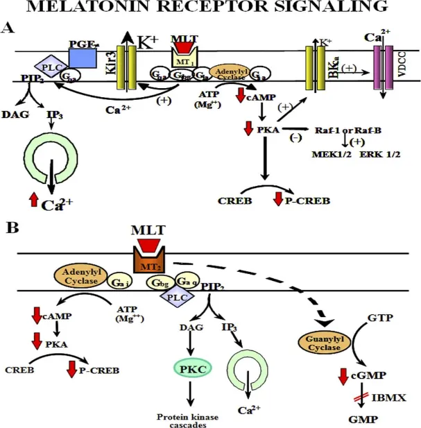
MT1 and MT2 are high-affinity melatonin receptors belonging to the G-protein-coupled receptor (GPCR) family. Both receptors are coupled to G-proteins and inhibit adenylyl cyclase activity, which plays an important role in cellular signalling. Structurally, MT1 and MT2 consist of seven transmembrane helices connected by intracellular and extracellular loops. MT1 and MT2 receptors contain 350 and 362 amino acids, respectively, with approximately 60% amino acid sequence similarity. They also possess different molecular structures and binding characteristics, allowing them to produce distinct cellular responses. MT1 is mainly involved in regulating circadian rhythms and the body's biological clock, whereas MT2 contributes to circadian phase shifting and sleep regulation. Both receptors mediate the physiological effects of melatonin by transmitting signals within cells. Their specific structural features and binding sites help them recognize melatonin and regulate its biological activities.[9]
Figure 2. Putative signalling pathways activated by MT1 and MT2 melatonin receptors. A: multiple signalling pathways for MT1 melatonin receptors coupled to Gi and Gq/11. B: signalling pathways coupled to MT2 melatonin receptor activation. PIP2, phosphatidylinositol 4,5-bisphosphate; PLC, phospholipase C; DAG, diacylglycerol; PKA, protein kinase A; CREB, cAMP responsive element binding protein; ER, endoplasmic reticulum; VSCC, voltage–dependent K+ channel; BKCa, calcium activated potassium channel; PGF2α, prostaglandin F2α,; IBMX, isobutylmethylxantine; ATP, adenosine triphosphate; MLT, melatonin; GTP, guanosine triphosphate; GMP, guanosine monophosphate. Modified from Masana and Dubocovich with permission.
3.3 Pharmacological Actions:
Melatonin receptor characterization and identification in native and/or heterologous expression systems requires knowledge of the pharmacological properties of the ligands, and radioligands in the particular receptor system under study. This knowledge is essential to characterize potential therapeutic targets. Here we will define ligand efficacy, potency, affinity, selectivity and specificity, and will use examples from the melatonin receptor literature to illustrate how each ligand can be characterized and used to discover functional receptors in native tissues.[9]
3.4 Role in regulation of circadian rhythm:
The photic signals from retina reach to pineal gland through SCN to regulate the synthesis and secretion of melatonin. The pineal melatonin gives feedback to SCN to control the circadian rhythm. SCN directly or indirectly controls hypothalamic–pituitary activity and the functions of autonomic nervous system throughout the circadian period. The SCN outputs at hypothalamic level regulate i) gonadotropin releasing hormone (GnRH) secretion, ii) pituitary prolactin release by controlling the activity of hypothalamic dopaminergic neurons, iii) ACTH-cortisol rhythm by modulating the functions of corticotrophin-releasing hormone (CRH) secreting neurons, and iv) the functions of autonomic nervous system for controlling sleep–wake cycle also other physiological activities of the peripheral organs. Melatonin receptors MT1 and MT2 are expressed in SCN. Melatonin gives an acute suppression of neuronal firing and phase shifting. MT1 receptor inhibits electrical activity and neuronal firing. This effect has been cancelled in MT1 receptor knockout mice without altering the phase shifting. MT2 receptor regulates phase shifting response. Pharmacological inhibition of MT2 receptor disturbs entrainment process. Moreover, Hablitz et al. (2015) reported that melatonin receptors in SCN are coupled with G-protein-coupled inwardly rectifying potassium (GIRK) channels. Melatonin affects the expression of clock genes in SCN. The feedback of melatonin modulates the activity of SCN during natural photoperiodic changes. Both MT1 and MT2 induce the expression of clock genes Period 1 and Period 2. So, SCN-melatonin feedback loop has a role in regulation of circadian rhythm.[10]
4. MELATONIN AND CIRCADIAN RHYTHM:
4.1 Relationship Between Melatonin and Circadian Clock:
Melatonin helps to regulate sleep by causing physiological changes that occur during the night. Melatonin acts upon regions of the brain that regulate sleep such as the SCN. This can affect how sleepy we feel and our sleep/wake cycle. Melatonin should not just be seen as a hypnotic hormone but instead a signal from our body to tell us what state our circadian rhythms are in and how long it has been dark. Whilst sleep is controlled by our circadian system and our drive to sleep, melatonin can cause us to fall asleep easier if taken under the right circumstances. However, depending on when it is taken it will affect our circadian phase. This means just because you find it easier to fall asleep does not mean your circadian clock has been reset.[11]
4.2 Melatonin as a Chronobiotic:
A chronobiotic is a substance that alters the timing or phase of biological rhythms and thereby facilitates their entrainment to the environment. Melatonin is one of the chronobiotic agents that have been most extensively studied in humans. Unlike a typical sedative, which primarily acts by decreasing arousal, melatonin has the ability to alter the timing of the circadian system when given at an appropriate circadian time.[11]
4.2.1 Mechanism of chronobiotic action:
Exogenous melatonin acts on melatonin receptors in the SCN and modifies the phase of the circadian pacemaker. Depending on the timing of administration it can have different effects. For example: In a delayed sleep phase individual, administration of melatonin at an appropriate time may advance the circadian system. Following trans meridian travel, appropriately timed administration of melatonin may shift the circadian rhythms to a new time zone.[11]
4.2.2 MELATONIN MAYBE MORE THAN A CHRONOBIOTIC:
Melatonin in addition to its main function as a chronobiotic agent, melatonin is considered to have protective effects in vivo. It has been established that at high concentrations, melatonin has antioxidative properties by acting as an electron donor and thereby being potentially able to scavenge hydroxyl radicals and to activate enzymes of the antioxidative defence system. Melatonin could also play a role as a putative anti-tumour drug. It has been shown that melatonin has antitumor action in human breast cancer cells in culture and that serum melatonin concentrations are significantly higher in melanoma patients compared with controls. This could be due to the significantly increased HIOMT activity that was measured post-mortem in the pineal glands of patients with malignant tumours. However, it is currently too soon to conclude that there is a direct connection in humans between an altered melatonin rhythm and pathophysiological conditions related to the immune system.[11]
4.2.3 Factors influencing chronobiotic efficacy:
The effectiveness of melatonin depends on:
Timing of administration: The most important determinant of the direction of the phase shift.
Dose and formulation: Immediate-release and prolonged-release preparations produce different exposure profiles.
Light exposure: Evening and morning light can reinforce or oppose the desired phase shift.
Individual circadian phase: People with the same bedtime may have different internal biological timings.
Consistency: A regular schedule may be necessary to maintain entrainment in some circadian disorders.[11]
4.3 Phase-Shifting Effect:
The phase-shifting effect is the ability of melatonin to change the timing of the circadian clock. This property is explained by the phase response curve (PRC), which describes how the effect of an intervention varies according to the biological time at which it is administered.[12]
4.3.1 Phase advances and phase delays:
A phase advance means that a circadian event occurs earlier than before. A phase delay means that it occurs later.
4.3.2 Mechanism of action:
Melatonin acts on MT1 and MT2 receptors in the SCN resulting in altered neuronal signals that modulate the phase of the central oscillator. The consequent changes in the rhythms of the central pacemaker led to phase advances or delays in downstream circadian rhythms and responses, including that of endogenous melatonin in the SCN, and the sleep-wake cycle. A given dose of exogenous melatonin does not consistently determine the extent of phase advance or delay. Larger doses of melatonin do not necessarily result in larger phase shifts and may have negative repercussions. Moreover, the timing of administration must be optimized to avoid unnecessary pharmacological effects.[12]
4.3.3 Clinical relevance:
Phase shifting is important in disorders in which the endogenous circadian clock is out of synchrony with desired sleep and wake times.
5. BUCCAL DRUG DELIVERY SYSTEM:
5.1 Definition:
Buccal drug delivery is an alternative to oral administration, particularly for drugs that undergo extensive hepatic first-pass metabolism. It involves drug absorption through the buccal mucosa, which consists of stratified squamous epithelium and underlying connective tissue.
Its major advantages include avoiding first-pass metabolism, reducing gastrointestinal degradation, ease of administration and relatively low enzymatic activity. Different dosage forms, such as mucoadhesive tablets, gels and patches, have been developed for this route.
However, the buccal epithelium acts as a major barrier, especially for hydrophilic drugs. To overcome this limitation, various approaches are used, including mucoadhesive polymers, permeation enhancers and modifications in drug properties. These strategies help improve drug penetration, absorption and residence time at the buccal site.[13]
5.2 Anatomy of Buccal Mucosa:
The oral mucosa surface area is around 100 cm2. It comes in three flavours: lining mucosa, specialized mucosa, and masticatory mucosa. The hard plate and gingiva are covered with thick (100-200 mm) masticatory mucosa that constitutes 25% of the oral mucosa. It is exposed to friction, shear stress, and abrasion during mastication and adheres firmly to the underlying structures. The lining mucosa comprises 60% of the oral mucosa. It is 500-800 mm thick and covers the floor of the oral cavity, cheek, soft palate, lower surface of the tongue, and lip. The dorsum of the tongue is covered with specialized mucosa, which is involved in taste and makes up 15% of the oral mucosa. Buccal refers to the mucosa lining the cheek and upper and lower lips, which constitutes approximately one-third of the oral mucosa; however, the term is sometimes incorrectly used to describe the mucosa of the whole mouth.
Buccal epithelium:
The buccal epithelium is a multilayered, non-keratinized tissue consisting of cells that undergo continuous growth, differentiation and shedding. Basal cells divide to maintain the epithelial layer, while mature cells gradually move towards the surface and are shed. The intermediate cells contain lipids and cytokeratin. Lamellar granules release lipids into the spaces between cells, contributing to the epithelial barrier. Unlike intestinal and nasal mucosa, the buccal epithelium lacks tight junctions. It contains gap junctions, desmosomes and hemidesmosomes that help maintain connections between cells. Importance in drug delivery: Its non-keratinized structure and cellular arrangement influence drug penetration and absorption through the buccal mucosa.[14]
5.3 Structure of Buccal Mucosa:
The oral mucosa is covered with a top layer of stratified squamous epithelium. Beneath this layer is a basement membrane followed by a lamina propria and the innermost layer called the submucosa. The epithelium found on the buccal mucosa is about 40-50 cell layers thick, while the sublingual epithelium is somewhat thinner. As the epithelial cells move from the basal layers to the outer layers, they enlarge and flatten. The estimated turnover time for the buccal epithelium is around 5-6 days, which may represent the general turnover time of the oral mucosa as well.
The thickness of the oral mucosa varies depending on the location: the buccal mucosa can be 500-800 μm thick, while the mucosa of the hard and soft palate, the floor of the mouth, the, and the gums can be between 100 and 200 μm thick. Moreover, the composition of the epithelium varies depending on the region. Thus, the most traumatized areas, such as the gums and hard palate, are covered with keratinized mucosa, typical of the skin. On the other hand, the mucosae of the soft palate, sublingual area, and buccal regions are not keratinized. The keratinized epithelia are rich in lipids such as ceramides and acyl ceramides which have been associated with the barrier function. These epithelia are fairly impermeable to water. On the other hand, non-keratinized epithelia, for instance the floor of the mouth and the buccal epithelia, lack acyl ceramides and hold only small amounts of ceramide. Additionally, they contain minor amounts of neutral but polar lipids, mostly cholesterol sulphate and glucosyl ceramides. These epithelia have been found to be significantly more permeable to water than keratinized epithelia.[13]
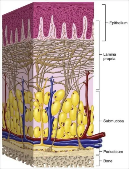
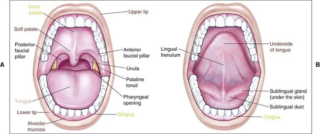
Figure 3: Anatomy of Oral Mucosa[13]
5.4 Mechanism of buccal drug absorption:
Permeability studies have shown that various substances can penetrate the oral epithelium at different rates, depending on the molecule's chemical nature and the histological characteristics of the tissue they pass through. There are considerable regional variations in the permeability pattern of oral mucosa. In general, the permeability of oral mucosa decreases progressively from the sublingual region through to the buccal and palatal mucosa. This rating is associated with the relative thickness and degree of keratinisation between these regions: sublingual mucosa is relatively thin and non-keratinized, the buccal is thicker and non-keratinized, and the palatal is in between thickness but keratinized. Compounds with different chemical properties penetrate the “barrier region” via different routes. Substances can be transported across the mucosal membrane through passive diffusion, carrier-mediated active transport, or endocytosis. Endocytosis is defined by the capacity of a large number of cells to take up solid particles (phagocytosis) or fluids (pinocytosis) from the external environment by engulfing the material inside membranous vesicles. However, pinocytosis or phagocytosis are less relevant here because of the stratified epithelium in the oral cavity.[15]
5.5 Pathways of Drug Transport:
Transcellular (intracellular) and paracellular (intercellular) are the two primary routes of drug penetration through the buccal mucosa. According to research conducted using microscopically visible tracers, such as dextrans and small proteins, intercellular penetration accounts for the majority of large molecule transfer through the stratified epithelium, which is associated with the altered intercellular substance in the superficial layers presenting a barrier to penetration.
It is well known that the extracellular space’s lipid matrix contributes to the paracellular pathway’s barrier function, particularly when the subject of the substances, such as peptides, is hydrophilic and has large molecular weights. The diffusant’s molecular weight and lipid solubility affect the buccal mucosa’s absorption capability. Certain medications are reported to be more absorbed through the buccal mucosa when the carrier pH is dropped and decreased by a rise in pH. Peptide drugs are traditionally thought to penetrate buccal epithelium via a para-cellular pathway through passive diffusion. It was recently found that drugs with a monocarboxylic acid residue might access the systemic circulation via the oral mucosa's carrier. A number of in vitro and in vivo models have been utilized to assess the permeability of oral mucosa and the efficacy of penetration enhancers. Permeability of oral mucosa has been evaluated using a variety of diffusion cell types including continuous flow perfusion chambers, Ussing chambers, Franz diffusion cells, and Grass-Sweetana.
In addition, cell lines of cultured epithelium have been developed as an in vitro model to study drug transport and metabolism at biological barriers and to elucidate potential penetration enhancer mechanisms of action. The TR146 cell culture model has recently been proposed as a useful in vitro model of human buccal mucosa for permeability and metabolism studies with enzymatically labile pharmaceuticals, such as leu-enkefalin, intended for buccal drug delivery.[16]
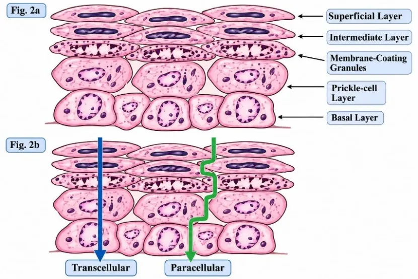
Fig. 4a: Ultra-structural features of oral buccal epithelium.
4b: Routes of drug transport across oral epithelium.[15]
5.6 Advantages of BDD:
1. The drug is easy to administer, and elimination therapy in emergency situations can be used.
2. The drug is released for a prolonged period of time.
3. In patients who are unconscious or have suffered trauma, the drug can be used.
4. Drugs avoid first-pass metabolism, increasing bioavailability.
5. Some drugs that are unstable in the acidic environment of the stomach were delivered through the buccal administration.
6. Drug absorption will occur by passive diffusion.
7. The absorption rate is maximized with the close contact with the absorbing membrane.
8. Fast onset of action.[13]
5.7 Disadvantages of BDD:
1. Small absorption area – the overall surface area of oral cavity membranes available for drug absorption is 170cm2 in which approximately 50cm2 comprises non keratinized tissue including buccal membrane.
2. Barriers such as saliva, mucus, membrane coating granules, basement membrane, and others reduce the rate and extent of drug absorption through the mucosa.
3. The danger of choking due to accidentally swallowed delivery system is a concern
4. Incompatible with buccal pH drugs are not appropriate for administration.
5. Drugs that have a bitter or unpleasant taste, an unpleasant odour or irritate the mucous membrane can’t be administered by this method.
6. Only drugs that require a small dose can be administered.
7. Only those drugs that are absorbed by passive diffusion can be administered through this way.[13,17]
5.8 Factors affecting Buccal Drug Absorption:
1. The epithelium's innate permeability: The permeability of the oral mucosal epithelium lies between that of the skin epithelium, highly specialized for barrier function, and the gut, highly specialized for an absorptive function. Within the oral cavity, the buccal mucosa is less permeable than the sublingual mucosa.
2. Epithelial thickness: The thickness of the oral epithelium differs considerably among sites within the oral cavity. The buccal mucosa is approximately 500-800μm thick.
3. Blood supply: An abundant blood supply and lymphatic system within the lamina propria supply the oral cavity, therefore drug moieties that pass through the oral epithelium are easily absorbed into the systemic bloodstream.
4. Metabolic activity: Drug moieties taken up through the oral epithelium are transported directly into the blood, bypassing the first-pass metabolism effect of the liver and the gut wall. Consequently, oral mucosal delivery could be especially appealing for the administration of enzymatically labile drugs like therapeutic peptides and proteins.
5. Saliva and mucous: The function of the salivary glands is such that the oral mucosal surfaces are continuously bathed in a flow of saliva, roughly 0.5-2L each day. Specifically, the sublingual region is exposed to a considerable amount of saliva that can enhance drug dissolution and therefore increase bioavailability.
6. Capacity to hold the delivery system: The buccal mucosa consists of a stretch of smooth and comparatively immobile surface which is properly suited for the use of retentive delivery systems.
7. Species-related differences: Rodents have a heavily keratinized epithelium and are thus not particularly suitable as animal models when investigating buccal drug delivery.
8. Transport pathways and mechanism: Drug permeation through the epithelial barrier occurs via two principal routes: The paracellular pathway: between neighbouring epithelial cells; The transcellular pathway: through the epithelial cells, which may occur through any of the following mechanisms: passive diffusion, carrier-mediated transport, and endocytic processes.[17]
6. MELATONINDELIVERU THROUGH BUCCAL ROUTE:
6.1 Rationale for buccal administration:
Buccal administration of melatonin is a promising alternative for treating sleep disorders it allows absorption through the cheek mucosa directly into systemic circulation, bypassing the gastrointestinal tract and hepatic first-pass metabolism.
Its main advantages include:
1. Improved bioavailability: Avoids extensive hepatic first-pass metabolism, potentially increasing systemic melatonin levels.
2. Rapid absorption: The rich blood supply of the oral mucosa facilitates drug absorption into the bloodstream.
3. Improved patient compliance: Buccal films are convenient, easy to administer and suitable for patients with swallowing difficulties because.
4. Controlled drug release: Mucoadhesive polymers can prolong drug residence time, while liposomes and niosomes may improve melatonin solubility and stability.
5. Therapeutic effectiveness: May help achieve therapeutic blood concentrations more rapidly and support circadian rhythm regulation.[18,19]
6.2 Potential improvement:
Enhancement of Melatonin Bioavailability through Buccal Route:
Buccal administration can enhance melatonin bioavailability by bypassing gastrointestinal absorption and hepatic first-pass metabolism. Since oral melatonin has limited bioavailability (9–33%) and considerable inter-individual variability, buccal delivery may improve systemic drug exposure and potentially reduce the required dose.[18]
The bioavailability of melatonin can be improved through the following strategies:
1. Mucoadhesive films and tablets: Prolong drug residence time in the oral cavity and enhance absorption.
2. Niosomes: Improve melatonin solubility and modify its release.
3. Gels: Provide sustained and prolonged drug release.
4. Permeation enhancers: Increase drug penetration through the buccal mucosa.[20,21]
Human studies have demonstrated that transmucosal melatonin delivery can produce different plasma concentration-time profiles. However, further comparative clinical trials are needed to establish its superiority over conventional oral administration and confirm its therapeutic effectiveness in treating insomnia.[18,22,23]
6.3 Rapid Onset of Action: The Promise of Buccal Route of Melatonin Administration:
Buccal administration of melatonin may provide faster absorption than oral delivery by allowing direct entry into systemic circulation and partially bypassing hepatic first-pass metabolism. Drug absorption depends on its physicochemical properties, permeability and formulation residence time. Mucoadhesive films, tablets and gels may prolong mucosal contact, while solubilizing agents can improve dissolution and bioavailability. Faster absorption may be beneficial for sleep disorders; however, it does not necessarily guarantee faster sleep onset. Therefore, further clinical studies are needed to establish its effectiveness compared with conventional oral melatonin.[20–22,24]
7. BUCCAL MELATONIN FORMULATION:
The oral mucosa is a promising route for melatonin delivery due to its ability to provide both local and systemic effects. Melatonin has demonstrated potential benefits in managing oral conditions such as cancer, mucositis and periodontitis, as well as promoting bone regeneration and osseointegration. Studies have shown that sublingual and transmucosal melatonin formulations can provide rapid and consistent systemic absorption with less variation than oral or transdermal administration.
Key clinical findings include:
1. Improved absorption: Transmucosal delivery produces rapid and consistent plasma melatonin concentrations, potentially mimicking its natural plasma profile.
2. Reduced anxiety: Sublingual melatonin (3 mg) administered before cataract surgery significantly reduced preoperative anxiety.
3. Sedative effects: Doses of 0.05 mg/kg melatonin reduced anxiety and increased sedation without impairing cognitive function or recovery.
4. Pain modulation: Studies involving 61 healthy participants demonstrated dose-dependent increases in plasma melatonin levels and changes in pain thresholds.
5. Perioperative benefits: Sublingual melatonin (5 mg) reduced anxiety and provided sedation before surgery without affecting postoperative psychomotor performance.
Overall, clinical studies support the potential of buccal and sublingual melatonin formulations for rapid absorption, anxiety reduction and sedation, with minimal adverse effects on cognitive and psychomotor functions.[25]
8. FORMULATION AGENTS OF BUCCAL MELATONIN:
8.1 Mucoadhesive polymers:
Mucoadhesive polymers play an important role in buccal drug delivery by adhering formulations to the cheek mucosa and prolonging drug residence time. Their adhesion depends on hydration, swelling, polymer-chain flexibility and interaction with mucin. The polymer selection varies according to the formulation, whether dry, partially hydrated or fully hydrated.
Commonly used mucoadhesive polymers include:
- Dry or partially hydrated formulations: Polyacrylic acid (PAA), polyvinyl alcohol (PVA), sodium carboxymethylcellulose (NaCMC), HPMC, HEC, HPC and sodium alginate.
- Fully hydrated formulations: PAA, chitosan and its derivatives, HPC, PVA, gelatin, carrageenan, NaCMC and hyaluronic acid.
- Other mucoadhesive materials: Glyceryl monooleate (GMO) and lectins, which can bind to specific sugar residues on the mucosal surface.
An ideal mucoadhesive polymer should possess the following characteristics:
1. Biocompatible, inert and non-toxic.
2. Rapid adhesion to moist mucosal surfaces with good site specificity.
3. Stable throughout the formulation's shelf life.
4. Non-toxic degradation products and suitable compatibility with the mucosal environment.
5. Easily available, economical and suitable for pharmaceutical formulation.[13,14]
8.2 Penetration enhancers:
Penetration enhancers are used in buccal drug delivery to improve drug absorption through the mucosal membrane and facilitate entry into systemic circulation. They should be non-irritating and have a reversible action, allowing the mucosal barrier to regain its normal function after drug absorption.
Common penetration enhancers include:
1. Fatty acids: Disrupt the arrangement of intercellular lipids to enhance drug penetration.
2. Surfactants and bile salts: Improve permeability by modifying membrane lipids and proteins.
3. Azone: Increases fluidity within intercellular lipid regions.
4. Alcohols: Alter lipid organization and protein structure to facilitate drug absorption.
5. Chitosan and its derivatives: Improve transmucosal absorption, potentially by temporarily widening tight junctions between cells.
Although chitosan is a promising penetration enhancer, its exact mechanism in buccal mucosa is not fully understood. Furthermore, existing in vitro and ex vivo studies may not accurately represent in vivo conditions. Therefore, standardized biological models are needed to evaluate and compare the effectiveness and safety of different penetration enhancers.[14]
8.3 Enzyme inhibitors:
Co-administration of enzyme inhibitors is an effective strategy for improving buccal absorption, particularly of peptide and protein drugs. These inhibitors, such as aprotinin, bestatin, puromycin and certain bile salts, protect drugs from enzymatic degradation by modifying enzyme activity or reducing drug exposure to enzymes.
Certain mucoadhesive polymers also exhibit enzyme-inhibitory properties:
1. Polyacrylic acid (carbomer): Binds calcium and zinc ions required for enzyme activity, leading to enzyme inactivation.
2. Chitosan–EDTA: Inhibits metallopeptidases, such as carboxypeptidase.
3. Thiol-modified polymers: Thiol-functionalized polyacrylates and chitosan derivatives enhance enzyme-inhibitory activity.[14]
8.4 Drug Profile:
Melatonin (N-acetyl-5-methoxytryptamine) is a hormone secreted by the pineal gland that regulates the circadian rhythm and sleep-wake cycle. It is commonly used for sleep disorders, particularly insomnia. However, its oral bioavailability is limited due to extensive hepatic first-pass metabolism and a short elimination half-life of approximately 45–60 minutes. Buccal delivery is a promising alternative to improve melatonin absorption and reduce first-pass metabolism.[22,24]
Various formulation components are used to enhance drug delivery:
1. HPMC, sodium alginate, PVP and chitosan: Improve mucoadhesion, flexibility and controlled drug release.
2. Glycerine: Enhances film softness and elasticity.
3. Propylene glycol: Acts as a permeation enhancer to improve melatonin absorption through the buccal mucosa.
4. Transmucosal gels and buccal films: May improve absorption and modify the pharmacokinetic profile of melatonin.[26]
9. EVALUATION OF BUCCAL MELATONIN FORMULATION:
9.1 Physical appearance:
Physical appearance serves as a key initial criterion when assessing melatonin-loaded buccal mucoadhesive and fast-dissolving films. The films that were prepared are inspected visually for colour, transparency, smoothness, flexibility, and homogeneity, as well as for the presence of cracks, air bubbles, or other surface defects. A film that is uniform and free of defects points to satisfactory film formation and may help ensure consistent drug distribution. Choosing appropriate polymers—for example hydroxypropyl methylcellulose (HPMC), sodium alginate, polyvinylpyrrolidone (PVP), and chitosan—is crucial in shaping the physical properties and overall quality of the films produced.[27]
9.2 Weight variation:
Weight variation is assessed in order to evaluate how uniform the prepared buccal films are. Films of the same dimensions are weighed individually on an analytical balance, after which the mean weight and standard deviation are computed. Marked variation could point to an uneven distribution of the formulation ingredients and, potentially, inconsistent drug loading. Because a uniform dose is essential for reproducible drug delivery in the management of sleep disorders, this test matters especially for melatonin-loaded films.[27,28]
9.3. Thickness:
Using a digital micrometre or a vernier caliper, film thickness is determined at several points. When thickness is uniform, it reflects consistent film casting and helps guarantee reproducible drug loading and release. For patient comfort and buccal administration, thin and uniform films are usually favoured.[27]
9.4. Drug Content Uniformity:
Drug content uniformity assesses how consistently melatonin is distributed within each individual film. Every film is dissolved in an appropriate solvent, diluted when needed, and then analysed with a validated UV-visible spectrophotometric or chromatographic method. Consistent drug content is critical for accurate dosing and dependable therapeutic performance in the management of sleep disorders.[27,29]
9.5. Mucoadhesive Strength:
Mucoadhesive strength is a key parameter in the evaluation of melatonin-loaded buccal films, as it governs how well they attach to the buccal mucosa and stay in place at the site of application. A texture analyser, using buccal mucosal tissue as the substrate, can be employed to measure it. The maximum force needed to detach the film from the mucosa is recorded as the mucoadhesive force, typically expressed in newtons (N). Adequate mucoadhesion can extend residence time and help support effective drug absorption. Adhesion strength is affected by polymer composition, especially the type and concentration of mucoadhesive polymers.[27,30]
9.6. Stability Study:
Stability studies are carried out to evaluate how the physical, chemical, and performance characteristics of melatonin buccal formulations hold up during storage. The films are kept under defined temperature and humidity conditions and assessed at set intervals for alterations in colour, appearance, flexibility, drug content, degradation, and in-vitro drug release. Stability testing should comply with the relevant ICH guidelines and use conditions suited to the intended packaging and storage environment. Preserving drug content and film integrity is essential for consistent melatonin delivery in the management of sleep disorders.[27,31]
10. RECENT DEVELOPMENT AND CLINICAL EVIDENCE:
Mucoadhesive Drug Delivery Systems:
Work on mucoadhesive drug delivery has recently concentrated on enhancing drug absorption, extending how long formulations remain on mucosal surfaces, and producing therapeutic effects that are more predictable. Buccal films, mucoadhesive patches, hydrogels, and fast-dissolving oral films are under investigation as alternatives to conventional oral dosage forms. To strengthen adhesion to the oral mucosa and control drug release, these formulations incorporate polymers including hydroxypropyl methylcellulose (HPMC), chitosan, sodium alginate, and Carbopol.[30]
11. RECENT DEVELOPMENT IN FORMULATION:
Nanotechnology-based carriers, polymeric nanoparticles, nanofibers, and amphiphilic mucoadhesive polymers are among the most promising recent developments. Such technologies could raise melatonin solubility, shield the drug from degradation, and improve its permeation across the oral mucosa. Fast-dissolving films are especially appealing because they need no water for administration, whereas mucoadhesive films can stay in prolonged contact with the mucosa to deliver drugs in a controlled manner.[30]
12. PROPECTS FOR THE FUTURE OF BUCCAL DRUG DELIVERY:
12.1. Individualized Melatonin Treatment:
Treatment may be personalized according to age, metabolism, circadian rhythms and sleep patterns. DLMO biomarkers may help determine the appropriate administration time.[32]
12.2. Innovative Buccal Formulations:
Nanocarrier-loaded films, mucoadhesive patches and fast-dissolving films may improve melatonin absorption, stability and drug release.[30,33]
12.3. Chronotherapeutic Drug Delivery:
Future formulations may provide immediate or controlled release according to the patient's biological clock and sleep–wake cycle.[32]
12.4. Requirements for Future Research:
Further studies should evaluate bioavailability, drug release, formulation stability, mucosal safety and clinical effectiveness compared with conventional oral melatonin.[30,33]
CONCLUSION:
Buccal delivery of melatonin is a promising alternative to conventional oral administration for managing sleep disorders and circadian rhythm disturbances. It may improve drug absorption by partially bypassing hepatic first-pass metabolism and reducing variability in bioavailability. Formulations such as mucoadhesive films, fast-dissolving films and nanocarrier-based systems may enhance drug absorption and controlled release.
Suitable polymers, including HPMC, chitosan, sodium alginate and Carbopol, can improve adhesion and drug-release properties. However, further research is needed to evaluate formulation quality, bioavailability, mucosal safety and clinical effectiveness. Overall, buccal melatonin delivery has potential as an alternative approach, but further clinical validation is required to establish its benefits over conventional oral formulations.
REFERENCES

Binith Raj, Snehal Padwal, Piyush Panchal, Sneha Nagbhidkar, Prajakta Nalawde, Shrutika Patil, Buccal Mucoadhesive and Fast-Dissolving Systems for Melatonin in Sleep Disorders: A Review, Int. J. of Pharm. Sci., 2026, Vol 4, Issue 11, 915-930. https://doi.org/10.5281/zenodo.23971871
 10.5281/zenodo.23971871