

We use cookies to ensure our website works properly and to personalise your experience. Cookies policy
1,2,4 Ezhuthachan college of Pharmaceutical Sciences, Marayamuttom, Neyyatinkara, Thiruvananthapuram
3 Consultant in General Medicine and Diabetology, Nims Medicity Neyyatinkara, Thiruvananthapuram.
Bullous pemphigoid is a rare autoimmune blistering disorder, predominantly affecting the elderly. It is characterized by autoantibodies directed against the hemidesmosomal proteins BP180 and BP230, triggering complement activation, inflammation in the dermis, and separation at the dermal-epidermal junction. A 60-year-old man was admitted with a two-week history of itchy blisters appearing on his arms and legs. He had a background history of type 2 diabetes mellitus. On examination, widespread large, taut bullae were observed across his body, on the lower limbs, with multiple eroded areas. Initial lab tests showed low sodium levels, increased neutrophils, reduced lymphocytes, and elevated eosinophils—abnormalities that mostly normalized by discharge. A skin biopsy revealed blister formation beneath the epidermis with a marked presence of eosinophils, confirming the diagnosis of bullous pemphigoid. Treatment began with intravenous corticosteroids, transitioning to oral prednisolone. Additional therapies included antibiotics, antihistamines, anti-inflammatory agents, protective skin creams, calcium and vitamin D supplements, proton pump inhibitors, parenteral nutrition, and strict glucose management. During hospitalization, the patient showed improvement: no new blisters developed, existing lesions healed, and both electrolyte imbalances and signs of inflammation resolved. He was discharged in stable condition, with instructions to continue prescribed medications and attend a follow-up visit with a dermatologist. This case underscores the diagnostic value of skin biopsy in confirming bullous pemphigoid. It also demonstrates the effectiveness of corticosteroid therapy in altering disease progression. Optimal outcomes depend on a coordinated, multidisciplinary approach and careful control of comorbid conditions such as diabetes.
Bullous pemphigoid is a chronic autoimmune subepidermal blistering disease that primarily affect the older population. It accounts for the majority of autoimmune blistering conditions that are recognized by dermatologists in their practice [1,2]. This disease is characterized by the production of pathogenic IgG autoantibodies targeting the hemidesmosomal proteins BP180 (type XVII collagen) and BP230 [3,4]. Autoantibodies cause complement activation, recruitment of inflammatory cells, and basement membrane zone destruction resulting in separation of the epidermis from the dermis [5,6].
The prevalence of bullous pemphigoid has increased significantly during the last 20 years due to the aging of the population, increased physician awareness, and enhanced diagnostic techniques [7,8]. An overall estimated incidence of 2-43 cases per 1 million persons was reported, with the highest prevalence in people aged over 60 years [9,10]. This autoimmune blistering disease affects both genders unequally, with slight variations reported in different studies [7,9].
Bullous pemphigoid (BP) typically presents with a prodromal stage characterized by severe itching and urticarial plaques, papules, or eczematous eruptions that evolve into tense bullae on normal-looking or erythematous skin [11,12]. These eruptions are commonly found on the trunk, abdomen, groin, axilla, and flexural areas of the extremities, with less frequent involvement of oral or genital mucosa, which is usually mild compared to pemphigus vulgaris [2,11].
The development of BP is multifactorial and complex, involving both cellular- and humoral-mediated mechanisms [13]. Autoantibodies recognizing the BP180 antigen trigger complement system activation, resulting in neutrophils, eosinophils, and mast cells accumulation, increased production of proteolytic enzymes, and subsequent basement membrane disruption [14]. Additionally, IL-5, IL-17, and eotaxin are elevated and promote eosinophil infiltration, enhancing inflammation and blister formation [14,15]. Advanced age, diabetes mellitus, chronic kidney disease, neurological conditions, malignancies, and certain pharmaceuticals are among the common risk factors for developing BP [16,17]. One of the most prevalent comorbidities associated with BP is diabetes mellitus, which may complicate the management of this autoimmune blistering disease as immunosuppressive agents such as glucocorticoids can aggravate glycemic control [17,18].
The diagnosis of bullous pemphigoid is established based on the clinical manifestation, histopathological, and immunopathological investigations [19,20]. A biopsy shows a subepidermal blister with a dense infiltration of eosinophils, whereas a direct immunofluorescence assay reveals linear deposits of IgG and C3 along the basement membrane [20]. An early diagnosis and timely treatment initiation are of paramount importance to avoid disease-associated mortality, morbidity, and impact on the patient’s quality of life [18,20].
The present case describes a biopsy-confirmed case of bullous pemphigoid in a 60-year-old male with type 2 diabetes mellitus who presented with widespread pruritic bullous lesions. Early diagnosis, prompt initiation of systemic corticosteroid therapy, comprehensive supportive care, and meticulous glycaemic control resulted in marked clinical improvement and successful recovery
CASE PRESENTATION
A 60-year-old man was referred to the Department of General Medicine with multiple fluid-filled blisters on his arms and legs, accompanied by intense itching that had lasted for two weeks. He initially developed red, itchy patches that progressed into large, taut blisters containing clear fluid. These blisters eventually broke open, resulting in painful sores and crusting. Despite prior treatment for these symptoms, there had been no noticeable improvement.
The patient had a known history of type 2 diabetes mellitus and was managing it with oral antidiabetic drugs. He reported no personal or family history of autoimmune blistering disorders, psoriasis, connective tissue diseases, or previous episodes of similar skin rashes. There was no history of injury, burns, exposure to chemicals, or drug allergies.
On examination, the patient was conscious, alert, and fully oriented, showing no signs of acute distress. Physical findings included numerous taut blisters and areas of skin erosion on the limbs. Some blisters had ruptured, leaving behind weeping sores covered with yellowish crusts, while surrounding skin appeared red and scratched from persistent itching. Mucous membranes were unaffected, and there were no signs of secondary infection, sepsis, or systemic involvement.The combination of tense blisters, severe pruritus, laboratory results, and histopathological analysis pointed toward bullous pemphigoid, a diagnosis later confirmed by skin biopsy.
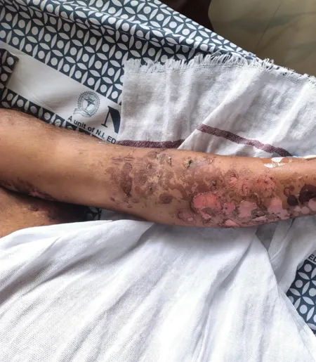
Figure 1A. Left forearm showing extensive erosions, ruptured bullae, crusting, and healing lesions.
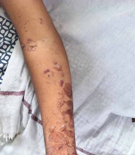
Figure 1B. Right forearm showing multiple intact tense bullae with post-inflammatory hyperpigmentation.
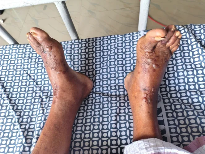
Figure 1C. Bilateral feet showing tense bullae, healed erosions, crusting, and post-inflammatory hyperpigmentation.
Laboratory Investigation
Initial lab results showed hyponatremia, with the patient’s serum sodium at 124 mmol/L upon admission. During hospitalization, sodium levels progressively improved, reaching 128, 132, and 134 mmol/L before returning to normal at discharge. Potassium levels remained within the normal range throughout the stay.The complete blood count revealed eosinophilia—a finding frequently seen in bullous pemphigoid. The patient had elevated eosinophil counts 15.7% on admission, which declined following treatment. Hemoglobin, total white blood cell count, and platelet levels were all within normal limits during the hospital course.
In addition, the blood test revealed that the C-reactive protein and neutrophils, markers linked to inflammation. These values gradually decreased with treatment. Glycated hemoglobin was measured at 7.5%, consistent with a pre-existing diagnosis of type 2 diabetes. Liver and kidney function tests remained normal throughout the hospitalization.Due to the presence of blister-like skin lesions, the patient was referred to dermatology for further assessment. A skin biopsy was performed to confirm the suspected diagnosis of an autoimmune blistering disorder.
HISTOPATHOLOGICAL FINDINGS
Histopathological examination of the skin biopsy demonstrated a subepidermal blister containing a mixed inflammatory cell infiltrate composed predominantly of eosinophils with occasional neutrophils. Eosinophils were observed lining the dermoepidermal junction, accompanied by mild spongiosis. The papillary dermis showed mild perivascular eosinophilic and lymphoplasmacytic infiltration, indicating an active inflammatory process. These histopathological findings are characteristic of bullous pemphigoid and, in conjunction with the patient's clinical presentation of pruritic tense bullae, confirmed the diagnosis.
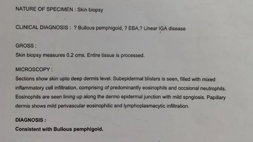
Figure 2. shows the histopathology report of the skin biopsy
TREATMENT
After being diagnosed with bullous pemphigoid, the patient received comprehensive treatment addressing the autoimmune condition, intense itching, potential secondary infections, wound recovery, electrolyte disturbances, blood sugar management, and adverse effects related to corticosteroid use.
Initial therapy included intravenous dexamethasone (10 mg) administered twice daily to control systemic inflammation and suppress the autoimmune response. Once the blistering symptoms improved, the treatment was transitioned to oral prednisolone (30 mg), given in divided doses throughout the day.
Given the increased risk of infection linked to skin lesions in bullous pemphigoid, the patient was treated with intravenous amoxicillin-clavulanate (1.2 g) twice daily during hospitalization. After discharge, this was changed to an oral formulation (625 mg) taken three times a day.To manage severe pruritus, the patient was prescribed nightly hydroxyzine sustained-release (30 mg), along with a daily dose of fexofenadine and montelukast (120/10 mg).Wound care involved twice-daily topical application of mupirocin cream to affected areas to support healing and prevent local infection.
Due to extensive blistering and the anticipated long-term corticosteroid use, calcium and vitamin D3 supplements were initiated to reduce the risk of osteoporosis. Additionally, pantoprazole (40 mg) was given intravenously initially, then switched to oral administration to protect against corticosteroid-related gastrointestinal ulcers.
The patient developed hyponatremia, which was addressed with tolvaptan (15 mg) once daily, combined with careful fluid regulation and ongoing electrolyte monitoring.
Nutritional support for tissue repair included Prohance-D, ascorbic acid, and zinc. Gabapentin (300 mg) was also administered at bedtime to help alleviate neuropathic pain and persistent itching.The patient had a history of type 2 diabetes mellitus, requiring close monitoring of blood glucose levels during corticosteroid therapy. The existing antidiabetic regimen—gliclazide (60 mg) and sitagliptin (100 mg, taken as half a tablet daily)—was maintained with regular capillary blood glucose checks to ensure glycemic stability.
HOSPITAL COURSE AND OUTCOME
During her hospital stay, the patient showed steady improvement following treatment with glucocorticoids and supportive measures. No new bullous lesions emerged, and existing ones started to heal. The combination of antihistamines and glucocorticoids helped reduce itching, which in turn limited skin scratching.After several days of therapy, blood tests indicated that her sodium levels had normalized, achieved through intravenous fluids and tolvaptan. Due to the glucocorticoid regimen, her glucose levels were closely monitored but remained within normal limits, managed when necessary with anti-diabetic medication.
Throughout her admission, the patient did not experience secondary bacterial infections, sepsis, or any adverse reactions related to glucocorticoid use. Her skin condition responded positively, with no further blister formation observed.
Upon discharge, she was prescribed oral prednisolone, antihistamines, calcium and vitamin D supplements, along with topical mupirocin. She was advised to maintain a regular diet, practice proper skin care, and continue glucose-lowering drugs. A follow-up visit with a dermatologist was recommended to assess her progress, gradually taper steroid use, check for possible relapse, and monitor for any treatment-related side effects.
DISCUSSION
Bullous pemphigoid is the most frequent autoimmune subepidermal blistering disease, which predominantly affects elderly people. Autoantibodies target BP180 and BP230, leading to complement activation, infiltration of eosinophils, and formation of tense bullae. A 60-year-old man came to our clinic with numerous bullous lesions that were itchy and widespread on his upper limbs and lower limbs. The histology of the skin lesion showed a blister located beneath the epidermis with eosinophilic infiltration, which allowed us to make a diagnosis of bullous pemphigoid. The patient was treated with systemic corticosteroids, antibiotics, antihistamines, wound care, correction of hyponatremia and glucose intolerance, and achieved an excellent outcome with no significant further complications. The positive outcome of the disease course was due to timely treatment and wound care preventing secondary infections or other serious complications.
The present case showed several similarities with previously reported cases by Miyamoto et al., Kridin et al., and Feliciani et al. [8,9,12]. Similar to Case 1 reported by Miyamoto et al., the present patient developed pruritic bullous lesions with subepidermal blister formation and eosinophilic infiltration on histopathology, and both cases responded favorably to corticosteroid therapy. However, the present patient was a 60-year-old male with type 2 diabetes mellitus, compared with a 74-year-old female with hypertension in Case 1. The present case also demonstrated more extensive laboratory abnormalities, including leukocytosis, eosinophilia, lymphopenia, elevated C-reactive protein, and hyponatremia, whereas Case 1 primarily showed peripheral eosinophilia. Compared with Case 2 reported by Kridin et al., both patients were male, had type 2 diabetes mellitus, and presented with tense pruritic bullae and subepidermal blistering. However, the present case had severe bullous involvement of both upper and lower limbs with eosinophil-rich inflammatory infiltrates, whereas Case 2 was characterized by a subepidermal blister and elevated inflammatory markers. Treatment also differed, as Case 2 was managed with oral prednisolone and doxycycline, while the present patient required intravenous dexamethasone followed by oral prednisolone, antibiotics, antihistamines, topical mupirocin, calcium and vitamin D supplementation, nutritional support, and optimized diabetic management. Compared with Case 3 reported by Feliciani et al., both cases demonstrated subepidermal blister formation with eosinophilic inflammation and responded well to corticosteroid-based therapy. However, Case 3 involved a 72-year-old female with chronic kidney disease and bullae over erythematous plaques, whereas the present case involved a younger male with diabetes and multiple tense bullae over the upper and lower limbs. Case 3 was treated with corticosteroids and azathioprine, while the present case received a broader supportive and pharmacological regimen. Overall, the present case was consistent with the clinical and histopathological characteristics of previously reported cases but was distinguished by its severe limb involvement, multiple hematological and inflammatory abnormalities, associated diabetes mellitus, and requirement for comprehensive multimodal management, with marked clinical improvement and no new blister formation at discharge.
CONCLUSION
Bullous pemphigoid is a chronic autoimmune disorder characterized by subepidermal blistering, primarily affecting older adults. Prompt diagnosis and appropriate management are crucial to minimizing health risks and preventing complications. This report presents a case of a 60-year-old male with type 2 diabetes who was diagnosed with bullous pemphigoid through biopsy and effectively treated with timely and targeted intervention.
Treatment began with systemic corticosteroids as part of an immunosuppressive regimen, along with supportive measures aimed at restoring tissue function. Histopathological analysis played a key role in confirming the diagnosis and guiding treatment decisions. The patient received intravenous steroids initially, followed by a transition to oral prednisolone. Additional interventions included antibiotics, antihistamines, topical antimicrobials, optimized nutritional intake, electrolyte balance, blood glucose regulation, and improved wound care. These combined efforts led to a favorable outcome, marked by complete healing of blisters, no new lesion development, and normal laboratory findings.
The case underscores the importance of coordinated, interprofessional care in achieving positive results. Dermatologists, internists, pharmacists, nurses, and dietitians collaborated closely throughout the treatment process. Ongoing communication among team members ensured consistent monitoring and adjustment of therapy. Regular evaluations were conducted to detect potential side effects of prednisolone and to assess wound progress. Follow-up visits enabled the healthcare team to track recovery and confirm that no further treatments were necessary, thereby reducing the risk of relapse or complications. Effective management of bullous pemphigoid relies on early detection, personalized treatment plans, diligent follow-up, and patient education.
REFERENCES


Jerin M, Dr. Grace N Raju, Dr. M. Shahbaz Zailu, Dr. Shaiju S Dharan, Bullous Pemphigoid : A Case Report, Int. J. of Pharm. Sci., 2026, Vol 4, Issue 9, 1702-1709. https://doi.org/10.5281/zenodo.22752512
 10.5281/zenodo.22752512