

We use cookies to ensure our website works properly and to personalise your experience. Cookies policy
PRES’s College of pharmacy (for women), Chincholi, Nashik.
Exosomes are nanosized extracellular vesicles, which are involved in intercellular communication and have been developed as promising nanocarriers for targeted drug delivery. Exosomes are 30 to 150 nm in diameter and contain a complex mixture of proteins, lipids, nucleic acids, and other bioactive molecules derived from their parent cells. They are generated by invagination of the endosomal membrane, multivesicular bodies formation and subsequent fusion of these bodies with the plasma membrane, which results in the release of exosomes in the extracellular environment. Exosomes have several potential advantages over traditional drug delivery methods due to their inherent biocompatibility, stability, low immunogenicity and their ability to interact and cross biological barriers . Several isolation and purification techniques have been developed including differential ultracentrifugation, density-gradient centrifugation, size-exclusion chromatography, ultrafiltration and immunoaffinity-based techniques. However, purity issues, scalability, reproducibility, heterogeneity and standardization still represent major hurdles for clinical translation. Exosomes can be naturally or engineered to encapsulate therapeutic drugs, proteins and nucleic acids and deliver them to specific tissues and cells. They are being explored for their therapeutic potential in oncology, neurological disorders, cardiovascular diseases, inflammatory conditions, and regenerative medicine. Here, we review the composition, biogenesis, isolation, characterization and drug-loading strategies of exosomes with emphasis on their applications as targeted drug-delivery systems, current limitations and future prospects for clinical development
Exosomes are small endosome-derived extracellular vesicles, typically between 30 and 150 nm in size . Exosomes can be secreted into the extracellular environment by almost all cell types. These vesicles have important roles in the normal homeostasis of cells and also in the development and progression of different illnesses .Exosomes consist of a complex and diverse mixture of lipids, proteins and various types of nucleic acids. The exact content of exosomes may vary depending on the cell type from which it is secreted, and the state of the cell (normal or diseased) [2].Exosomes are known to mirror the biological status of the parent cells and play an important function in various physiological and pathological processes related to different diseases [4] . Exosomes are considered as promising drug delivery carriers due to many advantages over many conventional delivery systems. They are coated with a variety of adhesion proteins that may aid in their interaction with and delivery of therapeutic agents to target cells. Some of their most important advantages are a good targeting ability, safety, stability and the ability to transport therapeutic molecules over relatively long distances. Exosomes are made by cells naturally and are usually less likely to cause strong immune responses than many synthetic delivery systems. Moreover, because of their small size and inherent biological properties, they can cross a variety of biological barriers, including the blood–brain barrier, which makes them especially useful for delivering drugs to otherwise inaccessible tissues [1] . One of the key advantages of exosomes is that they are surrounded by a lipid bilayer that protects their internal cargoes from enzyme breakdown. This protection makes exosomes available and stable in body fluids like blood, saliva and cerebrospinal fluid. In addition, the molecules in exosomes can reflect the normal or disease state of the cells they originate from. Therefore, exosomes are a useful and relatively non-invasive source of information to study changes and disorders in the central nervous system (CNS) [5] . In the past few decades, liposomes and polymer-based nanoparticles have been extensively studied as potential carriers for anticancer, antifungal drugs, analgesics and other biologically important molecules [6].
Concept of Exosomes
In the last decade, researchers have found that cells from higher animals and Micro-organisms can secrete extracellular vesicles (EVs)[3] . Research into the cell biology and physiological functions of exosomes started before 1990, and developed steadily in the following years, with the peak of interest around 2008–2010 [16] . Exosomes were first detected by P. Stahl and R. Johnstone in experiments on mature rat and sheep erythrocytes where they observed the release of small globular structures from the cells into the extracellular environment .However, with the development of sophisticated analytical techniques in the early 2000s, particularly mass spectrometry and next-generation sequencing, it became apparent that extracellular vesicles (EVs) bear a variety of biomolecules such as proteins, lipids, metabolites and distinct forms of DNA and RNA[1] . This discovery changed the notion of exosomes entirely, it demonstrated that EVs are involved in intercellular communication and not just a means of cellular waste disposal Since then the research on EVs has been rapidly increasing especially because of their emerging potential as diagnostic and therapeutic tools for a wide-range of diseases including neurodegenerative and cardiovascular diseases, metabolic diseases, immune-related diseases, and cancer [17] .
Biogenesis of Exosomes
Exosomes are small extracellular vesicles derived from the endosomal compartment of eukaryotic cells and are important mediators of intercellular communication. They are initiated by inward budding of the endosomal membrane and the formation of intraluminal vesicles (ILVs) in multivesicular bodies (MVBs). These MVBs can either fuse with lysosomes and be degraded or fuse with the plasma membrane and release the ILVs as exosomes into the extracellular environment [7,12] . At first, exosomes were believed to be released only from neoplastic cell lines. However, later studies have shown that neural cells are also able to produce and release exosomes. The formation of exosomes is distinct from other cell organelles in that it does not involve proteins secreted by the Golgi apparatus or mitochondria[5] .Exosomes can be released from various kinds of healthy cells, and diseased cells may secrete exosomes in pathological conditions. The biogenesis of exosomes is mainly regulated by the endosomal sorting complex required for transport (ESCRT) machinery, including ESCRT-0, ESCRT-I, ESCRT-II and ESCRT-III. Other important proteins such as VPS4, VTA1 and ALIX also participate in the formation and release of exosomes. In addition, several endogenous factors such as GTPase enzymes regulate the biogenesis and precise trafficking of these vesicles to coordinate their movement and release within cells [6] .
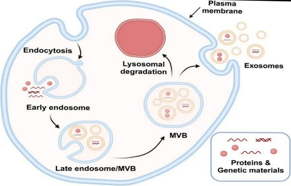
Figure:- Biogenesis of Exosome [28]
Classification of Exosomes
Exosomes are generally divided into three types: natural, modified and artificial exosomes [8] .According to the 2018 guidelines of the international Society for Extracellular Vesicles (MISEV2018), extracellular vesicles (EVs) can be broadly classified according to the size into medium/large EVs (>200 nm) and small EVs (sEVs) (<200 nm) [18] . Natural exosomes can be roughly divided into animal-derived and plant-derived exosomes. Animal-derived exosomes can be further subdivided into normal exosomes secreted by normal cells under normal physiological conditions and tumor-derived exosomes secreted by cancer cells under pathological conditions, depending on their cellular origin and physiological conditions [20] . Exosomes of animal origin are secreted mainly by various types of cells including immune cells, lymphocytes, red blood cells, platelets, dendritic cells, and tumor cells . They are present in different biological fluids such as urine, milk and plasma. Bovine milk exosomes are one of the most studied exosomes from animals due to their promising application in drug delivery and other biomedical fields [22] . Also, food-derived exosomes have been emerging as promising candidates for future biomedical applications. Recent studies suggest that plant-derived exosome-like nanoparticles (ELNs) bear some structural and functional similarities with mammalian exosomes. For example, ginger-derived nanoparticles have shown potential in preventing the progression of liver-related disorders. Also, ELNs derived from grapes, carrot, grapefruit and ginger have shown anti-inflammatory properties and may help to keep the intestine homeostasis [21].Natural exosomes can be engineered for specific therapeutic applications by loading them with drugs or other therapeutic agents and modifying their surface properties to improve cellular uptake. Various natural sources, including fruit and vegetable juices and mammalian biological fluids, have been used to obtain and modify exosomes in an effort to explore their potential in biomedical applications. These modifications can be classified into two major strategies, i.e. internal modification and surface modification. Internal modification refers to the alteration or incorporation of cargo inside the exosome, while surface modification refers to the alteration of properties or structure of the exosome outer membrane to improve targeting and therapeutic delivery [22].
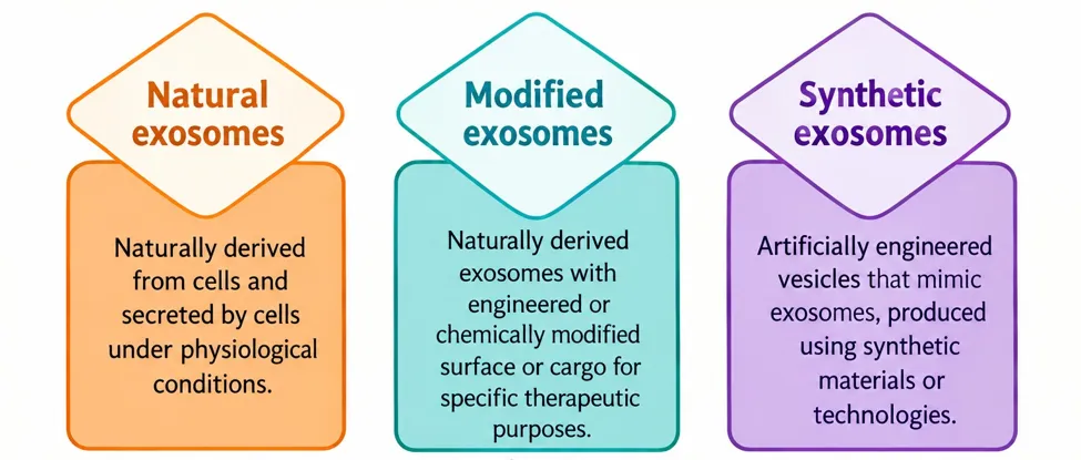
Figure: Schematic representation of exosomes classification according to their origin—natural, modified, and synthetic exosomes [22] .
Composition of Exosomes
Exosomes are derived from endosomal compartments and thus they carry a variety of biological molecules from their parent cells. These are a variety of components including surface proteins, heat shock proteins, lysosomal proteins, tumor related genes, fusion proteins, and nucleic acids. These components contribute to the unique biological functions of exosomes and to their cell-to-cell communication properties [13]. Some databases such as ExoCarta , EVpedia and Vesiclepedia are available with detailed information of the molecular contents of exosomes. Exosomes are heterogeneous in size and molecular cargo, even when they originate from the same cell type. However, exosomes from different cellular origins may contain some common cargo components, which reflects some similarity of their composition and biological functions [24] .
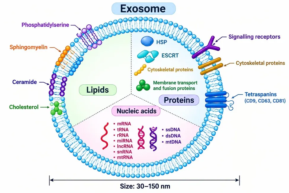
Figure:-Composition of Exosomes. [29]
Molecular cargo (proteins, nucleic acids, lipids) relevant to regeneration
Exosomal Protein
The exosomal proteins are a functionally diverse repertoire that governs vesicle biogenesis, targeting and regenerative signaling. Classical markers CD9, CD63, CD81, TSG101 and ALIX are found to assist in vesicle formation and intracellular trafficking. New proteomic studies, however, identify an extended protein environment rich in growth factors, bioactive enzymes, ECM (extracellular matrix) regulators and immunomodulatory molecules [2]. The cells of the donor are transfected with a gene for the protein of interest. The cell thus produces proteins encoded by the inserted genes, which are then secreted into exosomes. Survivin, an antiapoptotic protein, is essential for the viability of many cancer cells. The inactive mutant survivin-T34A inhibits this survivin and triggers the mitochondrial apoptotic pathway in cancer cells. It was shown that exosomes from melanoma cells that overexpress survivin-T34A by plasmid transfection cause apoptosis and increase sensitivity to gemcitabine in different pancreatic adenocarcinoma cell lines [4].
Exosomal Nucleic acid
Exosomes naturally transport nucleic acids like DNA and RNA to target cells and induce genetic modifications in biological and pathogenic processes as we discussed previously. The inherent ability to carry genetic material has made exosomes of great interest in drug delivery strategies involving genetic therapy that alter gene expression in certain diseases and improve genetic therapy [10].Regarding nucleic acids, exosomes are enriched with short RNAs, however, these vesicles have been reported to contain virtually any form of RNA . The RNA load in the exosomes is distributed very differently, however, from the whole cell. Likewise, the comparison of the various secretomes revealed that exosomes from bone marrow-derived mesenchymal stromal cells (MSCs) promote neurite formation to a greater extent than microvesicles or the total secretome [5] . This diverse composition of genetic material is found in EVs. A few cases have been demonstrated to contain DNA , including genomic and mitochondrial DNA . But in general, EVs are mainly enriched in small RNAs, the vast majority of which come from ribosomal 18S and 28S rRNAs and tRNA . Using a variety of techniques, including next generation sequencing, many small RNAs have been characterized. Besides the known RNA species, like mRNAs, miRNAs, rRNAs, long and short non-coding RNA, tRNA fragments, piwi-interacting RNA , vault RNA and Y RNA have been identified in EVs [26].
Exosomal lipids
Lipids are essential components in all cell types and are enriched in EVs. Sphingomyelin, phospholipids, ganglioside GM3 and cholesterol are lipid classes that are ubiquitously present in cell membranes and thus in exosomes .However, the relative abundance of these lipids in the exosomal membranes may vary depending on the producer cell type, the physiological stage of the producer cell, and the fate and function of the exosome. Several studies have shown in this respect that the composition in lipids and metabolites of exosomes produced under different conditions is changed to modulate their biological function [19].Different types of exosomes containing different types of lipids. The lipid bilayer of exosomes is mainly constituted of cell plasma membrane types of lipids such as sphingomyelin, phosphatidylcholine, phosphatidylethanolamine, phosphatidylserine, monosialotetrahexosylganglioside (GM3), and phosphatidylinositol. Sphingomyeline and GM3 are responsible for determining the exosome’s rigidity while phosphatidylserine is expressed on the plasma membrane of exosomes through different types of phospholipid transportation enzymes. It is involved in docking the outer proteins, allowing the signaling and fusion of the exosome to the plasma membrane. Other types of lipids that can form exosomes are cholesterol, ceramide, and phosphoglycerides , along with saturated fatty-acid chains. Additional constituents of exosomes include nucleic acids such as micro RNA (miRNA), messenger RNA (mRNA), and non-coding RNAs [25] .
Isolation and separation
Ultrafiltration
Ultrafiltration (UF), also called membrane filtration with a molecular weight cutoff (MWCO) value of 10, 50 and 100 kDa is used as a basic step for extracting exosomes from large amounts of original material, such as cell culture, into concentrated small volumes that can be used in further purification processes or analysis . The principle of this method is that exosomes can be isolated from all other sample components by membranous filters based on their sizes and molecular weights [30].One challenge with ultrafiltration is the clogging and trapping of vesicles (and therefore loss of exosomes) on the filter unit . While this can be minimized by starting with larger MWCO filters and moving to smaller ones, it leads to low Isolation efficiency, and the exosomes lost on the membrane cannot be used in downstream analysis . While ultrafiltration is less time consuming than ultracentrifugation and requires no special instrumentation, it can still lead to particle deformation and lysis of exosomes due to the shear force, though this can be reduced by monitoring and regulating transmembrane pressure [13].
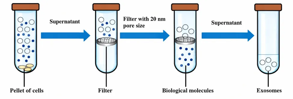
Figure:-Ultrafiltration method to isolate exosome [30]
Ultracentrifugation based separation
An analysis of the exosome literature before 2015 shows that 81% of studies used ultracentrifugation for their isolation method [31].UC is the most used exosome separation method and considered the gold standard. UC is used in 60% of exosome processing and is the most efficient way to isolate exosomes from different biological samples. The conventional UC method is based on the sedimentation principle for exosome isolation from complex biological samples such as blood serum or plasma, breast milk, cerebrospinal fluid, amniotic fluid, urine, aqueous humor, and cell culture lines . It consists of a series of low speed centrifugations to separate cells, microvesicles and apoptotic bodies, followed by high speed ultracentrifugation at 100,000x g to precipitate exosomes [30].Ultracentrifugation generally utilizes centrifugal forces in the order of 100,000 to 120,000 × g . For human plasma/serum, usually a cleaning step is done before the start of the isolation to remove large bioparticles in a sample. The sample is spiked with protease inhibitors to avoid degradation of exosomal proteins. The supernatant is aspirated after each run during exosome isolation and depending upon the centrifugal force used the supernatant or the pellet is re-suspended in an appropriate medium such as phosphate buffered saline and subjected to subsequent runs of centrifugation with increasing centrifugal force. The isolated exosomes are again re-suspended and kept at -80⁰C for further analysis. This exosome isolation method is also called pelleting method or simple ultracentrifugation method [14] .
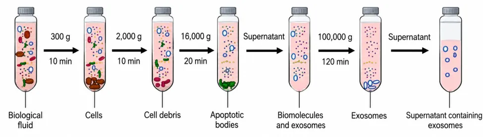
Figure:-Ultracentrifugation based separation to isolate exosome [30].
Immunoaffinity capture
Exosome membranes are known to contain large amounts of protein. Immunoaffinity methods are appropriate for exosome isolation because of the interactions of these proteins (antigens) with their antibodies and specific interactions of receptors with their ligands [32]. Immunoaffinity capture is particularly suitable for isolating EV subtypes based on markers rather than the isolation of all EVs at a single time with a relatively low yield but high purity. Immunoaffinity capture can be used to efficiently isolate specific EV populations in studies of specific EV subpopulations after UC or SEC to isolate sEVs. For example, melanoma-derived exosomes can be immunocaptured from plasma. First, mini-size exclusion chromatography (miniSEC) can be applied to get morphologically intact and biologically active sEVs. Next, specific tumor-targeted antibodies, antigen peptide epitope chondroitin sulfate peptidoglycan4 (CSPG4) monoclonal antibody can be used to precisely isolate MTEX [33] .
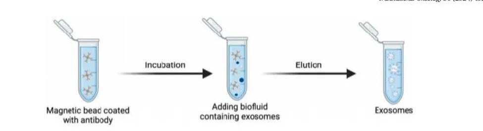
Figure-
Size-exclusion Chromatography (SEC)
The technique uses a porous gel filtration polymer as a stationary phase and a starting biofluid as a mobile phase . The stationary phase’s character permits differential elution, with larger particles eluting first, followed by smaller vesicles and then non-membrane-bound proteins. The larger the particle, the fewer pores it can traverse and thus the shorter path it will traverse to the end of the column and thus elute faster compared to its smaller counterparts The stationary phase or chromatography column can be packed with a number of gel polymers including cross linked dextrans (Sephadex), agarose (Sepharose), polyacrylamide (Biogel P), or allyldextran (Sephacryl) [31].Recent studies describe the development of an assay for comparing multiple isolation techniques (UC, precipitation and SEC), and demonstrated that SEC is the best method for sEVs purification from cerebrospinal fluid and plasma [33] .
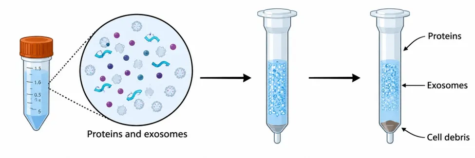
Figure -Size-exclusion Chromatography (SEC) [30].
Precipitation
The separation technique of precipitation depends on the dispersibility of the buffer where the sEVs are suspended .Hydrophilic polymers such as polyethylene glycol (PEG) are usually used as a highly hydrophilic polymers that interact with the environment to form a hydrophobic microenvironment, thus enabling the precipitation of the sEVs [34].Exosome precipitation relies on the use of polymers such as PEG (MW: 8 kDA) that bind to water molecules surrounding the exosomal membrane due to their negative charge [85]. PEG pulls water molecules in and pushes less soluble particles out of the exosome. This method involves overnight coincubation of samples with 8–12% of 6‑kDa PEG solution at 4◦C. It has been established experimentally that the addition of positively charged protamine molecules may stimulate exosome aggregation during the incubation period [30].
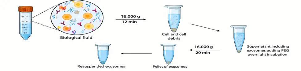
Figure -Precipitation method [30].
Microfluidics‑based techniques (MF)
New sophisticated methods using MF can be effectively applied for isolation of exosomes from samples with a minute volume in a time and cost effective way , offering also highly accurate/reliable results [34]. The microfluidic platform is also widely used for the isolation of DNA, protein and viruses .Currently, there are two main types of EV isolation strategies in microfluidics: label-based and label-free isolation technologies [33].This technique utilizes the classical separation determinants such as size, density and immunoaffinity, but also novel sorting mechanisms such as acoustic, electrophoretic, and electromagnetic manipulations; nanowire-based traps (NTs); nano-sized deterministic lateral displacement (nano-DLD); and viscoelastic flow [32].
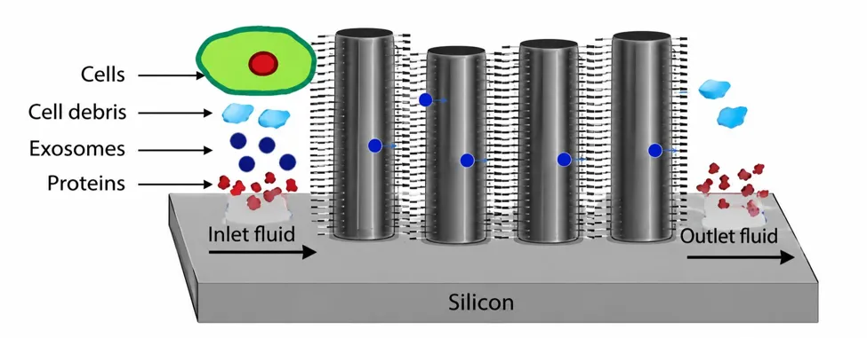
Figure -Microfluidics‑based techniques (MF)[30].
EXOSOMES FOR THERAPEUTIC AGENT DELIVERY
Exosomes are naturally involved in the transportation of various biological molecules between cells, and have potential to be applied in delivery of therapeutic agents [23] .In addition, exosomes are biocompatible and biodegradable, which minimizes the possibility of immune responses or toxicity commonly seen with synthetic drug delivery mechanisms. The small size of these vesicles allows them to cross biological barriers that larger carriers cannot cross and their stability in the bloodstream increases the chance of reaching their targets. Interestingly, exosomes have an innate ability to target specific cells or tissues and are capable of being used to deliver drugs directly to diseased cells thus increasing efficacy and minimizing side effects [25].Exosomes are versatile and can carry cargoes including proteins, nucleic acids and small drug molecules; thus, multiple therapeutic agents can be delivered simultaneously, and complex therapies with various types of molecules can be designed [35] .
Proteins
Protein drugs, which include substances such as therapeutic recombinant proteins, monoclonal antibodies, and hormones, can interact with specific targets within the body to treat diseases or restore normal functions. However, they can lose their effectiveness through immune responses, clearance from the bloodstream, degradation by enzymes, or binding to other molecules. To mitigate these challenges, carriers such as nanoparticles and exosomes can be used. Carriers can increase the stability and bioavailability of these drugs, control their release for sustained effects, enable targeted delivery, facilitate cell penetration, promote elevated therapeutic effects, and prevent immune responses. Thus, carriers play an indispensable role in improving the effectiveness and efficiency of protein delivery [28].Exosomes can also be used to deliver large molecules such as proteins in addition to small molecules. For example, a recent study demonstrated successful delivery of exosomes loaded with the antioxidant protein catalase across the BBB, resulting in an improved disease state in PD [26].
Nucleic acid
Exosomes can also naturally shuttle nucleic acids such as DNA and RNA to target cells, causing genetic changes in biological and pathogenic processes. These features of exosomes became a major interest in treatment strategies involving genetic therapy. Studies using exosomes as delivery systems for drugs have been conducted to deliver therapeutic genetic materials that alter gene expression in certain diseases and improve genetic therapy [26].
Genetic materials
Exosomes can also naturally shuttle nucleic acids such as DNA and RNA to target cells, causing genetic changes in biological and pathogenic processes. These features of exosomes became a major interest in treatment strategies involving genetic therapy. Studies using exosomes as delivery systems for drugs have been conducted to deliver therapeutic genetic materials that alter gene expression in certain diseases and improve genetic therapy [36]. Various delivery systems present problems such as off-target effects, limited tissue penetration, and rapid clearance, which can reduce therapeutic efficacy and cause unintended side effects. Therefore, targeted delivery of siRNAs, miRNAs, or ASOs to specific tissues or cells in the body remains a significant challenge. In efforts to overcome these limitations, exosomes have attracted significant attention as potential carriers for gene therapy. Sun et al. constructed a small RNA, anti-H19, that targets H19 lncRNA. They confirmed that overexpressed anti-H19 could be loaded into exosomes. They loaded anti-H19 into mouse plasma exosomes by injection These exosomes ameliorated colorectal cancer in a nontoxic, nonimmu- nogenic, and biocompatible manner. Their therapeutic effect was better than that of 5-Fu, one of the most commonly used cancer drugs [37].
Therapeutic Applications of Exosomes
Therapeutic Applications of Exosomes in Cancer
Many chemotherapeutic drugs have poor aqueous solubility and specificity, which necessitates the use of specialized delivery vehicles for their administration. Conventional therapies such as chemotherapy and radiotherapy are still the first choice for treating cancer, but they are not fully effective due to the increase in drug resistance. Thus, new therapies based on extracellular vesicles, immunotherapy and nanotechnologies represent the new generation of cancer treatment. In this case, exosomes seem to be a promising way for the specific delivery of drugs to tumor cells. Exosomes originating from tumor cells and exosomes secreted from other immune cells have been proven to regulate tumor microenvironment [1] .Exosomes, due to their endocytic origin, are loaded with RNA, DNA, proteins, lipids, metabolites and other molecules of their origin cells. These molecules allow exosomes to transfer information between cells that could be involved in the development of drug resistance, cancer progression and metastasis [38].
Glaucoma
Exosomes derived from stem cells can be a powerful neuroprotective and regenerative therapy. Using bone marrow MSC-exos, Mead et al. showed that exosomes can maintain RGCs function, promote RGCs survival and axonal regeneration through miRNA-dependent mechanisms. Exosomes deliver miRNA to RGCs for expression and subsequently translation of new proteins. Exosomes inhibit microglial activation and neuroinflammation, and up-regulate synapse-associated protein and brain-derived neurotrophic factor protein expression. The therapeutic effect of exosomes is weakened after knocking out the key effector molecule Argonaute-2 of miRNA. Bone marrow MSC-exos may act collectively as neuroprotective agents in glaucoma by promoting RGCs survival, inhibiting neuroinflammatory damage, and delivering neurotrophic proteins. Besides, umbilical cord MSC-exos and human embryonic stem cell-derived exosomes demonstrated neuroprotective effects [29].
Diabetes
EVs are also used for the treatment of diabetes mellitus in addition to cancer therapy. For instance, intravenous injection of exosomes derived from human urinary stem cells decreased urinary albumin and podocyte apoptosis, and increased the proliferation of glomerular endothelial cells in streptozotocin-treated rats. This study shows that EVs might be a new therapeutic approach for the treatment of diabetic nephropathy. Neurorestorative effects of bone marrow stem cell-derived exosomes including reduction of blood brain barrier leakage and hemorrhage and increase of axon and myelin density in rats with type II diabetes. Mesenchymal stem cell-derived exosomes ameliorate cognition in cognitively impaired diabetic animals through restoration of oxidative damage in neurons and astrocytes [32].
Neurological disorders
MVBs fuse with the plasma membrane releasing exosomes into the circulation or extracellular environment. Exosomes cross the BBB to reach the CNS and deliver the therapeutic agent to the target site where it acts. After they enter the central nervous system, exosomes may interact with neurons, astrocytes, microglia and oligodendrocytes via receptor-ligand interactions, membrane fusion or endocytosis. Their cargo consists of proteins, lipids, mRNA, microRNA and various regulatory chemicals, which can alter gene expression, impact synaptic plasticity, control inflammatory responses, and promote neuroprotection or regeneration [7].
Osteoarthritis
Osteoarthritis is a common chronic joint disease characterized by secondary bone hyperplasia and degenerative changes in the articular cartilage. With an aging population and increasing obesity rates linked to socio-economic development, the incidence of osteoarthritis is gradually rising. This condition significantly impacts patients’ habits and lifestyles, interfering with their daily activities. Non-steroidal anti-inflammatory drugs (NSAIDs) are widely used in clinical practice primarily for pain relief, reducing patient discomfort but not promoting cartilage regeneration. As a result, joint deterioration continues. Advances in regenerative medicine have revealed that stem cells, particularly stem cell-derived exosomes, hold promise for tissue and organ repair, offering potential new treatments for osteoarthritis [3].
PERSPECTIVES
Exosomes can transfer encapsulated proteins and genetic information to recipient cells and function as information messengers between cells. The surface molecules anchored on exosomes from different cell sources are different, which makes them selective for certain recipient cells. The purpose of surface engineering is to increase the local concentration of exosomes at the diseased site, thus reducing toxicity and side effects and maximizing therapeutic efficacy. Recent large pharmaceutical transactions signal the industry’s expectation for the use of exosomes in delivering drugs to hard-to-reach tissues [15] . Exosomes are natural vesicles secreted by many cells in the body and are important carriers of information between cells. Exosomes are natural carriers of functional genetic information, which have low toxicity and a wide tissue distribution in vivo. They also have a cellular uptake more than 30 times higher than alternative delivery systems such as nanoparticles or liposomes [17].Hypoxic cancer cells probably take up exosomes in acidic microenvironments. Exosomes can be directed to recipient cells by secreted molecules. The tetraspanin CD9 is expressed on the surface of exosomes and enhances drug delivery by promoting rapid membrane fusion with recipient cells.CD47 interacts with regulatory proteins on the surface of exosomes .To produce a signal that inhibits phagocytosis by the MPS and greatly reduces immunogenicity [9] . Exosomes can be also mixed with other biomaterials or inorganic materials for biomedical applications. Exosomes carrying metal-organic framework nanoparticles have shown improved drug-loading efficiency and release properties and protection from enzyme-mediated protein degradation [15].
REFERENCES

Dhanashri Shivdas Ghotekar, Diksha Jadhav, Asmita Bhoasale, Rajeshwari Gangurde, Exosomes as Smart Nanocarriers for Precision Drug Delivery, Int. J. of Pharm. Sci., 2026, Vol 4, Issue 10, 332-346, https://doi.org/10.5281/zenodo.23120258
 10.5281/zenodo.23120258