

We use cookies to ensure our website works properly and to personalise your experience. Cookies policy
East Point College of Pharmacy, Karnataka 560049, Affiliated to Rajiv Gandhi University of health sciences, Bengaluru, Karnataka- 560041
Osteoarthritis (OA) is a progressive degenerative joint disorder characterized by cartilage degradation, synovial inflammation, pain, and impaired joint function, affecting millions of individuals worldwide. Current therapeutic approaches, including oral non-steroidal anti-inflammatory drugs (NSAIDs), primarily provide symptomatic relief but are associated with significant gastrointestinal, renal, and cardiovascular adverse effects during long-term use. Topical drug delivery systems have emerged as an effective alternative by providing localized drug action while minimizing systemic exposure. Among NSAIDs, meloxicam is a selective cyclooxygenase-2 (COX-2) inhibitor with potent anti-inflammatory and analgesic activity; however, its poor aqueous solubility limits its therapeutic performance. This review highlights recent advances in cellulosic microsponge technology as a promising strategy for the sustained topical delivery of meloxicam in osteoarthritis management. The review discusses osteoarthritis pathophysiology, disease burden, current therapeutic limitations, physicochemical and pharmacokinetic characteristics of meloxicam, and challenges associated with transdermal drug delivery. Particular emphasis is placed on microsponge drug delivery systems, including their preparation techniques, structural characteristics, advantages, evaluation parameters, and controlled drug release behavior. The role of cellulosic polymers, including ethyl cellulose, hydroxypropyl methylcellulose, hydroxypropyl cellulose, cellulose acetate, and carboxymethyl cellulose, in improving formulation stability and sustained drug release is comprehensively reviewed. Furthermore, recent advances in microsponge-based topical formulations and their potential applications in arthritis therapy are summarized. Overall, cellulosic microsponge-based meloxicam gels represent a promising platform for enhancing topical drug delivery by improving drug retention, prolonging therapeutic action, reducing dosing frequency, and minimizing systemic adverse effects. Continued research and clinical translation of these systems may contribute to more effective and patient-friendly osteoarthritis management strategies.
Current management of osteoarthritis (OA) is primarily directed toward symptom control, with the goals of pain relief, improvement of joint function, and enhancement of patients’ quality of life. Common treatment modalities including nonsteroidal anti-inflammatory drugs (NSAIDs), analgesics, physical therapy, and joint replacement surgery are largely palliative in nature and do not effectively prevent, halt, or reverse disease progression. This symptom-focused approach reflects the incomplete understanding of OA pathophysiology and has significantly limited the development of effective targeted therapies.(1)
Emerging research indicates that OA pathogenesis is driven by multiple interconnected pathological processes, including progressive cartilage degradation, subchondral bone remodeling, synovial inflammation, and metabolic dysregulation. Importantly, many of these changes occur during the early stages of the disease; however, currently available treatments fail to adequately address these underlying mechanisms. Consequently, there remains a critical lack of disease-modifying osteoarthritis drugs (DMOADs) capable of altering the natural course of OA by slowing or preventing disease progression. This therapeutic gap has far-reaching clinical and socioeconomic consequences. The rising prevalence of OA, fueled by population aging and increasing rates of obesity, has led to significant declines in patient quality of life and has imposed a substantial economic burden on healthcare systems worldwide. Therefore, the development of effective DMOADs targeting the fundamental pathological processes of OA is essential not only to reduce patient morbidity but also to alleviate the broader societal and economic impact of the disease. (2)
Future DMOAD development should focus on inhibiting cartilage degeneration, regulating synovial inflammation, correcting metabolic imbalances, and promoting joint tissue regeneration. The successful advancement of such therapies holds the potential to revolutionize OA treatment and markedly improve long-term outcomes for affected patients. Osteoarthritis (OA) is the most prevalent degenerative joint disease and predominantly affects middle-aged and elderly individuals, particularly those aged 45 years and above . Patients with OA commonly experience symptoms such as joint pain, swelling, restricted mobility, and progressive joint deformity Prolonged disease duration leads to a significant deterioration in quality of life and may contribute to the development of cardiovascular and psychological disorders.
Numerous factors are known to increase the risk of OA, including genetic predisposition, advancing age, and increased body weight (Figure 1). In addition, OA may develop secondary to joint trauma, infection, or mechanical instability. In recent years, the rising prevalence of obesity and increased participation in high-impact physical activities have contributed to a noticeable shift toward younger age groups being affected by OA.
Projections indicate that by 2050, the global population living with knee OA will reach approximately 642 million individuals imposing a substantial burden on healthcare systems and society as a whole. (1,2)
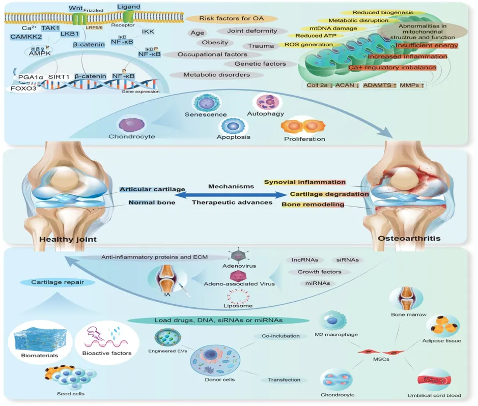
FIGURE: 1 A schematic illustration of the pathogenesis and emerging therapeutic strategies for OA, including: risk factors and molecular mechanisms of OA, joint changes between healthy and OA conditions, and current emerging therapeutic approaches for OA. Abbreviations: IA: intra-articular injection; EVs: extracellular vesicles.
Transdermal drug delivery systems have gained significant attention in the pharmaceutical field because of their advantages over conventional routes such as oral and injectable administration. The skin allows the passage of small, lipophilic molecules; however, it presents a barrier to larger polymeric substances and hydrophilic drugs. Drug permeation is particularly restricted by the stratum corneum, the outermost layer of the epidermis, which serves as the primary barrier to diffusion. In regenerative medicine, efforts are focused on overcoming this skin barrier to reduce pain and promote faster wound healing. Uncontrolled inflammation is a major contributor to pain and tissue damage. To address these challenges, non-steroidal anti-inflammatory drugs (NSAIDs) are commonly employed. Appropriate use of these medications can effectively alleviate discomfort and significantly improve the patient’s quality of life.
Meloxicam (MX) is an enolic acid derivative of non-steroidal anti-inflammatory drugs (NSAIDs) that selectively inhibits cyclooxygenase-2 (COX-2) over COX-1. It is well known for its potent analgesic, antipyretic, and anti-inflammatory effects. MX is considered pharmaceutically valuable due to its effectiveness at relatively low therapeutic doses and its comparatively reduced side effects. However, its poor aqueous solubility has prompted extensive research to incorporate it into suitable delivery systems. Among these, gel formulations have shown promise for both human and veterinary applications. Oral administration of MX may cause gastrointestinal complications and can negatively impact long-term patient health, particularly in conditions like rheumatoid arthritis. In contrast, transdermal delivery offers advantages such as sustained plasma drug levels, reduced gastrointestinal side effects, and improved localized pain relief. Effective skin permeation depends largely on selecting appropriate carriers with optimal properties. (2, 3)
This review focuses on advancements in gel-based formulations designed for prolonged MX release through minimally invasive approaches. It also highlights emerging systems such as stimuli-responsive gels, vesicular carriers, and nanostructured lipid systems. Additionally, the physicochemical properties of MX and the potential benefits and risks of its combination with other therapeutic agents are discussed.
Physicochemical properties and pharmaceutical profile of Meloxicam
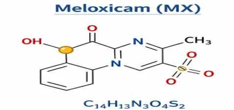
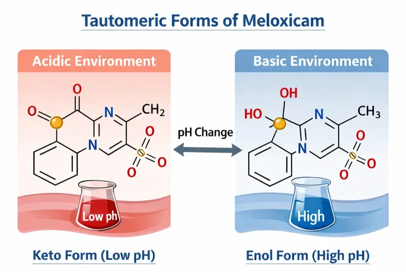
Meloxicam (MX) is an active pharmaceutical compound belonging to the oxicam class, with the molecular formula C₁₄H₁₃N₃O₄S₂ and the IUPAC name 4-hydroxy-2-methyl-N-(5-methyl-2-thiazolin)-2H-1,2-benzothiazine-3-carboxamide-1,1-dioxide. Its structure can exist in different tautomeric forms depending on the pH and the polarity of the surrounding solvent. (4)
Solubility:
Meloxicam (MX) is practically insoluble in water and only slightly soluble in solvents such as propylene glycol and liquid paraffin. Its solubility can be improved by using surfactants like Span 20 and Tween 20.
MX reacts with bases to form salts, which enhances its solubility under physiological conditions. For instance, it forms ammonium and sodium salts when combined with ammonia or sodium hydroxide. In contrast, acidic pH conditions are not suitable for MX incorporation, as they reduce its solubility in aqueous or alcohol-based systems. MX can also recrystallize from tetrahydrofuran in its enolic form. Structurally, it exists as a zwitterion in aqueous sodium hydroxide solutions, while a cationic form predominates under acidic conditions.
The solubility behavior and the balance between ionized and non-ionized forms of NSAIDs significantly influence gastrointestinal side effects, as they affect drug reabsorption in the gastrointestinal tract. Lower pH conditions promote ionization, which in turn reduces the solubility of the drug. (5)
Pharmacokinetic profile of Meloxicam:
Intravenous administration of meloxicam (MX) produces a plasma concentration of about 3.7 µg/mL within 12 hours, which is comparable to intramuscular administration. MX exhibits high bioavailability (approximately 89%) and shows extensive plasma protein binding (around 90%). It has a relatively long half-life of 20–24 hours and is capable of achieving therapeutic levels in synovial fluid. The drug becomes detectable within one hour, with peak concentrations reached after about six hours. Its pharmacokinetics remain largely unaffected in elderly patients or those with mild renal or hepatic impairment. Intramuscular administration provides a faster onset of action compared to oral dosing, as it bypasses gastrointestinal absorption delays. This makes it particularly useful for rapid symptom control, such as in acute pain, achieving therapeutic effects within about 1.5 hours with complete bioavailability. MX is mainly metabolized in the liver and eliminated through both biliary and urinary pathways. Pharmacokinetic characteristics play a key role in determining the onset of drug action. In a double-blind study comparing MX with piroxicam for periarticular shoulder pain, both drugs were administered orally. MX demonstrated a faster onset of pain relief than piroxicam, likely due to its quicker attainment of peak plasma concentration.
Current problems and limitations in osteoarthritis (OA) Treatment:
Prevalence:
Osteoarthritis (OA) is one of the most prevalent musculoskeletal disorders worldwide, affecting multiple joints, including the knee, hip, hand, ankle, and temporomandibular joint (TMJ) Among these, the knee, hand, and hip are the most commonly affected sites . Over the past century, the prevalence of OA has increased substantially, largely due to rising life expectancy and increasing body weight. A large U.S. cohort study reported a 2.1-fold increase in knee OA prevalence since the 1950s , and projections estimate that overall OA prevalence will rise from 26.6% to 29.5% by 2032. OA prevalence varies across studies depending on diagnostic criteria, with radiographic OA generally more common than symptomatic OA Data from the Global Burden of Disease (GBD) study showed that the global prevalence of OA increased from approximately 300 million in 2017 to 530 million in 2019. Between 1990 and 2019, the overall prevalence rose by more than 13%, with age-standardized rates increasing from 6,173.38 to 6,348.25 per 100,000 population. In 2019, China reported the highest number of OA cases (132.81 million), followed by India (62.36 million) and the United States (51.87 million), with prevalence increases of 156.58%, 165.75%, and 79.63%, respectively, since 1990 . OA was more prevalent in females than males and increased markedly with age, peaking in individuals aged 60–64 years Additionally, a recent retrospective cohort study reported an increase in hip OA prevalence from 4.03% in 2008 to 7.34% in 2019.
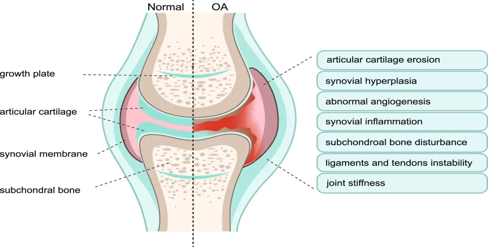
FIGURE: 2 Phenotypes of Osteoarthritis (OA). Clinic evidence shows that the majority of OA patients have a diversity of OA phenotypes, including articular cartilage erosion, synovial hyperplasia, abnormal angiogenesis, synovial inflammation, subchondral bone disturbance, ligaments and tendons instability, and joint stiffness. Left-half side shows the structure of the normal synovial joint. Right-half side showed the possible alterations of synovial joint structure and symptoms in osteoarthritis (7)
NEW THERAPEUTIC TARGETS IN OSTEOARTHRITIS
Four major therapeutic strategies for the management of osteoarthritis (OA). Agents classified as symptom modifiers primarily provide symptomatic relief and improvement in clinical outcomes (Table 1). In contrast, disease-modifying agents (Table 2) have demonstrated potential roles in promoting cartilage regeneration and remodeling of the subchondral bone. In addition, anti-obesity therapies and genicular nerve–targeted interventions, including nerve blocks and radiofrequency ablation, which have shown encouraging results in OA management, are discussed separately. (7,8)
The four therapeutic strategies include:
• symptom-modifying agents;
• disease-modifying agents;
• anti-obesity therapies;
• genicular nerve block and radiofrequency ablation.
Table: 1 symptom-modifying agents
|
drugs |
Mechanism of action |
Study phase and trial registration |
Target tissue |
Route of administration |
|
The liposome formulation of dexamethasone sodium phosphate |
Anti-inflammatory |
Phase 2/3; NCT03005873 |
Inflamed synovium |
IA |
|
Microsphere-based, extended-release formulation of triamcinolone acetonide |
Anti-inflammatory |
Phase-3; NCT03046446 |
Inflamed synovium |
IA |
|
Tanezumab |
Monoclonal antibody, inhibits NGF |
Phase-3; NCT02528188 |
Peripheral nociceptors |
SC |
|
Tanezumab |
Monoclonal antibody against NGF |
Phase-3; NCT02709486 |
Peripheral nociceptors |
SC |
Table: 2 Disease-modifying agents
|
Drugs |
Mechanism of action |
Study phase and trial registration |
Target tissue |
ROA |
|
OLP-1002 (SCN9A antisense peptide nucleic acid) |
Selective inhibition of Nav 1.7 sodium channel |
Phase-2; NCT05216341 |
Neuronal cells |
SC |
|
PTP-001 (allogenic placental tissue particulate) |
Release anti-inflammatory cytokines, promote synovial cell proliferation, and inhibit MMP-13 |
Phase-2; NCT05100225 |
Articular cartilage, inflamed synovium |
IA |
|
ROCCELLA (GLPG1972/S201086) (anti-ADAMTS-5) |
Aggrecanase-2 inhibition; inhibit TGF-beta mediated SMAD3 inhibition |
Phase 2; NCT03595618 |
Articular cartilage |
Oral |
|
QUC398 (anti-ADAMTS-5) |
Aggrecanase-2 inhibition; inhibit TGF-beta mediated SMAD3 inhibition |
Phase-2; NCT05462990 |
Articular cartilage |
SC |
|
X-STEM OA/allogenic MSC |
Chondrocyte proliferation, exosomes mediated |
Phase-1/2a; NCT05344157 |
Articular cartilage |
IA |
|
Lorecivivint (SM04690) |
Chondrogenesis, anti-inflammation |
Phase-3; NCT05603754 |
Articular cartilage, synovium |
Intra-articular |
|
Cymrus MSC (induced pluripotent stem cells) |
Chondrogenesis, anti-inflammation |
Phase 2b; ACTRN12620000870954 |
Articular cartilage, SCB, and synovium |
IA |
|
Pentosan polysulfate sodium (PPS) |
Chondrogenesis, reducing serum cholesterol and in cartilage, anti-thrombolytic |
Phase 2a; ACTRN12621000654853 |
Synovium, articular cartilage |
Oral |
|
Pentosan polysulfate sodium (PPS) |
Chondrogenesis, reducing serum cholesterol and in cartilage, anti-thrombolytic |
Phase 2/3; NCT04809376 |
Synovium, articular cartilage |
SC |
|
LNA043 (angiopoietin-like protein) |
Differentiation of endogenous MSC to chondrocytes |
Phase 2; NCT04864392 |
Articular cartilage |
IA |
|
GSK3858279 (blocks CCL17 receptor) |
Blocks the activity of a protein called CCL17 |
Phase 2; NCT05838742 |
Inflamed synovium |
IV |
|
ICM-203 (recombinant adenovirus associated vector) |
Inhibits inflammation and promotes cartilage repair |
Phase 1/2; NCT04875754 |
Inflamed synovium and articular cartilage |
IA |
TOPICAL DRUG DELIVERY SYSTEMS:
Topical NSAIDs are available in a wide range of dosage forms, including gels, foams, creams ointments, sprays, and patches or plasters. The formulation plays a critical role in determining the extent of skin penetration and therapeutic effectiveness. Optimal drug permeation requires a careful balance between lipid and aqueous solubility, and the use of prodrug esters has been proposed as a strategy to enhance transdermal permeability. Evidence suggests that cream-based formulations are generally less effective than gels or sprays; however, advanced delivery systems, such as microemulsions, have demonstrated greater potential for improving drug penetration and overall efficacy. Topical drug delivery is therefore most effective for treating pain in superficial joints, such as the knees, fingers, hands, ankles, and shoulders, which are located close to the skin surface in osteoarthritis (OA). In contrast, topical formulations are not suitable for managing pain in deeper joints, including the hip or spine, nor are they indicated for deep visceral pain or headaches. Additionally, topical NSAIDs are particularly preferred in patients with a limited number of painful joints, as this approach minimizes the risk of exceeding recommended dosage limits that may occur when multiple application sites are treated simultaneously. (8)
Topical therapies primarily exert their effects by achieving high drug concentrations within joint tissues while allowing only minimal amounts to enter the systemic circulation. Following topical administration, plasma drug levels are typically reported to be between 0.2% and 8% of those observed after oral dosing. Although limited systemic exposure is advantageous in reducing adverse effects on the circulatory system and other organs, it remains essential that adequate concentrations of the active agent reach the target joint tissues to produce a meaningful anti-inflammatory response.
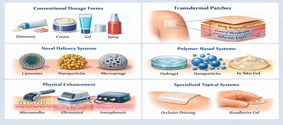
Figure: 3 Different types of topical drug delivery systems
Therefore, effective topical NSAIDs must be capable of penetrating the skin and permeating underlying tissues in sufficient quantities to achieve therapeutic efficacy. Measurement of drug concentrations at the site of action has been proposed as an important indicator of their clinical effectiveness. The pharmacological activity of topical formulations depends on their ability to penetrate the stratum corneum and subsequently diffuse into the deeper layers of the skin. As the stratum corneum serves as a protective barrier for underlying tissues, passive drug penetration is often limited. To overcome this barrier, topically applied drugs may exhibit a depot effect, whereby the drug accumulates within the stratum corneum, epidermis, dermis, and subcutaneous adipose tissue, forming a reservoir that enables sustained release into adjacent tissues over an extended period. (9)
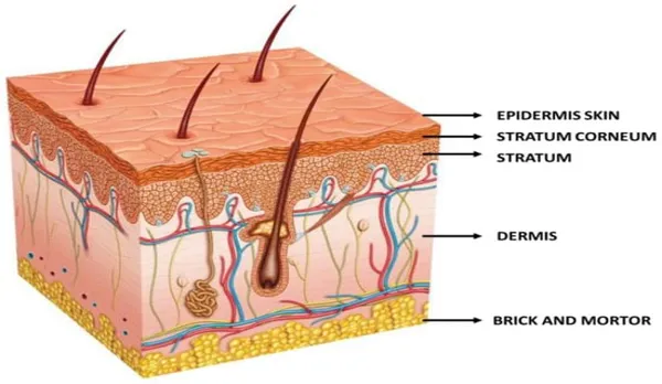
Figure: Schematic representation of the multi-layer composition of skin
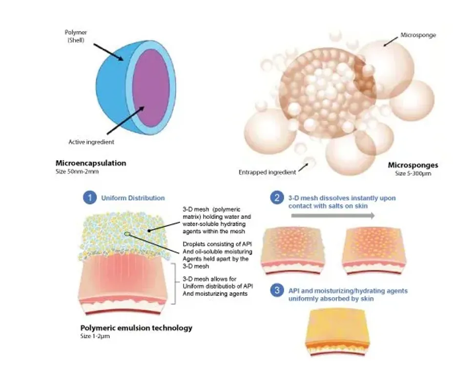
Figure:4 Advanced topical drug delivery systems in use in dermatology API, Active pharmaceutical ingredient.
Patients with OA commonly experience symptoms such as joint pain, swelling, restricted mobility, and progressive joint deformity. Prolonged disease duration leads to a significant deterioration in quality of life and may contribute to the development of cardiovascular and psychological disorders.
SKIN PENETRATION CHALLENGES
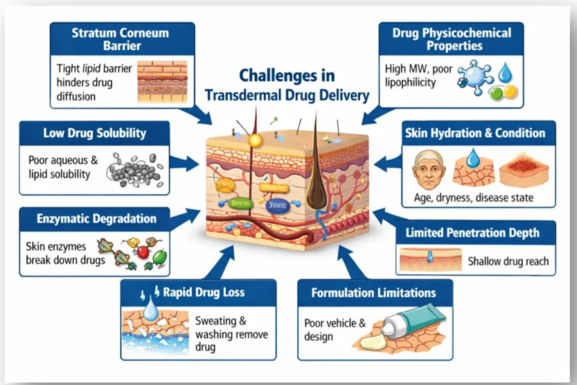
Table: 3 Skin penetration challenges in topical drug delivery.
|
Challenge |
Description |
|
Stratum corneum barrier |
The outermost skin layer acts as the main barrier, limiting passive diffusion of drugs due to its dense lipid–protein structure. |
|
Drug physicochemical properties |
High molecular weight, inappropriate lipophilicity, or polarity reduces skin permeation efficiency. |
|
Low drug solubility |
Poor solubility in aqueous or lipid phases decreases drug availability and concentration gradient at the skin surface. |
|
Skin hydration and condition |
Variations in skin thickness, hydration, age, and disease state influence drug absorption and penetration. |
|
Enzymatic degradation |
Cutaneous enzymes may metabolize drugs before reaching target tissues, lowering therapeutic efficacy. |
|
Limited penetration depth |
Topical formulations are mainly effective for superficial tissues and joints, with limited access to deeper targets. |
|
Rapid drug loss from skin |
Washing, sweating, and mechanical friction reduce drug residence time at the application site. |
|
Formulation limitations |
Inadequate vehicle selection or poor formulation design can fail to enhance permeation and sustained drug release. |
MICROSPONGE DRUG DELIVERY:
Microsponges are macroporous particulate delivery systems with particle sizes typically ranging from 10 to 25 µm. When applied to the skin, they release the entrapped active pharmaceutical ingredient in a controlled and sustained manner. Microsponges are capable of encapsulating a wide range of substances and can be incorporated into various semisolid and solid dosage forms. The porous nature of the microsponge surface facilitates prolonged drug release, thereby enhancing therapeutic efficacy while improving the safety profile of topically administered agents. (10)
Topical gels are semisolid dosage forms that contain an active pharmaceutical ingredient and are intended for application to the skin or mucous membranes. According to the United States Pharmacopeia (USP), gels are defined as semisolid systems composed of a dispersion of either small inorganic particles or large organic molecules that form a three-dimensional network, enclosing and interpenetrated by a liquid phase.
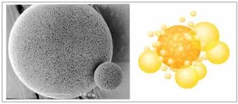
Figure: 4 Microsponge drug deliveries
Table:2 Microsponge drug delivery system and Topical gel.
|
Parameter |
Microsponge Drug Delivery System |
Topical Gel |
|
Definition |
Microporous particulate system designed for controlled and sustained drug release |
Semisolid dosage form intended for application to skin or mucous membranes |
|
Particle size |
Typically ranges from 10–25 µm |
No specific particle size; drug is dispersed or dissolved |
|
Structure |
Highly porous outer surface with interconnected channels |
Three-dimensional network formed by polymers enclosing a liquid phase |
|
Drug release |
Controlled and sustained release over a prolonged period |
Immediate or short-duration release |
|
Drug loading |
Can entrap a wide variety of active substances |
Limited drug-loading capacity |
|
Mode of action |
Releases drug gradually at the site of application |
Delivers drug rapidly to the application site |
|
Safety profile |
Reduced irritation and systemic side effects due to controlled release |
Higher risk of irritation due to rapid drug release |
|
Formulation flexibility |
Can be incorporated into semisolid and solid dosage forms |
Mainly limited to semisolid preparations |
|
Stability |
Provides improved stability to entrapped drugs |
Drug stability depends on gel composition |
|
Therapeutic efficacy |
Enhanced efficacy due to prolonged drug residence time |
Moderate efficacy with frequent application required |
|
Application in topical therapy |
Ideal for chronic conditions like osteoarthritis, acne, and dermatitis |
Commonly used for acute or short-term skin conditions |
Advantages of microsponge drug delivery
TECHNIQUES OF MICROSPONGES PREPARATION:
Quasi-Emulsion Solvent Diffusion Method:
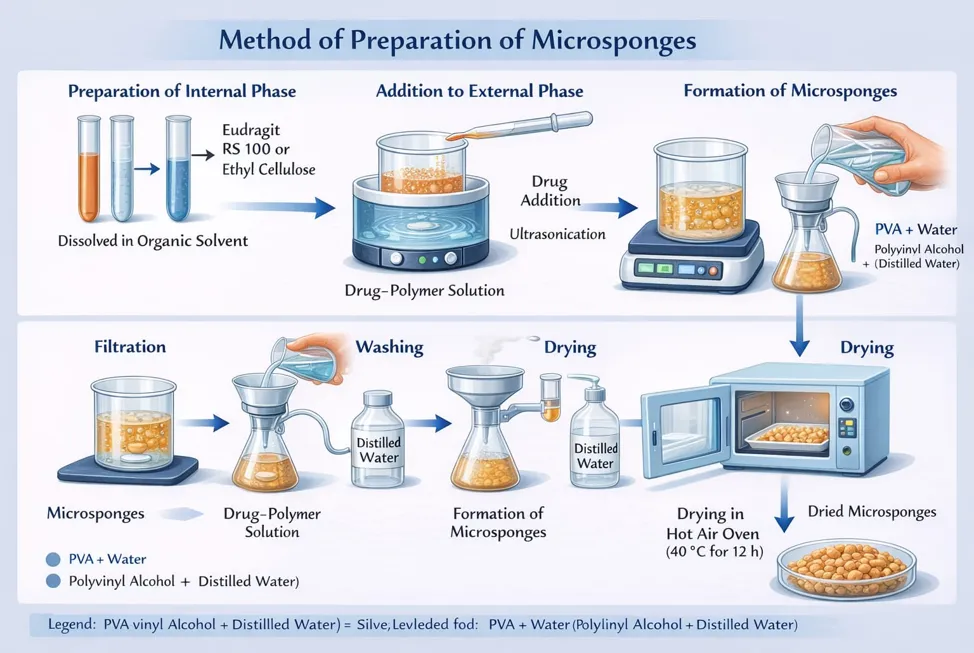
FIGURE 5: PREPARATION OF MICROSPONGES
Microsponges were prepared using the quasi-emulsion solvent diffusion method. In this process, the internal phase—consisting of the drug dissolved in a polymer solution—was added dropwise to the external aqueous phase containing polyvinyl alcohol (PVA) at room temperature. The resulting dispersion was continuously stirred for 3 hours to facilitate microsponge formation.
The drug-loaded microsponges were then dispersed in propylene glycol and incorporated into pre-soaked Carbopol, followed by thorough mixing to obtain a uniform gel. The final formulation pH was adjusted using triethanolamine.
The prepared microsponge-based gel was evaluated for various parameters, including particle size, percentage entrapment efficiency, percentage production yield, surface morphology, drug content, in vitro drug release, rheological behavior, and in vitro skin permeation characteristics. (11)
CELLULOSIC POLYMERS
Cellulose is one of the most widely recognized biodegradable polymers and is the primary structural component of plant fibers. It offers several advantageous properties, including low cost, excellent biocompatibility, and high mechanical and thermal stability, making it a highly promising material for a wide range of applications. However, its inherently low solubility limits its direct use, particularly in biomedical and pharmaceutical applications. This limitation can be effectively addressed through chemical modification processes such as esterification, etherification, and oxidation, which yield a variety of cellulose derivatives with improved solubility and functionality. As a result, cellulose and its derivatives are extensively employed in the pharmaceutical industry as excipients to regulate drug release rates and achieve optimal drug concentrations. (11,12)
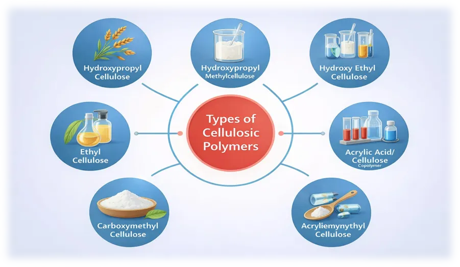
FIGURE: 5 TYPES OF CELLULOSIC POLYMERS.
CELLULOSE ACETATE
Cellulose acetate is a significant ester derived from cellulose, valued for its versatility across a wide range of applications. Depending on how it is processed, it can be used in the production of films, membranes, and fibres, among other materials. One particularly notable application is the synthesis of porous, spherical particles known as cellulose beads.
ETHYL CELLULOSE
Ethyl cellulose (EC) is a cellulose derivative in which a portion of the hydroxyl groups on the repeating anhydroglucose units are converted into ethyl ether groups, making it widely recognized as a non-ionic ethyl ether of cellulose. Drug delivery systems based on microencapsulated ethyl cellulose are currently being investigated for their potential to achieve sustained drug release while also shielding the core substance from degradation.
HYDROXYPROPYL CELLULOSE (HPC)
Hydroxypropyl cellulose (HPC) is a cellulose derivative distinguished by its solubility in both water and organic solvents. It serves multiple purposes, including functioning as a lubricant and being used in the treatment of various ocular conditions such as keratoconjunctivitis sicca, corneal erosions, and neuroparalytic keratitis. Additionally, it is commonly employed as a lubricant for patients fitted with artificial eyes. (12)
HYDROXYPROPYL METHYLCELLULOSE (HPMC)
HPMC has gained widespread use in the pharmaceutical industry due to its excellent safety profile, non-toxic characteristics, and its inability to be absorbed through oral consumption, meaning it does not contribute to the caloric value of food. It performs a diverse range of functions across various dosage forms, serving as a film-forming agent, thickener, binder, sustained-release agent, blending agent, and suspending agent. These capabilities allow for the consistent and reliable production of pharmaceutical preparations that maintain their structural integrity, deliver drugs in a controlled and sustained manner, and form stable emulsions that resist phase separation. (13)
In addition, HPMC is frequently incorporated as a matrix material, adhesive, and framework component in pharmaceutical formulations. It also plays a key role in the development of sustained and controlled-release microcapsules and pellets, cementing its status as a vital and versatile excipient in contemporary pharmaceutical manufacturing. (13,14)
CARBOXYMETHYL CELLULOSE (CMC)
Carboxymethyl cellulose (CMC), also commonly known as cellulose gum, is a cellulose derivative characterized by the attachment of carboxymethyl groups (-CH₂-COOH) to certain hydroxyl groups present on the glucopyranose monomers that form the cellulose backbone. It is most frequently utilized in its sodium salt sform, known as sodium carboxymethyl cellulose.
The introduction of carboxylic acid groups onto the cellulose backbone imparts water solubility to CMC, a property that natural cellulose does not possess. This distinctive characteristic makes it highly suitable for a broad spectrum of food and pharmaceutical applications where the use of water-soluble polymers is essential. As a result, CMC has become a valuable and widely adopted ingredient across these industries, serving functions such as thickening, stabilizing, and binding in various formulations and products. (14)
EVALUATION OF MICROSPONGE DRUG DELIVERY SYSTEM:
Drug entrapment efficiency of the microsponges was determined by accurately weighing 10 mg of drug-loaded microsponges and dispersing them in 50 mL of ethanol to extract the encapsulated drug. The dispersion was vortex-mixed for 1 hour to ensure complete drug extraction, followed by centrifugation at 3000 rpm for 10 minutes to separate the undissolved particulate matter. The resulting supernatant was collected and analyzed for Meloxicam content using high-performance liquid chromatography (HPLC). All measurements were performed in triplicate, and the results were expressed as mean ± standard deviation.(15)
The particle size distribution of the prepared microsponges was determined using a particle size analyzer (Malvern Master sizer Hydro 2000, Malvern, UK). Prior to analysis, the microsponges were dispersed in double-distilled water to obtain an appropriate concentration, ensuring that the light-scattering signal (indicated by particles count per second) remained within the optimal sensitivity range of the instrument. (16)
The surface morphology of prepared microsponges can be examined using scanning electron microscopy (SEM). Prior to analysis, the microsponges are coated with a gold-palladium layer under an argon atmosphere at room temperature to enhance imaging quality. In addition to surface examination, SEM can also be performed on fractured microsponge particles to provide a detailed visualization of their internal ultrastructure. This technique offers valuable insights into both the external surface characteristics and the internal architectural features of the microsponges, enabling a comprehensive morphological evaluation of the prepared particles.
The loading efficiency of microsponges can be evaluated using the following equation:
Loading Efficiency (%) = (Actual Drug Content in Microsponges / Theoretical Drug Content) × 100 …….. (1)
This calculation provides a measure of how effectively the drug has been incorporated into the microsponge system relative to the expected theoretical amount. The production yield of the microparticles, on the other hand, is determined by precisely measuring the initial weight of all raw materials used in the formulation process and comparing it to the final weight of the microsponges obtained after preparation. This is expressed using the following equation: (17)
Production Yield (%) = (Practical Mass of Microsponges / Theoretical Mass (Polymer + Drug)) × 100 …….. (2).
The pore volume and diameter of microsponges are critical parameters that play a significant role in regulating both the intensity and duration of the active ingredient's effectiveness. Furthermore, pore diameter directly influences the rate at which active ingredients migrate from the microsponges into the vehicle in which they are dispersed.
Mercury intrusion porosimetry is a widely employed technique for investigating the relationship between pore diameter, pore volume, and the rate of drug release from microsponges. This analytical method is capable of determining a comprehensive range of porosity-related parameters, including:
By providing detailed information on all these parameters, mercury intrusion porosimetry serves as an invaluable tool for thoroughly characterizing the pore structure of microsponges and understanding how these structural features influence drug release behavior and overall formulation performance.
In vitro drug release studies were performed using vertical Franz diffusion cells fitted with a cellophane membrane (molecular weight cut-off: 83). The membrane was soaked overnight prior to the experiment and positioned between the donor and receptor compartments. The formulation was placed in the donor compartment over an effective diffusion area of 4.52 cm², while the receptor compartment was filled with 8 mL of phosphate buffer (pH 6.8). The receptor medium was maintained at 32 ± 0.5 °C and continuously stirred at 100 rpm using a magnetic stirrer. (17,18)
At predetermined time intervals (1, 2, 4, 6, 8, 10, and 12 hours), 1 mL samples were withdrawn from the receptor compartment and immediately replaced with an equal volume of fresh buffer to maintain sink conditions. The withdrawn samples were analyzed at 275 nm using a UV–Visible spectrophotometer.
Applications of Microsponges
Microsponges are regarded as an highly effective drug delivery system capable of administering pharmaceutical active ingredients efficiently at minimal doses. Beyond their delivery capabilities, they offer several additional advantages, including enhanced formulation stability, reduced side effects, and improved control over drug release profiles.
The practical utility of microsponge-based drug delivery systems extends to a wide range of over-the-counter products. These include various moisturizing formulations, specialized skin rejuvenation products, and sunscreens, all of which leverage the unique properties of microsponges to improve product performance and consumer experience. The incorporation of microsponge technology into these everyday products highlights its versatility and growing significance in both pharmaceutical and cosmetic industries (18,19)
Microsponges for Topical Delivery
Recent Advances in Microsponge Drug Delivery System
Significant progress has been made in microsponge technology through the development of nanosponges, nanoferrosponges, and porous microbeads. Notably, β-cyclodextrin (β-CD) nanosponges were developed by crosslinking β-CD with biphenyl carbonate, making them suitable for both hydrophilic and hydrophobic drugs. These advanced systems have been investigated for the oral delivery of various drugs including dexamethasone, flurbiprofen, doxorubicin hydrochloride, itraconazole, and serum albumin. Research has also demonstrated that nanosponges can serve as effective carriers for gas delivery and that incorporating cytotoxic agents into nanosponge carriers can enhance drug potency, suggesting their potential use in cancer cell targeting.
Several studies have further explored microsponge applications across different therapeutic areas. Betamethasone microsponges prepared using the quasi-emulsion solvent diffusion method and incorporated into gels demonstrated effective topical anti-inflammatory activity. Risperidone-based microsponges using ethyl cellulose and Eudragit RS100 were developed for controlled antipsychotic drug release, improving patient compliance. Famotidine microsponges loaded into gels showed promising results for ulcer treatment, while diclofenac diethylamine microsponges were formulated for prolonged arthritis therapy. Optimization studies utilizing factorial designs and characterization techniques such as SEM, DSC, and FT-IR further enhanced understanding of encapsulation efficiency, particle size, and drug release profiles across these formulations. (20)
REFERENCES

Bapanapalli Vasundhara, Jyothi M, Cellulosic Microsponge Technology for Sustained Topical Delivery of Meloxicam in Osteoarthritis- Formulation and Evaluation Insights, Int. J. of Pharm. Sci., 2026, Vol 4, Issue 9, 3754-3772. https://doi.org/10.5281/zenodo.23022781
 10.5281/zenodo.23022781