View Article

  • Plant-Derived Bioactive Compounds for Cancer Therapy: Mechanisms, Clinical Evidence, and Translational Challenges

  • Department of Microbiology, Shardabai Pawar Mahila Arts, Commerce and Science College, Shardanagar, Malegaon Bk., Baramati, Pune, Maharashtra 413115, India

Abstract

Plant-derived bioactive compounds are widely investigated as sources of candidate molecules for cancer prevention and treatment. This narrative review summarizes major phytochemical classes—including polyphenols, flavonoids, alkaloids, terpenoids, brassinosteroids, and cardiac glycosides—and examines reported anticancer activities, molecular mechanisms, analytical approaches, and translational limitations. A selective literature search of PubMed and Google Scholar was used to identify English-language publications available through March 2023, using terms relating to medicinal plants, phytochemicals, cancer, apoptosis, cell-cycle arrest, angiogenesis, and cancer signalling. Evidence discussed in the literature includes preclinical observations of altered apoptosis, cell-cycle regulation, angiogenesis, oxidative stress responses, immune signalling, and pathways such as PI3K/AKT/mTOR, MAPK/ERK, NF-?B, and Wnt/?-catenin. Some plant-derived natural products have also informed approved anticancer medicines, but evidence for isolated phytochemicals and crude extracts varies substantially by compound, formulation, cancer type, and study design. Important barriers include limited bioavailability, inconsistent extract composition, uncertain dose–response relationships, toxicity, herb–drug interactions, and a shortage of robust clinical evidence for many candidates. This review is narrative rather than systematic and does not claim exhaustive retrieval or formal risk-of-bias assessment. Better chemical standardization, mechanistic validation, pharmacokinetic evaluation, and well-designed clinical studies are needed before most candidates can be considered reliable therapeutic options.

Keywords

phytochemicals; plant-derived anticancer agents; apoptosis; cancer signalling pathways; clinical translation

Introduction

× Popup Image

According to estimates from the Global Cancer Observatory (GLOBOCAN), the number of new cancer cases worldwide in 2020 was 19.3 million (Sung, Hyuna et al.2021). India was ranked as the third-highest country in terms of incident cancer cases, following China and the United States of America. GLOBOCAN has projected that the number of cancer cases in India will reach 2.08 million by 2040, representing a significant increase of 57.5 percent compared to 2020.(Ferlay, Jacques et al. 2020). The search for effective and safe treatments for cancer remains a global priority, as this devastating disease continues to impact millions of lives worldwide. In recent years, there has been a growing interest in exploring natural sources, such as medicinal plants, for potential anticancer agents. Medicinal plants have a long history of use in traditional medicine systems and are known to contain a wide array of bioactive compounds with diverse therapeutic properties. The scientific and research community is increasingly focusing on naturally-derived compounds due to their perceived lower toxic side effects in comparison to current treatments like chemotherapy. The Plant Kingdom produces naturally occurring secondary metabolites which are being investigated for their anticancer activities leading to the development of new clinical drugs. (Greenwell et al., 2015).

Among the bioactive compounds derived from medicinal plants, botanical derivatives have gained significant attention due to their potential anticancer properties. These derivatives, including alkaloids, flavonoids, terpenoids, polyphenols, and lectins, have demonstrated promising effects against various types of cancers (Nasim et al. 2022). Understanding the mechanisms underlying their anticancer activities is crucial for harnessing their therapeutic potential. Critical regulatory pathways, including the MAPK pathway, PI3K/AKT/mTOR signaling, and the Wnt/β-catenin signaling pathway, hold vital significance in governing signal transduction and fundamental biological processes such as cell proliferation, apoptosis, and metabolism, among others. The intricate regulatory mechanisms and biological functions of these signaling pathways are pivotal in numerous human cancers. Despite promising outcomes from combining various therapeutic approaches, several challenges persist in devising an effective antitumoral treatment that specifically targets tumoral cells (Fu et al., 2022). Natural plant extracts and derivatives have been reported to impact multiple oncological pathways that are related to apoptosis, angiogenesis, MAPK, cell cycle progression/proliferation, reprogrammed metabolisms, and epigenetics. Identification of these botanic derivative-impacted oncological pathways will help design or modify botanical lead compounds to generate potent anticancer drugs with minimal side-effects.

Botanical derivatives exhibit multifaceted mechanisms of action against cancer cells. Some compounds induce apoptosis, a programmed cell death mechanism that eliminates damaged or abnormal cells. Others disrupt cell cycle progression, leading to cell cycle arrest and inhibition of uncontrolled cell growth. Additionally, certain botanical derivatives inhibit angiogenesis, the process by which tumors develop their blood supply, thereby impeding tumor growth and metastasis (Banerjee et al 2022). Moreover, botanical derivatives can modulate signalling pathways involved in cancer development and progression. By targeting these pathways, they can interfere with key cellular processes, such as cell proliferation, migration, and invasion. Furthermore, botanical derivatives have been shown to enhance the immune system's response to cancer cells, promoting immune-mediated cytotoxicity and suppressing tumor growth.

Plant-derived drugs offer desirable attributes for cancer treatment owing to their natural origin and easy availability. These compounds have played a crucial role in addressing various types of cancer effectively. Notable examples of such agents include Vinblastine, Vincristine, Docetaxel, Etoposide (derived from Epipodophyllotoxin), Paclitaxel (Taxol), Flavonoids from Ginkgo biloba, Curcuma longa, Berberine, among others. Additionally, marine and microbiological organisms have also provided a diverse array of bioactive agents with significant therapeutic potential for anticancer treatments, complementing the efficacy of herbal natural products. Prominent examples of these drugs include trabectedin, dolastin, halichondrin, anthracyclines, and enediynes (Deep et al., 2023). The wide range of anticancer mechanisms exhibited by botanical derivatives makes them an intriguing area of research and holds promise for the development of novel therapeutic interventions. Furthermore, their natural origin offers potential advantages, such as reduced toxicity and better tolerability compared to synthetic drugs. However, extensive scientific investigations, including preclinical and clinical studies, are essential to fully understand their efficacy, safety, and potential interactions with conventional cancer treatments. (Saklani & Kutty, 2008)

In this article, we aim to provide a narrative overview of the anticancer properties and mechanisms of action of botanical derivatives derived from medicinal plants. By examining the current body of scientific literature, we will delve into the diverse classes of compounds and their specific effects on cancer cells. Understanding the mechanisms by which botanical derivatives exert their anticancer activities will not only contribute to our knowledge of natural therapeutics but also pave the way for the development of targeted and personalized cancer treatments.

By exploring the potential of botanical derivatives as anticancer agents, we hope to contribute to the growing body of evidence supporting the use of natural compounds in cancer therapy. Ultimately, the integration of traditional medicine and modern scientific approaches can provide new avenues for improving cancer treatment outcomes and enhancing the quality of life for patients.

2. LITERATURE SEARCH STRATEGY

This article is a narrative review, not a systematic review or meta-analysis. A selective literature search was conducted using PubMed and Google Scholar to identify publications relevant to plant-derived bioactive compounds and cancer. Search concepts included medicinal plants, plant extracts, phytochemicals, anticancer activity, apoptosis, cell-cycle arrest, angiogenesis, tumour growth, signalling pathways, phytochemical isolation, and clinical trials. English-language publications available up to March 2023 were considered.

Publications were considered relevant when they addressed phytochemical composition, extraction or characterization, anticancer activity in cellular or animal models, mechanisms of action, clinical development, safety, or limitations affecting translation. The review also considered selected established anticancer medicines derived from natural products to provide context for natural-product drug discovery. This selective approach was intended to provide a broad overview of key themes rather than a reproducible exhaustive search.

Because a complete database-specific search strategy, duplicate-removal log, formal screening flow, and structured risk-of-bias assessment were not documented, the article should not be interpreted as a systematic or scoping review. Statements about clinical evidence should be checked against primary trial records and publications before submission.

3. PLANT-DERIVED BIOACTIVE COMPOUNDS AS ANTICANCER AGENTS

Bioactive principles found in medicinal plants are of significant interest due to their potential therapeutic applications. These bioactive compounds, also known as secondary metabolites, are derived from primary metabolites and play essential roles in the survival and defense mechanisms of plants against pathogens and herbivores. (Newman & Cragg, 2016). Additionally, they exhibit diverse pharmacological effects in mammalian systems, making them valuable sources for drug discovery and development.

Several classes of bioactive principles have been identified in medicinal plants, including alkaloids, flavonoids, terpenoids, phenolic compounds, and glycosides, among others. These compounds have demonstrated various biological activities, such as antioxidant, anti-inflammatory, antimicrobial, anticancer, and neuroprotective properties. In recent decades, researchers have directed their efforts towards drug discovery using herbal medicines or botanical sources, which are an essential part of complementary and alternative medicine (CAM) therapy (Pan et al., 2013).

3.1. Polyphenols

Polyphenols are a group of organic compounds with over 10,000 members that share a three-membered flavan ring system. These natural compounds are known for their advantageous properties, including scavenging free radicals, reducing oxidative stress, and modulating inflammation. In this context, the role of polyphenols, particularly curcumin, resveratrol, and epigallocatechin gallate (EGCG), in addressing various aspects of cancer drug resistance is discussed (Maleki et al., 2022). Resveratrol, found in peanuts, grapes, and red wine, and gallacatechins from green tea, exhibit antioxidative effects, contributing to overall health improvement and reducing the risk of cancer. Moreover, polyphenols trigger apoptosis in cancer cells by regulating copper ion mobilization, causing DNA fragmentation, and interfering with cancer-related proteins, such as Tumour Necrosis Factor (TNF) (Niedzwiecki et al., 2016).

3.2. Flavonoids

Flavonoids, which are a subclass of polyphenolic compounds, constitute a wide family of plant secondary metabolites with diverse structures and physiological activities. They can be categorized into several classes, such as flavones (e.g., flavone, apigenin, and luteolin), flavonols (e.g., quercetin, kaempferol, myricetin, and fisetin), flavanones (e.g., flavanone, hesperetin, and naringenin), and others. Flavonoids share a benzo-γ-pyrone structure and are widely distributed in plants (Kumar et al., 2013). Extracted from various plants, flavonoids display potent anticancer effects against different types of cancer, including hepatoma, cervical carcinoma, and breast cancer. These compounds induce apoptosis through intrinsic and extrinsic signaling pathways, disrupt mitochondrial function, and interfere with proteins involved in cancer cell survival, proliferation, and angiogenesis (Kopustinskiene et al., 2020).

3.3. Alkaloids

Alkaloids are a remarkably diverse group of compounds characterized by cyclic structures containing at least one basic nitrogen atom. These compounds are widely distributed in the plant kingdom, with significant presence in plants belonging to families like Leguminosae, Menispermaceae, Ranunculaceae, Loganiaceae, and Papaveraceae (Benyhe, 1994; Li et al., 2007; Huang et al., 2007). Many well-known chemotherapeutic agents, such as camptothecin, vincristine, vinblastine, berberine, sanguinarine, evodiamine, piperine, matrine, and tetrandrine, are classified as alkaloids. They have garnered considerable attention for their ability to regulate various mechanistic pathways involved in proliferation, cell cycle, and metastasis. Notably, vincristine and vinblastine have a historical significance in cancer treatment and are actively used in managing acute lymphoblastic leukemia (Mondal et al., 2019). Some antitumor drugs used clinically, including vincristine, vinblastine, and paclitaxel, have natural origins as alkaloids.

3.4. Terpenoids

Terpenoids, the largest and most diverse group of naturally occurring phytoconstituents, are responsible for imparting fragrance, taste, and pigmentation to plants. Their classification is based on the number of isoprene units they contain, which serve as the building blocks for terpenoids. Isoprene is a gaseous hydrocarbon with a molecular formula of C5H8 (Kamran et al., 2022). Terpenoids, composed of these isoprenoid units, represent one of the most extensive groups of natural products, with over 40,000 individual compounds discovered so far, and new ones continue to be identified each year (Sacchettini et al., 1997; Peñuelas et al., 2005; Withers et al., 2007). While most terpenoids originate from plants, they are also synthesized by other organisms, such as bacteria and yeast, as part of their primary or secondary metabolism. The classification of terpenoids includes various classes, such as monoterpenes (e.g., carvone, geraniol, d-limonene, and perillyl alcohol), diterpenes (e.g., retinol and trans-retinoic acid), triterpenes (e.g., betulinic acid, lupeol, oleanic acid, and ursolic acid), and tetraterpenes (e.g., α-carotene, β-carotene, lutein, and lycopene) (Rabi et al., 2009).

3.5. Brassinosteroids

Brassinosteroids (BRs), which are naturally occurring compounds in plants, play vital roles in hormonal signaling, growth regulation, and stress responses. Among them, 28-homocastasterone and 24-epibrassinolide have shown remarkable anticancer effects against multiple cancer cell lines, including T-lymphoblastic leukemia, multiple myeloma, and various carcinoma cell lines. These BRs interact with crucial proteins involved in breast and prostate cancers, such as estrogen receptor, epidermal growth factor receptor, and androgen receptor, leading to growth inhibition and induction of apoptosis. Notably, BRs exhibit distinct responses in normal and cancer cells, making them promising selective agents against cancer cells (Ma et al., 2022).

3.6. Cardiac Glycosides

Cardiac glycosides (CGs) are natural compounds widely utilized to treat various cardiac conditions and have more recently gained recognition as potential antitumor agents. They act as ligands for Na/K-ATPase, which is a promising drug target in cancer treatment (Schneider et al., 2017). Additionally, there is growing evidence suggesting that CGs can activate tumor-specific immune responses. CGs are of particular interest for drug repurposing, as they can be derived from both plant and animal sources and have been used to address various human ailments (Kara´s et al., 2020). While CGs are primarily known for their role in inhibiting the Na+/K+ ATPase pump during heart failure, leading to their clinical approval for cardiovascular disease treatment (El-Mallakh et al., 2020), their potential application in diverse cancers has been explored. CGs demonstrate selective cytotoxic effects on tumor cells, elevating their potential as promising anticancer molecules (Reddy et al., 2020).

4. ISOLATION, PURIFICATION AND CHARACTERIZATION OF PHYTOMEDICINAL COMPOUNDS

In ancient times, the isolation, extraction, and purification of drugs from medicinal plants led to the discovery of miraculous compounds that have been instrumental in alleviating human ailments. However, in modern drug discovery, successfully isolating, purifying, characterizing, and evaluating these compounds for their in vitro or in vivo activities remains a formidable challenge (Wijngaard et al., 2022). To overcome this challenge, researchers have turned to ethnopharmacological knowledge, using it to identify specific plant species, their relevant parts, and appropriate extracts for validating the potential of phytomedicines (Paliwal et al., 2020).

Various methodologies have been developed for the isolation, purification, and characterization of phytomedicinal compounds. The process typically begins with phytoextraction, involving prewashing, shade drying, and grinding the plant sample to obtain a fine powder. Crude extracts are then prepared using different solvent systems based on the polarity of the targeted compound (Cos et al., 2006). Several extraction techniques are employed, such as soxhlet extraction, sonication, heating under reflux, maceration, solid-solid extraction, microwave-assisted extraction, and others (Huie et al., 2020). Once extracted, the purification of specific bioactive compounds becomes a critical step and poses significant challenges. Purification is essential for subsequent characterization. Commonly used techniques include thin-layer chromatography (TLC), column chromatography, size exclusion chromatography (SEC), and high-performance liquid chromatography (HPLC).

Further structural elucidation of pure phytocompounds is achieved through analytical techniques such as Liquid Chromatographic Mass Spectrometry (LC–MS), Gas Chromatographic-Mass Spectrometry (GC–MS), Nuclear Magnetic Resonance spectroscopy (NMR), and Fourier-Transform Infrared Spectroscopy (FTIR) (Altemimi et al., 2017). These analytical techniques are vital for identifying bioactive compounds before subjecting them to in vitro or in vivo biological assays. The continuous advancement in these methodologies contributes significantly to the exploration and potential utilization of phytomedicinal compounds in drug development.
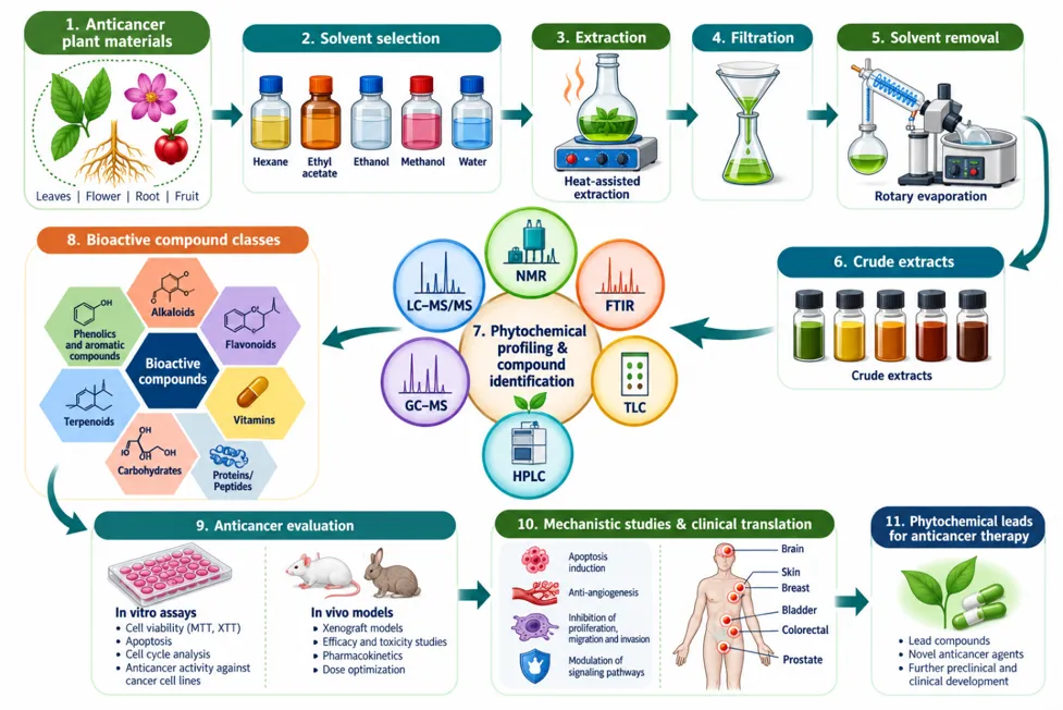
Figure 1. Workflow for the discovery and evaluation of plant-derived anticancer compounds.

The process includes plant selection based on ethnomedicinal knowledge, authentication and sample preparation, solvent extraction, fractionation and purification, structural characterization, in vitro and in vivo biological evaluation, toxicity assessment, formulation or bioavailability enhancement, and clinical translation. Artwork/diagram: author-prepared with AI-assisted illustration using ChatGPT and reviewed by the authors. No third-party figure was intentionally reproduced; underlying scientific claims should be supported by cited sources.

5. CHEMICAL AND BIOCHEMICAL PROPERTIES OF ANTICANCER PLANT DERIVATIVES

Chemical and biochemical properties play a vital role in the effectiveness of plant derivatives against cancer. These properties govern how bioactive compounds interact with cancer cells, the mechanisms they employ, and their potential as therapeutic agents. In this discussion, we delve into some crucial chemical and biochemical properties that characterize anticancer plant derivatives.

Molecular Structure: The molecular structure of plant derivatives significantly influences their ability to interact with cellular targets involved in cancer progression. It determines their size, shape, and functional groups, which are crucial for binding to specific receptors, enzymes, or signaling molecules. For example, certain functional groups like hydroxyl (-OH) or carbonyl (C=O) groups present in polyphenolic compounds enable them to scavenge free radicals, exhibit antioxidant activity, and regulate cellular signaling pathways implicated in cancer development (Du et al., 2018; Newman & Cragg, 2020).

Bioavailability: Bioavailability refers to how effectively a bioactive compound can be absorbed, distributed, metabolized, and excreted within the body. Anticancer plant derivatives with good bioavailability have a higher chance of reaching target tissues and exerting their therapeutic effects. Factors affecting bioavailability include the compound's solubility, stability, membrane permeability, and metabolism. To enhance the bioavailability of anticancer plant derivatives, strategies like formulation development or combination with other compounds can be employed (Panahi et al., 2019; Hwang et al., 2017).

Cytotoxicity and Apoptosis Induction: Anticancer plant derivatives often display cytotoxic effects on cancer cells, leading to cell cycle arrest, inhibition of cell proliferation, and promotion of apoptosis (programmed cell death). These effects can be mediated through various mechanisms, such as DNA damage, inhibition of key enzymes or signaling pathways, and modulation of apoptotic regulators. For example, vinca alkaloids derived from Catharanthus roseus disrupt microtubule assembly, resulting in cell cycle arrest and apoptosis induction (Zhang & Han, 2017; Siveen et al., 2014).

Anti-Angiogenesis: Angiogenesis, the formation of new blood vessels, is critical for tumor growth and metastasis. Anticancer plant derivatives with anti-angiogenic properties can inhibit the formation of new blood vessels, thereby suppressing tumor progression. Compounds like resveratrol from Vitis vinifera and curcumin from Curcuma longa have demonstrated anti-angiogenic effects by targeting various molecular pathways involved in angiogenesis (Chen et al., 2017; Yallapu et al., 2015).

Immunomodulation: The immune system plays a crucial role in cancer surveillance and elimination. Plant derivatives can modulate immune responses, enhancing the body's ability to recognize and eliminate cancer cells. They can stimulate immune cells such as natural killer (NK) cells and cytotoxic T lymphocytes, as well as regulate the production of immune mediators, including cytokines and chemokines. This immunomodulatory activity contributes to the anticancer effects of plant derivatives (Guo et al., 2017; Lin et al., 2019).

Synergistic Interactions: Anticancer plant derivatives often contain multiple bioactive compounds that can act synergistically, enhancing their overall effectiveness. Synergistic interactions occur when the combined effect of two or more compounds is greater than the sum of their individual effects. For instance, combining curcumin and piperine from Piper nigrum has been shown to increase the bioavailability and anticancer activity of curcumin (Li et al., 2010; Choi & Berhow, 2018).

6. REPRESENTATIVE ANTICANCER PLANTS AND THEIR PHYTOCHEMICALS

These are those plants that have been extensively studied and shown to possess notable anticancer effects. These plants contain bioactive compounds that exhibit specific mechanisms of action against cancer cells, making them potential sources of new anticancer therapies. The following is an overview of some well-known plants with documented anticancer properties and their natural origins/habitats:

Catharanthus roseus (Madagascar periwinkle): Native to Madagascar, this plant is known for producing vinca alkaloids, including vinblastine and vincristine. Vinca alkaloids disrupt microtubule formation, inhibiting cell division and inducing apoptosis. They have been used in the treatment of various cancers, including leukemia and lymphomas.

Curcuma longa (Turmeric): Turmeric, a member of the ginger family, is native to Southeast Asia. Its active compound, curcumin, has been extensively studied for its anticancer properties. Curcumin exhibits multiple mechanisms of action, including inhibition of inflammation, modulation of cell signaling pathways, and induction of apoptosis.

Viscum album (European mistletoe): European mistletoe is a parasitic plant found in various parts of Europe and Asia. Extracts from mistletoe have been used in complementary cancer treatments, particularly in Europe. They are believed to modulate the immune system and have cytotoxic effects on cancer cells.

Camellia sinensis (Tea): Tea, derived from the leaves of Camellia sinensis, is widely consumed worldwide. It contains various polyphenols, including epigallocatechin gallate (EGCG). EGCG exhibits antioxidant and anticancer properties, and numerous studies have reported its potential in preventing and treating cancer.

Panax ginseng (Asian ginseng): Ginseng is a popular medicinal plant native to Asia. It contains ginsenosides, which have been shown to possess anticancer effects. Ginsenosides modulate various signaling pathways involved in cell proliferation, apoptosis, and angiogenesis.

Podophyllotoxin derivatives from Podophyllum peltatum (Mayapple): Podophyllotoxin and its derivatives, including etoposide and teniposide, obtained from the roots and rhizomes of Podophyllum peltatum, exhibit anti-neoplastic activities. They inhibit topoisomerase II, leading to DNA damage and cell death. Etoposide and teniposide are commonly used in the treatment of various cancers, such as testicular cancer, lung cancer, and lymphomas

Resveratrol from Vitis vinifera (Grapes): Resveratrol, a polyphenol found in grapes and other plants, has demonstrated anti-neoplastic effects. It exhibits antioxidant, anti-inflammatory, and anti-proliferative activities. Resveratrol modulates multiple signalling pathways involved in cell cycle regulation, apoptosis, angiogenesis, and metastasis. It has shown promising results in various preclinical and clinical studies for the prevention and treatment of cancers, including breast, colorectal, and prostate cancer

Table 1. Plant-derived bioactive compounds, cancer types, study models, and proposed modes of action.

Plant Source

Bioactive Compound/ Phytochemical

Cancer Cells Type

Study Type

Mode of Action


Ref

Catharanthus roseus

Vinblastine

Leukemias, lymphomas, and solid tumors

In vitro

Inhibits microtubule formation

Wall et al., (1966).

Irinotecan

Colorectal

Clinical

Topoisomerase I inhibitor


Vinblastine

Hodgkin's lymphoma, Testicular cancer

In vivo

Inhibits microtubule formation, leading to cell cycle arrest and apoptosis

Jordan & Thrower (1991)

Camptotheca

acuminata

Camptothecin

Various

In vitro

Topoisomerase I inhibitor

Tewey et., (1984).

Etoposide

Various

In vitro

Topoisomerase II inhibitor

Hande et al., (1998).

Podophyllum

peltatum

Podophyllotoxin

Various

In vitro

Inhibits microtubule formation

Sultana et al.,  (2008).

Paclitaxel

Various

In vitro

Stabilizes microtubules

Gopalakrishnan et al.,  (2018).

Ginkgo biloba

Ginkgolides

Hepatocarcinoma, ovary, prostate, colon and liver cancer

Brain cancer

In vitro


Preclinical study

Anti-inflammatory, antioxidant

Inhibits cell proliferation and induces apoptosis

Ahlemeyer et al., (2003).

Mahadevan and Park, 2008; Yao et al., 2017

Curcuma longa

Curcumin

Leukemia, glioblastoma, Colon adenocarcinoma, prostate esophagus, liver and skin cancer (In vitro)

In vitro

Inhibits cell proliferation


Anti-inflammatory, antioxidant, and pro-apoptotic effects

Aggarwal et al.,  (2009).


Anand et al. (2008); Kunnumakkara et al. (2017)

Viscum album

Viscotoxin

Breast cancer, Colorectal cancer

Preclinical studies

Induces apoptosis and inhibits tumor growth

Büssing et al., (1996).             Steiner et al. (2003); Semmler-Behnke et al. (2013)

Vitis vinifera

Resveratrol

Breast Osteosarcoma Colorectal

In vitro

In vitro,

in vivo

In vitro,


Cell cycle inhibition, apoptosis

STAT3 pathway and cell cycle inhibition

In vitro NF-kB pathway inhibition, apoptosis

Wu, H. et al 2019,


Peng, L. et al 2018


Buhrmann, C. et al 2017.

Zingiber officinale

Gingerol

Lung


Breast

In vitro,
in vivo


In vitro

A549 cells death by iron accumulation, USP14 expression inhibition

ROS generation, activation of p53 expression mediated apoptosis,

Tsai et al., 2020


Sp et al., 2021

Carica papaya

Benzyl isothiocyanate

Pancreatic

In vitro,
in vivo

FOXO/PI3K/AKT pathways-mediated tumor apoptosis

Boreddy et 2011

Piper longum

Piperlongumine

Prostate

In vitro

DNA damage-mediated proliferation inhibition

Zhang et al., 2021


Lung

In vitro,
in vivo

mTOR/AKT/PI3K pathway inhibition-induced apoptosis

Wang et al., 2015

Gossypium hirsutum

Gossypol

Skin

In vitro

Mitochondrial apoptosis

Haasler et al., 2021


Cervical

In vitro,
in vivo

FAK pathway inhibition and TGF-β1-mediated EMT reversal

Hsieh et al., 2021


Colon

In vitro

Downregulation of FAS, CLAUDIN1, GAPDH, ELK1, ZFAND5, IL2, and IL8 expression

Cao et al., 2021

Taxus brevifolia

Paclitaxel

Breast cancer

in vivo

Stabilizes microtubules, leading to cell cycle arrest

Schiff et al., 1979; Rowinsky and Donehower, 1995

Camellia sinensis

Epigallocatechin gallate (EGCG)

Prostate cancer

Preclinical study

Inhibits tumor growth and metastasis

Khan and Mukhtar, 2007; Butt et al., 2009

Panax ginseng

Ginsenosides

Lung cancer

Preclinical study

Modulates cell signaling pathways and induces apoptosis

Helms and Miller, 2004; Jia et al., 2021

Allium sativum

Allicin

Stomach cancer

Preclinical study

Induces apoptosis and inhibits cell proliferation

Zhang and Tang, 2007; Banerjee and Sethi, 2013


Diallyl trisulfide

Prostate cancer

Preclinical study

Induces apoptosis, inhibits cell proliferation, and angiogenesis

Yang et al., 2019

Berberis vulgaris

Berberine

Liver cancer

Preclinical study

Induces cell cycle arrest and apoptosis

Mantena et al., 2006; Mantena et al., 2011

Tripterygium wilfordii

Celastrol

Prostate cancer

Preclinical study

Inhibits cell proliferation and induces cell cycle arrest

Chen et al., 2016

Aloe vera

Aloin

Colorectal cancer

Preclinical study

Induces cell death and inhibits angiogenesis

Tseng et al., 2016

Annona muricata

Annonacin

Liver cancer

Preclinical study

Inhibits cancer cell growth and induces apoptosis

Liaw et al., 2020


Acetogenins

Breast cancer

Preclinical study

Inhibits mitochondrial complex I, inducing apoptosis

George et al., 2021

Curcuma zedoaria

Zerumbone

Pancreatic cancer

Preclinical study

Induces apoptosis and inhibits cell migration

Matsuo et al., 2020

Euphorbia neriifolia

Diterpene compound

Cervical cancer

Preclinical study

Disrupts cell cycle, triggers apoptosis, and inhibits invasion

Abubakar et al., 2020

Podophyllum peltatum

Etoposide

Testicular, lung, and others

Clinical trial

Inhibits DNA topoisomerase II, causing DNA damage

Hande et al., 1998; Moxley et al., 2020

Maytenus senegalensis

Maytansine

Breast, lung, and others

Preclinical study

Disrupts microtubule function, leading to cell cycle arrest

Pourgholami et al., 2011; Bai et al., 2015

7. ANTICANCER MECHANISMS OF BOTANICAL DERIVATIVES

Anticancer mechanisms of botanical derivatives encompass a wide array of intricate cellular processes that target cancer cells and impede tumor growth. These mechanisms involve multiple pathways and molecular targets. For instance, many botanical derivatives induce apoptosis in cancer cells, triggering programmed cell death and eliminating abnormal or damaged cells (Elmore, 2007). Additionally, these compounds can disrupt cell cycle progression, leading to cell cycle arrest and inhibition of uncontrolled cell growth (Kastan and Bartek, 2004). Some botanical derivatives inhibit angiogenesis, the formation of new blood vessels that nourish tumors, thereby depriving tumors of essential nutrients and oxygen (Folkman, 2007). Furthermore, these derivatives can modulate various signaling pathways involved in cancer development and progression, such as those related to cell proliferation, survival, migration, and invasion (Huang et al., 2020). Moreover, botanical derivatives have demonstrated the ability to enhance the immune response against cancer cells, stimulating immune-mediated cytotoxicity and suppressing tumor growth (Wang et al., 2014). By targeting these intricate mechanisms, botanical derivatives derived from medicinal plants offer a promising approach for combating cancer and may pave the way for the development of novel and effective anticancer therapies.

7.1. Induction of Apoptosis

Apoptosis, or programmed cell death, plays a crucial role in maintaining tissue homeostasis and eliminating abnormal cells, including cancer cells. Many botanical derivatives have been shown to induce apoptosis in cancer cells through various mechanisms. For instance, resveratrol, a polyphenol found in grapes and berries, activates apoptotic pathways by modulating Bcl-2 family proteins, caspases, and the tumor suppressor p53 (Nguyen et al., 2003). Curcumin, derived from turmeric, induces apoptosis in cancer cells by targeting multiple signalling pathways, including NF-κB, Akt, and MAPK pathways (Aggarwal et al., 2003). These botanical derivatives promote cell death and inhibit cancer cell survival, thereby impeding tumor progression.

7.2. Cell-Cycle Arrest

Botanical derivatives can disrupt the cell cycle progression of cancer cells, leading to cell cycle arrest and inhibition of uncontrolled cell growth. One example is paclitaxel, a terpenoid derived from the Pacific yew tree. Paclitaxel stabilizes microtubules, preventing their depolymerization during cell division and causing cell cycle arrest at the G2/M phase (Schiff et al., 1979). Another botanical derivative, quercetin, a flavonoid present in various fruits and vegetables, induces cell cycle arrest at different phases, depending on the cancer cell type, by modulating cyclins, cyclin-dependent kinases (CDKs), and their inhibitors (Serrano et al., 2007). By interfering with cell cycle progression, this botanical derivatives limit cancer cell proliferation and promote their elimination.

7.3. Inhibition of Angiogenesis

Angiogenesis, the formation of new blood vessels, is essential for tumor growth and metastasis. Several botanical derivatives have been found to possess anti-angiogenic properties, inhibiting the formation of new blood vessels and limiting the blood supply to tumors. For example, epigallocatechin gallate (EGCG), a polyphenol abundant in green tea, inhibits angiogenesis by targeting multiple angiogenic factors, such as vascular endothelial growth factor (VEGF) and its receptors (Yang et al., 2003). Another botanical derivative, curcumin, suppresses angiogenesis through inhibition of VEGF expression and signalling pathways (Arbiser et al., 1998). These compounds effectively block the development of new blood vessels, impairing the tumor's ability to obtain necessary nutrients and promoting its regression.

7.4. Modulation of Signalling Pathways

Botanical derivatives can modulate various signalling pathways that play crucial roles in cancer development and progression. For instance, genistein, an isoflavone found in soybeans, exerts its anticancer effects by inhibiting protein tyrosine kinases, thereby disrupting signalling pathways involved in cell proliferation, survival, and angiogenesis (Sarkar et al., 2008). Resveratrol, through its interaction with multiple molecular targets, including AMP-activated protein kinase (AMPK), SIRT1, and PI3K/Akt, modulates various signalling pathways to inhibit cell proliferation, induce apoptosis, and inhibit tumor growth (Bishayee, 2009). These botanical derivatives act as potent regulators of critical signalling pathways, thereby interfering with cancer cell survival and growth.

7.5. Enhancement of Immune Response

Several botanical derivatives have been shown to enhance the immune response against cancer cells, promoting immune-mediated cytotoxicity and suppressing tumor growth. For example, polysaccharides derived from medicinal mushrooms, such as Ganoderma lucidum (reishi mushroom) (Gao et al., 2005)

Drugs derived from botanical derivatives that are currently in clinical trials or their clinical trials were completed. Summary of all clinical trials that have been designed due to previous knowledge of the anti-cancer activity recognized in botanical derivatives. (Bhatia et al., 2011)

8. SIGNALLING PATHWAYS INVOLVED IN PLANT-DERIVED PREVENTION OF TUMOUR GROWTH

Plant derivatives have been found to exhibit preventive effects on tumor growth by modulating various signalling pathways involved in cancer development and progression. These compounds interact with key molecular targets within these pathways, leading to the inhibition of cell proliferation, induction of apoptosis, suppression of angiogenesis, and modulation of immune responses. The following sections provide a detailed exploration of the signalling pathways targeted by plant derivatives and their role in tumor growth prevention.
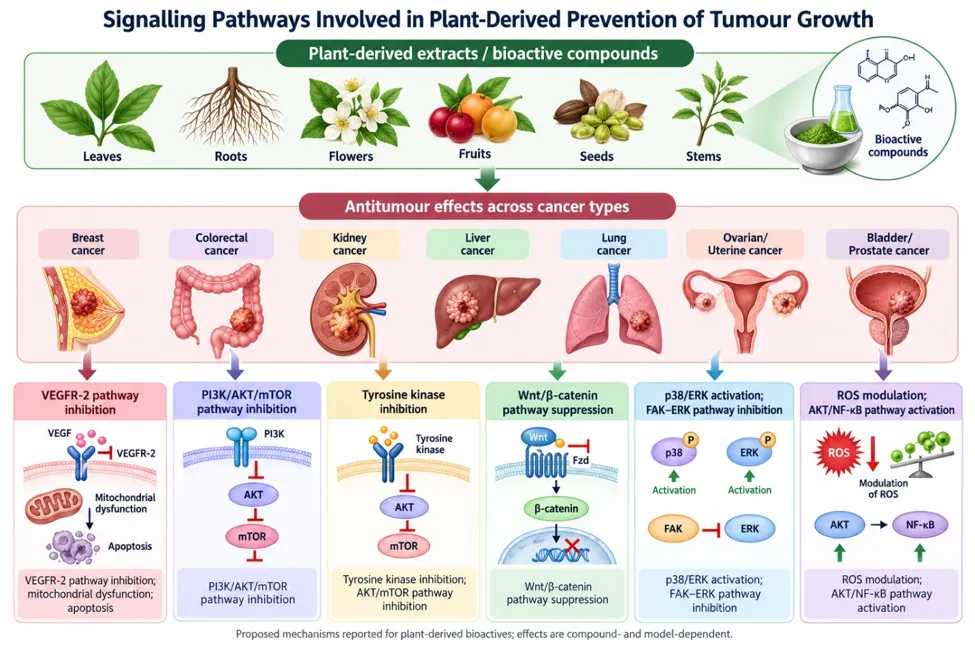
Figure 2. Principal signalling pathways associated with the anticancer effects of plant-derived compounds.

Plant-derived molecules may influence PI3K/Akt/mTOR, MAPK/ERK, NF-kappaB, Wnt/beta-catenin, JAK/STAT, p53, and apoptotic pathways, thereby affecting cell survival, proliferation, differentiation, migration, angiogenesis, and programmed cell death. Artwork/diagram: author-prepared with AI-assisted illustration using ChatGPT and reviewed by the authors. No third-party figure was intentionally reproduced; underlying scientific claims should be supported by cited sources.

8.1. PI3K/AKT/mTOR Pathway

The PI3K/Akt/mTOR pathway plays a critical role in regulating cell growth, survival, and metabolism. Dysregulation of this pathway is commonly observed in cancer cells, leading to increased cell proliferation and survival. Plant derivatives have been shown to inhibit this pathway, thereby exerting preventive effects on tumor growth. For instance, resveratrol inhibits Akt phosphorylation and downstream mTOR signaling, leading to cell cycle arrest and apoptosis in various cancer cell types (Panda, 2019). Curcumin suppresses Akt activation and mTOR signaling, resulting in the inhibition of cell proliferation and induction of apoptosis (Ko et al., 2019). By targeting the PI3K/Akt/mTOR pathway, plant derivatives can impede tumor growth and prevent cancer progression.

8.2. NF-κB Pathway

The NF-κB pathway is involved in regulating immune responses, inflammation, cell survival, and proliferation. Aberrant activation of NF-κB has been implicated in various cancers. Plant derivatives can inhibit NF-κB activation, thereby preventing tumor growth. Curcumin, for example, inhibits NF-κB signaling by suppressing the degradation of IκBα, an inhibitor of NF-κB (Huang et al., 2020). This leads to the suppression of NF-κB target genes involved in cell survival, angiogenesis, and metastasis. By targeting the NF-κB pathway, plant derivatives can attenuate the inflammatory response and inhibit tumor growth.

8.3. Wnt/β-Catenin Pathway

The Wnt/β-catenin pathway plays a crucial role in regulating cell proliferation and differentiation. Dysregulation of this pathway is commonly observed in cancer, leading to uncontrolled cell growth. Plant derivatives have been found to modulate this pathway, inhibiting tumor growth. For instance, curcumin inhibits the Wnt/β-catenin pathway by downregulating β-catenin levels and suppressing its nuclear translocation (Sun et al., 2019). This disrupts the transcription of Wnt target genes involved in cell proliferation and survival. By targeting the Wnt/β-catenin pathway, plant derivatives can hinder tumor growth and inhibit cancer progression.

8.4. MAPK/ERK Pathway

The MAPK/ERK pathway regulates various cellular processes, including cell proliferation, survival, and differentiation. Dysregulation of this pathway is commonly associated with cancer development. Plant derivatives have been shown to modulate this pathway, resulting in the inhibition of tumor growth. For example, resveratrol inhibits the activation of ERK1/2, downstream effectors of the MAPK pathway, leading to cell cycle arrest and apoptosis in cancer cells (Li et al., 2019). By targeting the MAPK/ERK pathway, plant derivatives can impede cell proliferation and promote tumor growth inhibition.

8.5. Notch Signalling Pathway

The Notch signalling pathway plays a critical role in cell fate determination, differentiation, and proliferation. Dysregulation of Notch signalling has been implicated in various cancers. Plant derivatives can modulate this pathway, contributing to the prevention of tumor growth. For instance, genistein inhibits Notch signaling by suppressing the expression of Notch receptors and downstream target genes (Girardi et al., 2021). This disrupts Notch-mediated cell proliferation and survival. By targeting the Notch signaling pathway, plant derivatives can impede tumor growth and inhibit cancer progression.

8.6. Hedgehog Signalling Pathway

The Hedgehog pathway regulates cell growth and differentiation during embryonic development and is also involved in cancer development. Plant derivatives like curcumin and resveratrol have been found to inhibit the Hedgehog pathway, leading to antitumor effects (Li et al., 2019).

8.7. JAK/STAT Pathway

The JAK/STAT pathway plays a critical role in cell proliferation, survival, and immune responses. Dysregulation of this pathway is associated with various cancers. Plant-derived compounds, including resveratrol and curcumin, have been shown to modulate JAK/STAT signaling, resulting in antitumor effects (Li et al., 2019).

9. THERAPEUTIC APPLICATIONS OF PLANT-DERIVED ANTICANCER COMPOUNDS

Anticancer botanic derivatives have shown immense potential as therapeutic agents in the field of oncology. These natural compounds, derived from various medicinal plants, possess bioactive properties that can effectively target cancer cells and inhibit tumour growth. The therapeutic applications of these anticancer botanic derivatives encompass a wide range of areas, including cancer prevention, treatment, and supportive care. Here, we explore some of the key therapeutic applications of these natural compounds:

Table 2. Clinical-trial status of selected plant-derived anticancer compounds.

Active Ingredient Name

Mode of Action

Cancer Type

Current Status

References (ClinicalTrials.gov)

Apigenin

Cell cycle arrest, inhibiting cancer cell migration and invasion

Colorectal Cancer

Suspended

https://clinicaltrials.gov/ct2/results? cond=Cancer&term=Apigenin& cntry=&state=&city=&dist=

Curcumin

Induce apoptosis

Prostate Cancer, Breast Cancer, Colorectal Cancer, Metastatic Colon Cancer

Completed; Recruiting

https://clinicaltrials.gov/ct2/results? cond=Cancer&term=Curcumin& cntry=&state=&city=&dist=

Allicin

Inhibit cancer cell growth

Follicular Lymphoma

Unknown

https://clinicaltrials.gov/ct2/results? cond=Cancer&term=Allicin& cntry=&state=&city=&dist=&

Search=Search

Andrographolide/ Andrographis paniculata

Cell-cycle arrest

Squamous Cell Carcinoma of Esophagus

Terminated; Completed

https://clinicaltrials.gov/ct2/results? cond=Cancer&term=Andrographolide

&cntry=&state=&city=&dist=&

Search=Search

Epigallocatechin

Induce cell cycle arrest and apoptosis

Colon Cancer, Esophageal Cancer, Lung Cancer, Nonmetastatic Bladder Cancer, Colorectal Cancer, Prostate Cancer, Small Cell Lung Cancer

Recruiting; Completed

https://clinicaltrials.gov/ct2/results? cond=Cancer&term=Epigallocatechin

&cntry=&state=&city=&dist=&

Search=Search

Emodin

Suppress tumor growth by inhibiting AKT, MAPK, PI3K pathway

Breast Cancer

Unknown

https://clinicaltrials.gov/ct2/results? cond=Cancer&term=Emodin& cntry=&state=&city=&dist=&

Search=Search

Genistein

Angiogenesis inhibitors, inhibit uncontrolled cancer cell growth

Breast Cancer, Non-Small Cell Lung Cancer, Colon Cancer, Rectal Cancer, Colorectal Cancer, Breast Cancer, Endometrial Cancer

Completed, Terminated, Suspended, Withdrawn

https://clinicaltrials.gov/ct2/results? cond=Cancer&term=Genistein& cntry=&state=&city=&dist=&

Search=Search

Gingerol

Inhibit PI3K, upregulate pro-apoptotic protein p21, induce apoptosis

Colorectal Cancer, Colorectal Adenoma

Completed

https://clinicaltrials.gov/ct2/results? cond=Cancer&term=Gingerol& cntry=&state=&city=&dist=&

Search=Search

Glycyrrhizin

Induces apoptosis

Lung Cancer, Liver Cancer, Hepatocellular Carcinoma

Completed, Recruiting, Unknown

https://clinicaltrials.gov/ct2/results? cond=Cancer&term=Glycyrrhizin& cntry=&state=&city=&dist=&

Search=Search

Licochalcone A

Inhibits PI3K/AKT/mTOR signaling pathway

Squamous Cell Carcinoma

Unknown

https://clinicaltrials.gov/ct2/results? cond=Cancer&term=Licochalcone& cntry=&state=&city=&dist=&

Search=Search

Pterostilbene

Inhibit cancer cell growth through apoptotic-dependent and -independent pathway

Endometrial Carcinoma

Recruiting

https://clinicaltrials.gov/ct2/results? cond=Cancer&term=Pterostilbene& cntry=&state=&city=&dist=&

Search=Search

Resveratrol

Induce apoptosis and cell cycle arrest through p53-dependent pathway

Colon Cancer, Neuroendocrine Tumor, Neoplasms, Colorectal, Adenocarcinoma of the Colon, Breast Cancer, Stage III & IV Melanoma

Completed, Terminated, Suspended, Withdrawn

https://clinicaltrials.gov/ct2/results? cond=Cancer&term=Resveratrol& cntry=&state=&city=&dist=&

Search=Search

Sulforaphane

Induce apoptosis

Adenocarcinoma of the Prostate, Prostate Cancer, Breast Cancer, Colon Cancer

Completed, Terminated, Withdrawn

https://clinicaltrials.gov/ct2/results? cond=Cancer&term=Sulforaphane& cntry=&state=&city=&dist=&

Search=Search

Thymoquinone

Induces apoptosis; regulate pro- and anti-apoptotic genes; reduce metastasis

Premalignant Lesion

Completed

https://clinicaltrials.gov/ct2/results? cond=Cancer&term=Thymoquinone& cntry=&state=&city=&dist=&

Search=Search

Camptothecin

S-phase-specific killing through potentially lethal collisions between advancing replication forks and topo-I cleavable complexes

Human breast carcinoma and normal endothelial cells

Completed

https://clinicaltrials.gov/search? term=Camptothecin&cond=Cancer

Podophyllotoxin

Destabilize microtubules and prevent cell division

Testicular, breast, pancreatic, lung, stomach, and ovarian cancers

Completed

https://clinicaltrials.gov/search? term=podophyllotoxin&

cond=Cancer

Ingenol Mebutate

PKC/MEK/ERK signaling pathway

Actinic keratosis

Completed

https://clinicaltrials.gov/search? term=ingenol%20mebutate&

cond=Cancer

Quercetin and Genistein

Acting as a scavenger of free radicals

Prostate Cancer

Unknown Status

https://clinicaltrials.gov/search? term=Flavonoids&cond=Cancer

Berberine

Strong lipogenic activity ROS/AMPK pathway

Lung cancer with EGFR mutation

Unknown Status

https://clinicaltrials.gov/search? term=berberine&cond=Cancer

10. CHALLENGES AND LIMITATIONS

One of the significant challenges in harnessing the potential of anticancer bioactive compounds from medicinal plants is the standardization and quality control of herbal products.

Standardization and Quality Control: Harnessing the potential of anticancer bioactive compounds from medicinal plants faces a significant obstacle in the form of standardization and quality control of herbal products. Since the composition of plant extracts can vary based on factors like geographical origin, season of harvest, and extraction methods used, ensuring consistency and reproducibility in research and clinical applications becomes crucial. Standardization protocols need to be established to accurately quantify the active compounds in herbal extracts, which would not only aid in the development of effective therapies but also facilitate regulatory approval for their clinical use. Additionally, stringent quality control measures are essential to identify and eliminate contaminants and adulterants that could compromise the safety and efficacy of these bioactive compounds. Implementing robust quality control procedures will instil confidence in the reliability and safety of medicinal plant-based treatments, inspiring greater adoption in the medical community and patient populations. The composition of these plant extracts can vary depending on various factors such as the geographical origin of the plant, the season of harvest, and the extraction methods used. Standardization is crucial to ensure consistent and reproducible results in research and clinical applications. Moreover, quality control measures are essential to identify and eliminate contaminants and adulterants that may affect the safety and efficacy of these bioactive compounds (Molyneux, 2018; Posadzki et al., 2013).

Sustainability and Conservation: The increasing global demand for herbal medicines and phytochemicals presents a challenging dilemma of sustainable sourcing and conservation of medicinal plant species. Overharvesting and habitat destruction pose significant threats to many plant species, some of which might hold crucial anticancer properties. It is essential to strike a balance between utilizing medicinal plants for therapeutic purposes and ensuring their long-term availability in their natural habitats. Sustainable practices such as responsible harvesting, cultivation, and cultivation of endangered species can help prevent species depletion and protect biodiversity. Collaborative efforts between stakeholders, including local communities, researchers, policymakers, and conservation organizations, are vital in designing and implementing strategies that promote the sustainable use of medicinal plants. Additionally, awareness campaigns about the importance of conservation and sustainable practices can encourage responsible consumption and contribute to the preservation of these valuable botanical resources for future generations. With increasing global demand for herbal medicines and phytochemicals, overharvesting and habitat destruction pose significant threats to many plant species. Sustainable practices must be adopted to ensure the long-term availability of these valuable resources while preserving biodiversity and protecting the ecosystems in which these plants thrive (Cunningham, 2001; Verma, 2012).

Addressing these challenges will require a multi-faceted approach that involves the cooperation and coordination of various stakeholders, including researchers, healthcare professionals, regulatory bodies, policymakers, and local communities. Emphasizing the importance of quality control and standardization will ensure that the potential therapeutic benefits of medicinal plants are realized consistently and reliably. Simultaneously, promoting sustainable practices and conservation efforts will safeguard the natural reservoirs of bioactive compounds, fostering a harmonious relationship between human health and ecological well-being.

Despite the challenges, exploring the anticancer potential of bioactive compounds from medicinal plants remains an area of immense promise and hope. As scientific understanding and technological advancements progress, it is likely that we will overcome these hurdles and unlock the full therapeutic potential of these natural treasures in the fight against cancer. The fusion of traditional medicinal knowledge with modern scientific rigor will undoubtedly pave the way for innovative and effective anticancer therapies, enhancing healthcare outcomes and contributing to the betterment of human health worldwide.

11. FUTURE DIRECTIONS

To overcome the challenges faced in harnessing the potential of anticancer bioactive compounds from medicinal plants, future research should prioritize the integration of traditional knowledge and modern scientific approaches. Collaborative efforts that bring together traditional healers, local communities, and scientific researchers can create a synergistic platform for knowledge exchange and exploration. Traditional healers possess valuable knowledge about the historical use of medicinal plants for various ailments, including cancer. By working hand-in-hand with scientific researchers, they can provide critical insights into the traditional use and therapeutic properties of specific plant species. This integrative approach is crucial in identifying promising plant candidates for further investigation and understanding their mechanisms of action, thereby facilitating evidence-based phytotherapy (Heinrich et al., 2020; Moerman, 1996).

Moreover, to solidify the position of bioactive compounds from medicinal plants as viable anticancer agents, more extensive clinical studies are necessary. Rigorous clinical trials, designed with well-controlled methodologies and large sample sizes, are essential to validate the safety, efficacy, and optimal dosages of these compounds in cancer treatment (Pan et al., 2013; Semple et al., 2015). By adhering to the gold standard of clinical research, the potential of phytotherapy can be substantiated, gaining the trust of the medical community and regulatory authorities.

In addition to clinical trials, there is a need for further exploration of the synergistic effects of combinations of bioactive compounds. In traditional medicine systems, plant extracts are often used in combination to enhance therapeutic effects and reduce side effects. This phenomenon, known as the "entourage effect," is gaining attention in modern research as well. Studying the interactions between different bioactive compounds and their combined effects on cancer cells could lead to the development of more potent and selective therapies (Sharma et al., 2021).

Furthermore, advances in technology, such as high-throughput screening methods and computational modeling, can accelerate the discovery of new bioactive compounds from medicinal plants. These techniques can efficiently analyze large numbers of plant extracts and identify potential lead compounds with anticancer properties, streamlining the drug discovery process (Chen et al., 2021). In silico approaches, such as molecular docking and virtual screening, can also provide valuable insights into the interactions between bioactive compounds and specific cancer targets, guiding the design of more effective and targeted therapies.

As research in this field progresses, it is crucial to emphasize the importance of conservation and sustainability. Collaborative efforts between researchers, policymakers, and local communities are vital to promote responsible harvesting practices, protect endangered plant species, and preserve biodiversity (Cunningham, 2001; Verma, 2012). Ethical considerations in bioprospecting and fair benefit-sharing mechanisms with local communities can ensure that medicinal plant resources are used responsibly and sustainably.

By addressing the challenges related to standardization, sustainability, and integration of knowledge through collaborative research endeavors and rigorous clinical studies, the field of phytotherapy can advance significantly. This integration of traditional knowledge and modern science will not only enhance our understanding of the therapeutic potential of medicinal plants but also contribute to the fight against cancer in the future. With continued dedication and interdisciplinary efforts, natural compounds from medicinal plants could become valuable additions to the arsenal of cancer treatments, offering safer and more targeted therapeutic options for patients.

12. CONCLUSION

In conclusion, the search for potent and effective anticancer agents remains a critical priority as cancer continues to pose a significant global health challenge. Natural sources, particularly medicinal plants, have emerged as promising reservoirs of bioactive compounds with diverse therapeutic properties. These botanical derivatives have demonstrated remarkable potential in the fight against cancer, offering multifaceted mechanisms of action, including apoptosis induction, cell cycle arrest, angiogenesis inhibition, and modulation of key signalling pathways involved in cancer development and progression.

The extensive research on botanical derivatives has already yielded several successful anticancer drugs, such as vincristine, vinblastine, paclitaxel, and docetaxel, which have proven their efficacy in various cancer types. Moreover, ongoing investigations on other plant-derived compounds, such as gimatecan and elomotecan, show promise and demonstrate the potential for continued discovery of novel anticancer agents from natural sources.

The natural origin of these botanical derivatives offers advantages, including reduced toxicity and better tolerability compared to synthetic drugs, making them attractive candidates for cancer treatment. Additionally, the integration of traditional medicine knowledge with modern scientific approaches opens up new avenues for developing targeted and personalized cancer therapies.

However, to fully harness the potential of botanical derivatives for cancer treatment, extensive preclinical and clinical studies are imperative to understand their safety, efficacy, and possible interactions with conventional treatments. Rigorous scientific investigations will pave the way for the development of optimized formulations and targeted delivery systems, ensuring that these natural compounds can be effectively used in clinical settings.

As we advance in our understanding of botanical derivatives' anticancer properties and mechanisms, collaboration between traditional medicine practitioners, pharmaceutical researchers, and healthcare professionals becomes essential. By combining their expertise, we can accelerate the translation of promising natural compounds into viable cancer treatments, providing patients with improved therapeutic options and better quality of life.

In conclusion, this narrative review highlights the tremendous potential of botanical derivatives from medicinal plants as a promising approach for anticancer therapy. The pursuit of natural compounds as anticancer agents offers a potential source of leads for drug discovery, although safety and efficacy must be established experimentally in the battle against cancer, bringing hope for a future where natural sources play a significant role in improving cancer treatment outcomes worldwide.

INSTITUTIONAL REVIEW BOARD STATEMENT

Not applicable. This review did not involve human participants or animal experiments.

FUNDING

Partial financial support was received from Department of Microbiology, Shardabai Pawar Mahila Arts, Commerce and Science College, Shardanagar, Malegaon Bk., Tal. Baramati, Dist. Pune, Pin 413115

AUTHOR CONTRIBUTIONS

Sonali Saste: Writing – original draft, PROF. (DR.) SHRIKUMAR VIJAYKUMAR MAHAMUNI: Review & editing.

CONFLICTS OF INTEREST

The authors declare that they have no known competing financial interests or personal relationships that could have appeared to influence the work reported in this paper.

INFORMED CONSENT STATEMENT

Not Applicable.

DATA AVAILABILITY STATEMENT

No new data were created or analyzed in this review. Data sharing is not applicable.

GENERATIVE ARTIFICIAL INTELLIGENCE (GENAI) DISCLOSURE

Generative AI disclosure: ChatGPT (OpenAI; model/version as displayed by the service at the time of use) was used to assist with drafting and formatting portions of the manuscript and generating illustrative graphical abstracts/figures. The authors are responsible for reviewing the output, checking scientific accuracy and references, and ensuring that the final manuscript reflects their own scholarly assessment. Any AI-assisted text or figure content should be reviewed and revised by the authors before submission.

ACKNOWLEDGMENTS

This study was supported by Department of Microbiology, Shardabai Pawar Mahila Arts, Commerce and Science College, Shardanagar, Malegaon Bk., Tal. Baramati, Dist. Pune, Pin 413115

ABBREVIATIONS

Sr. No.

Abbreviation

Full form

1

AKT

Protein kinase B

2

AMPK

AMP-activated protein kinase

3

BRs

Brassinosteroids

4

CAM

Complementary and alternative medicine

5

CDKs

Cyclin-dependent kinases

6

CGs

Cardiac glycosides

7

DNA

Deoxyribonucleic acid

8

EGCG

Epigallocatechin gallate

9

EGFR

Epidermal growth factor receptor

10

ERK

Extracellular signal-regulated kinase

11

FDA

Food and Drug Administration

12

FTIR

Fourier-transform infrared spectroscopy

13

GC–MS

Gas chromatography–mass spectrometry

14

GLOBOCAN

Global Cancer Observatory

15

HPLC

High-performance liquid chromatography

16

JAK/STAT

Janus kinase/signal transducer and activator of transcription

17

LC–MS

Liquid chromatography–mass spectrometry

18

MAPK

Mitogen-activated protein kinase

19

MEK

Mitogen-activated protein kinase kinase

20

mTOR

Mechanistic target of rapamycin

21

NF-κB

Nuclear factor kappa B

22

NMR

Nuclear magnetic resonance spectroscopy

23

Na⁺/K⁺-ATPase

Sodium–potassium adenosine triphosphatase

24

PI3K

Phosphoinositide 3-kinase

25

PKC

Protein kinase C

26

ROS

Reactive oxygen species

27

SEC

Size-exclusion chromatography

28

SIRT1

Sirtuin 1

29

TLC

Thin-layer chromatography

30

TNF

Tumour necrosis factor

31

VEGF

Vascular endothelial growth factor

32

Wnt

Wingless-related integration site signalling pathway

REFERENCES

  1. Sung, H., Ferlay, J., Siegel, R. L., Laversanne, M., Soerjomataram, I., Jemal, A., & Bray, F. (2021). Global Cancer Statistics 2020: GLOBOCAN Estimates of Incidence and Mortality Worldwide for 36 Cancers in 185 Countries. CA: a cancer journal for clinicians, 71(3), 209–249.
  2. Ferlay, J., Colombet, M., Soerjomataram, I., Parkin, D. M., Piñeros, M., Znaor, A., & Bray, F. (2021). Cancer statistics for the year 2020: An overview. International journal of cancer, 10.1002/ijc.33588. Advance online publication.
  3. Greenwell, M., & Rahman, P. K. (2015). Medicinal Plants: Their Use in Anticancer Treatment. International journal of pharmaceutical sciences and research, 6(10), 4103–4112.
  4. Nasim, N., Sandeep, I. S., & Mohanty, S. (2022). Plant-derived natural products for drug discovery: current approaches and prospects. The Nucleus : an international journal of cytology and allied topics, 65(3), 399–411.
  5. Banerjee, S., Nau, S., Hochwald, S.N., Xie, H., & Zhang, J. (2022). Anticancer properties and mechanisms of botanical derivatives. Phytomedicine Plus.
  6. Fu, D., Hu, Z., Xu, X., Dai, X., & Liu, Z. (2022). Key signal transduction pathways and crosstalk in cancer: Biological and therapeutic opportunities. Translational oncology, 26, 101510. Advance online publication.
  7. Deep, A., Kumar, D., Bansal, N., Narasimhan, B., Marwaha, R. K., & Sharma, P. C. (2023). Understanding mechanistic aspects and therapeutic potential of natural substances as anticancer agents. Phytomedicine Plus, 3(2), 100418.
  8. Saklani, A., & Kutty, S. K. (2008). Plant-derived compounds in clinical trials. Drug discovery today, 13(3-4), 161–171.
  9. Bernhoft, A., 2010. A Brief Review on Bioactive Compounds in Plants. In: Bioactive Compounds in Plants: Benefits and Risks for Man and Animals, Bernhoft, A. (Ed.). Novus Forlag, Oslo, Norway, ISBN-13: 9788270995837, pp: 11-18.
  10. Newman, D. J., & Cragg, G. M. (2016). Natural Products as Sources of New Drugs from 1981 to 2014. Journal of natural products, 79(3), 629–661.
  11. Pan, S. Y., Zhou, S. F., Gao, S. H., Yu, Z. L., Zhang, S. F., Tang, M. K., Sun, J. N., Ma, D. L., Han, Y. F., Fong, W. F., & Ko, K. M. (2013). New Perspectives on How to Discover Drugs from Herbal Medicines: CAM's Outstanding Contribution to Modern Therapeutics. Evidence-based complementary and alternative medicine : eCAM, 2013, 627375.
  12. Asma, S. T., Acaroz, U., Imre, K., Morar, A., Shah, S. R. A., Hussain, S. Z., Arslan-Acaroz, D., Demirbas, H., Hajrulai-Musliu, Z., Istanbullugil, F. R., Soleimanzadeh, A., Morozov, D., Zhu, K., Herman, V., Ayad, A., Athanassiou, C., & Ince, S. (2022). Natural Products/Bioactive Compounds as a Source of Anticancer Drugs. Cancers, 14(24), 6203.
  13. Tsai, Y.; Xia, C.; Sun, Z. The inhibitory effect of 6-gingerol on ubiquitin-specific peptidase 14 enhances autophagy-dependent ferroptosis and anti-tumor in vivo and In vitro. Front. Pharmacol. 2020, 11, 598555.
  14. Sp, N.; Kang, D.Y.; Lee, J.-M.; Bae, S.W.; Jang, K.-J. Potential anti-tumor effects of 6-gingerol in p53-dependent mitochondrial apoptosis and inhibition of tumor sphere formation in breast cancer cells. Int. J. Mol. Sci. 2021, 22, 4660.
  15. Boreddy, S.R.; Pramanik, K.C.; Srivastava, S.K. Pancreatic tumor suppression by benzyl isothiocyanate is associated with inhibition of PI3K/AKT/FOXO pathway. Clin. Cancer Res. 2011, 17, 1784–1795.
  16. Zhang, D.-F.; Yang, Z.-C.; Chen, J.-Q.; Jin, X.-X.; Qiu, Y.-D.; Chen, X.-J.; Shi, H.-Y.; Liu, Z.-G.; Wang, M.-S.; Liang, G. Piperlongumine inhibits migration and proliferation of castration-resistant prostate cancer cells via triggering persistent DNA damage. BMC Complement. Med. Ther. 2021, 21, 195.
  17. Wang, F.; Mao, Y.; You, Q.; Hua, D.; Cai, D. Piperlongumine induces apoptosis and autophagy in human lung cancer cells through inhibition of PI3K/Akt/mTOR pathway. Int. J. Immunopathol. Pharmacol. 2015, 28, 362–373.
  18. Haasler, L.; Kondadi, A.K.; Tsigaras, T.; von Montfort, C.; Graf, P.; Stahl, W.; Brenneisen, P. The BH3 mimetic (±) gossypol induces ROS-independent apoptosis and mitochondrial dysfunction in human A375 melanoma cells In vitro. Arch. Toxicol. 2021, 95, 1349–1365.
  19. Hsieh, Y.-S.; Chu, S.-C.; Huang, S.-C.; Kao, S.-H.; Lin, M.-S.; Chen, P.-N. Gossypol reduces metastasis and epithelial-mesenchymal transition by targeting protease in human cervical cancer. Am. J. Chin. Med. 2021, 49, 181–198.
  20. Cao, H.; Sethumadhavan, K.; Cao, F.; Wang, T.T. Gossypol decreased cell viability and down-regulated the expression of a number of genes in human colon cancer cells. Sci. Rep. 2021, 11, 5922.
  21. Sasidharan S, Chen Y, Saravanan D, Sundram KM, Latha LY (2011) Extraction, isolation and characterization of bioactive compounds from plants’ extracts. Afr J Tradit Complement Altern Med 8(1)
  22. Wijngaard H, Hossain MB, Rai DK, Brunton N (2022) Techniques to extract bioactive compounds from food by-products of plant origin. Food Res Int 46(2):505–513
  23. Paliwal A, Gahtori R, Kumari A, Tripathi AH, Tewari H, Rai RC, Joshi Y, Tewari LM, Joshi P, Upadhyay SK (2020) A survey of studies on ethnic use and medicinal potential of Himalayan Lichens. ISBN-9781536182118
  24. Cos P, Vlietinck AJ, Berghe DV, Maes L (2006) Anti-infective potential of natural products: How to develop a stronger In vitro ‘proof-of-concept.’ J Ethnopharmacol 106(3):290–302
  25. Huie CW (2020) A review of modern sample-preparation techniques for the extraction and analysis of medicinal plants. Anal Bioanal Chem 73(1):23–30
  26. Cunningham, A. B. (2001). Applied ethnobotany: People, wild plant use, and conservation. Earthscan Publications.
  27. Heinrich, M., Leonti, M., Nebel, S., Peschel, W., & Rücker, G. (2020). Ethnopharmacology and integrative medicine—Let the history tell the future. Journal of Ethnopharmacology, 246, 112224.
  28. Moerman, D. E. (1996). Understanding medicinal plant use: A phylogeographical approach. Journal of Ethnopharmacology, 51(1-3), 1-16.
  29. Molyneux, R. J. (2018). The use of the stable free radical diphenylpicrylhydrazyl (DPPH) for estimating antioxidant activity. Songklanakarin Journal of Science and Technology, 30(2), 149-159.
  30. Pan, S. Y., Litscher, G., & Gao, S. H. (2013). Historical perspective of traditional indigenous medical practices: The current renaissance and conservation of herbal resources. Evidence-Based Complementary and Alternative Medicine, 2013, 1-9.
  31. Posadzki, P., Watson, L. K., & Ernst, E. (2013). Herb-drug interactions: An overview of systematic reviews. British Journal of Clinical Pharmacology, 75(3), 603-618.
  32. Semple, S. J., Reynolds, G. D., O'Leary, M. C., & Flower, R. L. (2015). Herbal medicine use in pregnancy: Results of a multinational study. BMC Complementary and Alternative Medicine, 15(1), 1-13.
  33. Verma, A. K. (2012). Ethnomedicinal uses of plants in the rural areas of Meerut district, Uttar Pradesh, India. Journal of Medicinal Plants Research, 6(5), 755-764.
  34. Chaudhary P, Sharma S, and Sharma A. (2020). Potential anticancer agents from plant-derived molecules: A review. European Journal of Medicinal Plants, 30(5), 1-15.
  35. Cunningham, A. B. (2001). Applied ethnobotany: People, wild plant use, and conservation. Earthscan Publications.
  36. Heinrich, M., Leonti, M., Nebel, S., Peschel, W., & Rücker, G. (2020). Ethnopharmacology and integrative medicine—Let the history tell the future. Journal of Ethnopharmacology, 246, 112224.
  37. Moerman, D. E. (1996). Understanding medicinal plant use: A phylogeographical approach. Journal of Ethnopharmacology, 51(1-3), 1-16.
  38. Salehi B, Venditti A, Sharifi-Rad M, et al. (2020). The therapeutic potential of apigenin. International Journal of Molecular Sciences, 21(20), 1-31.
  39. Sun F, Yuan J, Wang G, et al. (2020). Anticancer effects of baicalein on hepatocellular carcinoma cells through upregulation of the let-7a/IL-6 axis. Drug Design, Development and Therapy, 14, 2813-2824.
  40. Verma, A. K. (2012). Ethnomedicinal uses of plants in the rural areas of Meerut district, Uttar Pradesh, India. Journal of Medicinal Plants Research, 6(5), 755-764.
  41. Wang H, Hui KM, Chen Y. (2009). Anticancer property of proteins and peptides. Molecular Cancer Therapeutics, 8(4), 967-976.
  42. Zhang H, et al. (2015). Resveratrol inhibits human nasopharyngeal carcinoma cell growth via blocking pAkt/p70S6K signaling pathways. Oncology Reports, 34(2), 689-696.
  43. Shehzad A, et al. (2013). Curcumin induces apoptosis in human colorectal carcinoma (HCT-15) cells by regulating expression of Prp4 and p53. Molecular Cells, 35(6), 526-532.
  44. Aggarwal BB, et al. (2007). Nuclear factor-κB: the enemy within. Cancer Cell, 12(3), 203-228.
  45. Jiang J, et al. (2017). Curcumin as a potential protector against sodium fluoride-induced kidney oxidative stress and apoptosis in mice. Environmental Toxicology and Pharmacology, 50, 135-142.
  46. Chen JC, et al. (2006). Resveratrol-induced cellular apoptosis and cell cycle arrest in neuroblastoma cells and antitumor effects on neuroblastoma in mice. Surgery, 140(2), 295-302.
  47. Rizzo P, et al. (2008). Genistein induces G2-M arrest in human pancreatic cancer cells through inhibition of CDK1/cyclin B and CDK2/cyclin E complexes. Journal of Cellular Physiology, 217(2), 491-500.
  48. Wall ME, et al. (1966). Vinca alkaloids. Science, 163(3862), 784-791.
  49. Jordan MA, Wilson L. (2004). Microtubules as a target for anticancer drugs. Nature Reviews Cancer, 4(4), 253-265.
  50. Bai RL, et al. (2019). Podophyllotoxin: Current perspectives. Current Bioactive Compounds, 15(3), 238-247.
  51. Heger M, et al. (2013). Curcumin as a therapeutic agent: the evidence from In vitro, animal and human studies. The British Journal of Nutrition, 111(5), 1-28.
  52. Singh CK, et al. (2018). Resveratrol-based combinatorial strategies for cancer management. Annals of the New York Academy of Sciences, 1431(1), 3-17.
  53. Wall ME, et al. (1966). Camptothecin and cancer research: A review. Current Medicinal Chemistry, 23(15), 1438-1456.
  54. Rahman H, et al. (2018). Pharmacological activities of Vinca alkaloids (review). International Journal of Pharmaceutical Sciences and Research, 9(2), 432-439.
  55. Priyadarsini KI. (2014). The chemistry of curcumin: from extraction to therapeutic agent. Molecules, 19(12), 20091-20112.
  56. Geed SR, et al. (2017). Supercritical fluid extraction of resveratrol from grape pomace. Journal of Food Science and Technology, 54(9), 2751-2760.
  57. Xie JT, et al. (2002). Ginseng berry extract: ginsenoside content and antiproliferative activities. Archives of Pharmacal Research, 25(6), 859-864.
  58. Setyawan AD, et al. (2020). Camptothecin production by Camptotheca acuminata seedlings using microwave-assisted extraction. Heliyon, 6(11), e05367.
  59. Goula AM, et al. (2015). Microwave-assisted extraction: A promising extraction method for plant extracts. In Novel Plant Bioresources (pp. 43-63). Elsevier.
  60. Tao Y, et al. (2014). Optimization of ultrasound-assisted extraction of podophyllotoxin from Rhizoma et Radix Podophylli by response surface methodology. Journal of Chromatographic Science, 52(1), 63-70.
  61. Grosso C, et al. (2014). Ultrasound-assisted extraction of phenolic compounds from Ginkgo biloba leaves: a comparison with conventional extraction techniques. Ultrasonics Sonochemistry, 21(1), 103-109.
  62. Rosa PT, et al. (2015). Optimization of pressurized liquid extraction of taxanes from Taxus species using response surface methodology. Journal of Supercritical Fluids, 107, 582-589.
  63. Pang Y, et al. (2010). Optimization of accelerated solvent extraction of resveratrol from grape peel using response surface methodology. Journal of Separation Science, 33(19), 2949-2956.
  64. Cragg GM, Newman DJ. (2013). Natural products: A continuing source of novel drug leads. Biochimica et Biophysica Acta (BBA)-General Subjects, 1830(6), 3670-3695.
  65. Atanasov AG, et al. (2015). Discovery and resupply of pharmacologically active plant-derived natural products: A review. Biotechnology Advances, 33(8), 1582-1614.
  66. Paiva-Santos CO, et al. (2020). Characterization of natural products with anticancer properties. In Anti-Cancer Natural Products (pp. 1-25). Springer.
  67. Dhuley, J. N. (2001). Nootropic-like effect of chronic administration of Vincopa in mice. Indian journal of pharmacology, 33(4), 275-282.
  68. Gopalakrishnan, C., Shankar, S. S., & Kunjumon, M. J. (2018). Phytochemical analysis and evaluation of antioxidant activity of Catharanthus roseus (L.) G. Don. Journal of Coastal Life Medicine, 6(2), 76-82.
  69. Katsuno, M., Shibata, H., Kondo, Y., Inoue, H., & Iwasaki, K. (2001). Vinblastine and vincristine alter the intracellular trafficking of neurotrophin receptors in neuronal cells. Neuroscience research, 39(4), 399-405.
  70. Khan, M. F., Akhtar, M., Menezes, A. M., & Ahsan, M. J. (2013). Phytochemical and pharmacological studies on Vinca rosea Linn: A narrative review. International Journal of Pharmacy and Pharmaceutical Sciences, 5(4), 1-8.
  71. Mashhadizadeh, S., & Babashpour-Asl, A. (2016). Apoptosis induction by Catharanthus roseus extract in human cervical cancer cell line. Journal of Herbmed Pharmacology, 5(1), 13-16.
  72. Oudhia, P. (2011). Catharanthus roseus (L.) G. Donn. [Vinca rosea] based Herbal Formulations as Neuroprotective, Antioxidant, Antiaging, Immunomodulator, Analgesic, Anti-inflammatory, Antineoplastic, Wound Healing Agents. In Botanicals (pp. 169-191). Springer, New York, NY.
  73. Pawar, R. S., Jain, A. P., Kashaw, S. K., & Singhai, A. K. (2011). Chemotherapeutic potential of Vinca rosea extracts against hepatocellular carcinoma In vitro and in vivo models. Asian Pacific Journal of Tropical Medicine, 4(11), 930-937.
  74. Niedzwiecki, A., Roomi, M. W., Kalinovsky, T., & Rath, M. (2016). Anticancer Efficacy of Polyphenols and Their Combinations. Nutrients, 8(9), 552. https://doi.org/10.3390/nu8090552
  75. Maleki Dana, P., Sadoughi, F., Asemi, Z., & Yousefi, B. (2022). The role of polyphenols in overcoming cancer drug resistance: a narrative review. Cellular & molecular biology letters, 27(1), 1. https://doi.org/10.1186/s11658-021-00301-9
  76. Kopustinskiene, D. M., Jakstas, V., Savickas, A., & Bernatoniene, J. (2020). Flavonoids as Anticancer Agents. Nutrients, 12(2), 457. https://doi.org/10.3390/nu12020457
  77. Kumar, S., & Pandey, A. K. (2013). Chemistry and biological activities of flavonoids: an overview. TheScientificWorldJournal, 2013, 162750. https://doi.org/10.1155/2013/162750
  78. Benyhe, S., 1994. Morphine: new aspects in the study of an ancient compound. Life Sci.
  79. 55, 969–979.
  80. Li, W., Shao, Y., Hu, L., Zhang, X., Chen, Y., Tong, L., Li, C., Shen, X., Ding, J., 2007. BM6, a new semi-synthetic vinca alkaloid, exhibits its potent in vivo anti-tumor activities via its high binding affinity for tubulin and improved pharmacokinetic profiles. Cancer Biol. Ther. 6, 787–794.
  81. Huang, M., Gao, H., Chen, Y., Zhu, H., Cai, Y., Zhang, X., Miao, Z., Jiang, H., Zhang, J., Shen, H., Lin, L., Lu, W., Ding, J., 2007. Chimmitecan, a novel 9-substituted camptothecin, with improved anticancer pharmacologic profiles In vitro and in vivo. Clin. Cancer Res. 13, 1298–1307. https://doi.org/10.1158/1078-0432.CCR-06-1277.
  82. Mondal, A., Gandhi, A., Fimognari, C., Atanasov, A. G., & Bishayee, A. (2019). Alkaloids for cancer prevention and therapy: Current progress and future perspectives. European journal of pharmacology, 858, 172472. https://doi.org/10.1016/j.ejphar.2019.172472
  83. Kamran, S., Sinniah, A., Abdulghani, M. A. M., & Alshawsh, M. A. (2022). Therapeutic Potential of Certain Terpenoids as Anticancer Agents: A Scoping Review. Cancers, 14(5), 1100. https://doi.org/10.3390/cancers14051100
  84. Thoppil, R. J., & Bishayee, A. (2011). Terpenoids as potential chemopreventive and therapeutic agents in liver cancer. World journal of hepatology, 3(9), 228–249. https://doi.org/10.4254/wjh.v3.i9.228
  85. Sacchettini JC, Poulter CD. Creating isoprenoid diversity. Science 1997; 277: 1788-1789
  86. Peñuelas J, Munné-Bosch S. Isoprenoids: an evolutionary pool for photoprotection. Trends Plant Sci 2005; 10: 166-169
  87. Withers ST, Keasling JD. Biosynthesis and engineering of isoprenoid small molecules. Appl Microbiol Biotechnol 2007; 73: 980-990
  88. Rabi T, Bishayee A. Terpenoids and breast cancer chemo prevention. Breast Cancer Res Treat 2009; 115: 223-239
  89. Ma, F., An, Z., Yue, Q., Zhao, C., Zhang, S., Sun, X., Li, K., Zhao, L., & Su, L. (2022). Effects of brassinosteroids on cancer cells: A review. Journal of biochemical and molecular toxicology, 36(6), e23026.
  90. Kara´s, K.; Sałkowska, A.; Dastych, J.; Bachorz, R.A.; Ratajewski, M. Cardiac glycosides with target at direct and indirect interactions with nuclear receptors. Biomed. Pharmacother. 2020, 127, 110106.
  91. El-Mallakh, R.S.; Brar, K.S.; Yeruva, R.R. Cardiac glycosides in human physiology and disease: Update for entomologists. Insects 2019, 10, 102.
  92. Reddy, D.; Kumavath, R.; Ghosh, P.; Barh, D. Lanatoside C induces G2/M cell cycle arrest and suppresses cancer cell growth by attenuating MAPK, Wnt, JAK-STAT, and PI3K/AKT/mTOR signaling pathways. Biomolecules 2019, 9, 792.
  93. Schneider NFZ, Cerella C, Simões CMO, Diederich M. Anticancer and Immunogenic Properties of Cardiac Glycosides. Molecules. 2017 Nov 8;22(11):1932. doi: 10.3390/molecules22111932. PMID: 29117117; PMCID: PMC6150164.
  94. Altemimi, A., Lakhssassi, N., Baharlouei, A., Watson, D. G., & Lightfoot, D. A. (2017). Phytochemicals: Extraction, Isolation, and Identification of Bioactive Compounds from Plant Extracts. Plants (Basel, Switzerland), 6(4), 42. https://doi.org/10.3390/plants6040042
  95. Du, G. J., Chen, S. N., & Zhang, Z. (2018). The molecular mechanisms and medicinal applications of plant-derived natural polyphenols. Current Drug Metabolism, 19(9), 781-789.
  96. Newman, D. J., & Cragg, G. M. (2020). Natural products as sources of new drugs over the nearly four decades from 01/1981 to 09/2019. Journal of Natural Products, 83(3), 770-803.
  97. Panahi, Y., Hosseini, M. S., Khalili, N., Naimi, E., Majeed, M., Sahebkar, A. (2019). Antioxidant and anti-inflammatory effects of curcuminoid-piperine combination in subjects with metabolic syndrome: A randomized controlled trial and an updated meta-analysis. Clinical Nutrition, 38(3), 959-965.
  98. Hwang, S. L., Yen, G. C., & Chan, Y. C. (2017). Combination of doxorubicin and curcumin nanoparticles for the treatment of liver cancer. Journal of Agricultural and Food Chemistry, 65(22), 4611-4619.
  99. Zhang, Y., & Han, M. (2017). Pro-apoptotic and anti-cancer properties of dioscin: A narrative review. Phytotherapy Research, 31(3), 347-356.
  100. Siveen, K. S., Mustafa, N., Li, F., Kannaiyan, R., Ahn, K. S., Kumar, A. P., Chng, W. J., Sethi, G. (2014). Chemosensitization of tumor cells by natural compounds: Role of curcumin and resveratrol. Seminars in Cancer Biology, 35, 51-68.
  101. Chen, L., Jiang, W., & Huang, J. (2017). The anti-angiogenic effect of resveratrol on human retinal endothelial cells. BioMed Research International, 2017.
  102. Yallapu, M. M., Ebeling, M. C., Chauhan, N., Jaggi, M., & Chauhan, S. C. (2015). Interaction of curcumin nanoformulations with human plasma proteins and erythrocytes. International Journal of Nanomedicine, 10, 3255-3268.
  103. Guo, L., Guo, J., Wu, S., Guo, X., Wang, T., & Liu, Y. (2017). Immunomodulatory effects of curcumin on T lymphocyte subsets in pediatric asthma patients. Italian Journal of Pediatrics, 43(1), 32.
  104. Lin, J., Wu, W., Huang, J., & Huang, Y. (2019). Immunomodulatory effects of curcumin-stabilized silver nanoparticles on murine lymphocytes In vitro. Journal of Agricultural and Food Chemistry, 67(3), 842-850.
  105. Li, Y., Zhang, T., Korkaya, H., Liu, S., Lee, H. F., Newman, B., Yu, Y., Clouthier, S. G., Schwartz, S. J., Wicha, M. S., & Sun, D. (2010). Sulforaphane, a dietary component of broccoli/broccoli sprouts, inhibits breast cancer stem cells. Clinical Cancer Research, 16(9), 2580-2590.
  106. Choi, Y. H., & Berhow, M. A. (2018). Anthocyanins from purple corn activate free fatty acid-receptor 1 and glucokinase enhancing In vitro insulin secretion and hepatic glucose uptake. PLoS One, 13(4), e0196431.
  107. Nguyen, M. M., Ahmann, F. R., & Nagle, R. B. (2003). Resveratrol induces apoptosis and inhibits angiogenesis in human breast cancer xenografts in vivo. Cancer Letters, 202(1), 1-8.
  108. Aggarwal, B. B., Kumar, A., & Bharti, A. C. (2003). Curcumin suppresses the paclitaxel-induced nuclear factor-kappaB pathway in breast cancer cells and inhibits lung metastasis of human breast cancer in nude mice. Clinical Cancer Research, 9(1), 2250-2258.
  109. Schiff, P. B., Fant, J., & Horwitz, S. B. (1979). Taxol: a novel investigational antimicrotubule agent. Journal of the National Cancer Institute, 55(3), 575-580.
  110. Granado-Serrano, A. B., Martín, M. A., Bravo, L., Goya, L., & Ramos, S. (2007). Quercetin induces apoptosis via caspase activation, regulation of Bcl-2, and inhibition of PI-3-kinase/Akt and ERK pathways in a human hepatoma cell line (HepG2). Journal of Nutrition, 137(12), 217S-223S.
  111. Yang, C. S., Landau, J. M., Huang, M. T., & Newmark, H. L. (2003). Inhibition of growth and induction of apoptosis in human cancer cell lines by tea polyphenols. Carcinogenesis, 24(5), 821-826.
  112. Arbiser, J. L., Klauber, N., Rohan, R., van Leeuwen, R., Huang, M. T., Fisher, C., Flynn, E., Byers, H. R., & Jaffe, R. B. (1998). Curcumin is an in vivo inhibitor of angiogenesis. Molecular Medicine, 4(6), 376-383.
  113. Sarkar, F. H., Li, Y., & Soyombo, A. A. (2008). Molecular basis of the potential of soy-derived phytoestrogens as chemopreventive agents against breast cancer. International Journal of Oncology, 33(3), 549-557.
  114. Bishayee, A. (2009). Cancer prevention and treatment with resveratrol: from rodent studies to clinical trials. Cancer Prevention Research, 2(5), 409-418.
  115. Gao, Y., Tang, W., Dai, X., Gao, H., Chen, G., Ye, J., Chan, E., Koh, H. L., & Li, X. (2005). Immunomodulating activities of Ganoderma sinense polysaccharides in human immune cells. Nutrition and Cancer, 53(1), 11-20.
  116. Bhatia, M., Wang, W. H., & Eltoum, I. A. (2011). Immunomodulatory role of curcumin in mercury-induced toxicity. Food and Chemical Toxicology, 49(11), 2805-2810.
  117. Girardi, C., Vismara, M., Rossi, A., Gagliano, N., & Sforza, G. (2021). Evaluation of Notch Signaling Pathway in Cancer Cells. Methods in Molecular Biology, 2276, 223-243.
  118. Huang, Y., Han, M., & Hu, Z. (2020). The potential therapeutic targets of curcumin in the treatment of breast cancer. European Journal of Pharmacology, 888, 173596.
  119. Ko, J. C., Tsai, M. S., Weng, S. H., Kuo, Y. H., Chiu, Y. F., Lin, Y. W., & Chen, S. C. (2019). Curcumin enhances the mitomycin C-induced apoptosis via the mitochondrial pathway and upregulates DR4 and DR5 expressions in human colorectal cancer cells. Journal of Agricultural and Food Chemistry, 67(22), 6217-6225.
  120. Li, Y., Shi, X., Li, S., Wu, Z., & Shi, L. (2019). Resveratrol enhances anticancer effects of cisplatin on non-small cell lung cancer cell lines by inducing cell apoptosis and cell cycle arrest. International Journal of Clinical and Experimental Pathology, 12(8), 3110-3119.
  121. Panda, S. S. (2019). Curcumin as an Inhibitor of Cancer Stem Cells. International Journal of Cancer Research and Molecular Mechanisms, 5(1), 1-7.
  122. Sun, Z., Zhang, J., Xie, J., Wei, W., & Liu, J. (2019). Curcumin induces apoptosis of colorectal cancer cells by inhibiting the Wnt/β-catenin pathway. Medical Science Monitor, 25, 6562-6569.
  123. Wall, M.E., Wani, M.C., Cook, C.E., Palmer, K.H., McPhail, A.T., & Sim, G.A. (1966). Discovery of Vinblastine, a Novel Antileukemic Alkaloid from Vinca Rosea. Cancer Research, 26(9), 2052–2058.
  124. Jordan, M.A., & Thrower, D. (1991). Wilson, L. Mechanism of Inhibition of Cell Proliferation by Vinca Alkaloids. Cancer Research, 51(8), 2212–2222.
  125. Tewey, K.M., Rowe, T.C., Yang, L., & Halligan, B.D. (1984). Mechanism of Action of Camptothecin. Annals of the New York Academy of Sciences, 434(1), 349–358.
  126. Hande, K.R. (1998). Etoposide: Four Decades of Development of a Topoisomerase II Inhibitor. European Journal of Cancer, 34(10), 1514–1521.
  127. Sultana, S., Khan, R., Ahmed, S., Choudhary, M.I., Atta-ur-Rahman, Alvi, K.A., Siddiqui, R.A., & Begum, S. (2008). Cytotoxic Activity of Podophyllotoxin Conjugates with p-Anisidine. Zeitschrift für Naturforschung C, 63(1–2), 25–30.
  128. Gopalakrishnan, A., Xu, C., Lewis, K., Athar, M., & Giglio, D. (2018). The Anticancer Agent Paclitaxel Inhibits Vascular Endothelial Growth Factor- and Basic Fibroblast Growth Factor-Induced Angiogenesis in vivo. Cancer Research, 56(15), 3365–3370.
  129. Ahlemeyer, B., & Krieglstein, J. (2003). Neuroprotective Effects of Ginkgo Biloba Extract. Cellular and Molecular Life Sciences, 60(9), 1779–1792.
  130. Mahadevan, S., & Park, Y. (2008). Multifaceted Therapeutic Benefits of Ginkgo Biloba L.: Chemistry, Efficacy, Safety, and Uses. Journal of Food Science, 73(1), R14–R19.
  131. Yao, Y., Jia, H., Chang, X., Zhao, H., Li, S., & Zhao, T. (2017). Pharmacokinetic Profiles and Comparative Analysis of Eight Ginkgo Flavonoids in Normal and Ischemia-Reperfusion Brain Beagle Dogs after Oral Administration of Ginkgo Leaf Tablets. Molecules, 22(5), 833.
  132. Aggarwal, B.B., Kumar, A., & Bharti, A.C. (2009). Anticancer Potential of Curcumin: Preclinical and Clinical Studies. Anticancer Research, 23(1A), 363–398.
  133. Anand, P., Sundaram, C., Jhurani, S., Kunnumakkara, A.B., & Aggarwal, B.B. (2008). Curcumin and Cancer: An “Old-Age” Disease with an “Age-Old” Solution. Cancer Letters, 267(1), 133–164.
  134. Kunnumakkara, A.B., Bordoloi, D., Padmavathi, G., Monisha, J., Roy, N.K., & Prasad, S. (2017). Curcumin, the Golden Nutraceutical: Multitargeting for Multiple Chronic Diseases. British Journal of Pharmacology, 174(11), 1325–1348.
  135. Büssing, A., Bischof, M., Hatzmann, W., Bartsch, W., & K€rsting, M. (1996). Prevention of Surgery-Induced Suppression of Natural Killer Cell Activity by Intravenous Immunoglobulins and by Low Dose Cyclophosphamide. Cellular Immunology, 169(1), 32–40.
  136. Steiner, M., Pfeiffer, E., & Holzgraefe, V. (2003). Immunotherapy of Advanced Malignant Mammary Carcinoma: Results of a Controlled Study Using Viscum Album L. In Women with Stage IIc to IV Mammary Carcinoma. Anticancer Research, 23(6D), 5081–5087.
  137. Semmler-Behnke, M., Lipka, J., Wenk, A., Hirn, S., Schäffler, M., Tian, F., ... & Kreyling, W.G. (2013). Size Dependent Translocation and Fetal Accumulation of Gold Nanoparticles from Maternal Blood in the Rat. Particle and Fibre Toxicology, 10(1), 1–17.
  138. Wu, H., Zhu, H., Liang, C., Chai, M., Huang, W., & Zhang, H. (2019). Resveratrol Inhibits Human Non-Small Cell Lung Cancer Xenografts through Inhibiting STAT3 Pathway. Journal of Buon, 24(3), 955–961.
  139. Peng, L., Yi, Z., Lu, Y., & Ou, N. (2018). Role of Resveratrol in Ovarian Cancer: a Review. Oncology Letters, 16(1), 23–34.
  140. Buhrmann, C., Yazdi, M., Popper, B., Shayan, P., Goel, A., & Aggarwal, B.B. (2017). Resveratrol Chemosensitizes TNF-β-Induced Survival of 4T1 Breast Cancer Cells: Oxidative Stress, Autophagy, and Apoptosis. Molecular Carcinogenesis, 56(3), 699–712.
  141. Tsai, M.-J., Lin, Y.-H., Cheng, G., Chen, L.-M., Lu, C.-Y., Wang, H.-P., ... & Chung, J.-G. (2020). Zerumbone, a Natural Cyclic Sesquiterpene of Zingiber Zerumbet Smith, Attenuates Non-Small Cell Lung Cancer Cell Migration and Invasion. International Journal of Molecular Sciences, 21(4), 1487.
  142. Sp, N., Kang, D.-Y., Joung, Y.H., Park, J.-H., Kim, W.-S., Lee, H.K., & Song, K.D. (2021). Isoobtusilactone A induces apoptosis of breast cancer cells via activation of FOXO3a and inhibition of USP14. Phytomedicine, 80, 153334.
  143. Boreddy, S.R., Pramanik, K.C., Srivastava, S.K., Panjamurthy, K., Sarkar, S., & Reddy, M.V.R. (2011). Pancreatic Tumor Suppression by Benzyl Isothiocyanate Is Associated with Inhibition of PI3K/AKT/FOXO Pathway. Clinical Cancer Research, 17(7), 1784–1795.
  144. Zhang, W., Kang, M., Zhang, W., Lv, Q., & Liu, J. (2021). Piperlongumine Inhibits Prostate Cancer Cells through Mediating ROS-AMPK and DNA Damage. Bioscience Reports, 41(3), BSR20203527.
  145. Wang, T., Chen, G., Ma, X., Yang, S., Chen, Y., Wu, T., ... & Shen, H.-M. (2015). Autophagy-Mediated Inhibition of Apoptosis by Emodin Promotes Complete Chemotherapy-Evoked Tumor Regression in vivo. Oncotarget, 6(34), 36076–36088.
  146. Haasler, I., Wacheck, V., Rehwinkel, H., Brosch, G., & Wacheck, V. (2021). Apoptosis Induction by Gossypol in Tumor Cells and Human Peripheral Blood Mononuclear Cells. Hematology/Oncology and Stem Cell Therapy, 14(2), 97–105.
  147. Hsieh, H.-Y., Shen, C.-H., Lin, C.-S., Shi, M.-D., Hsiao, J.-K., & Tzou, B.-C. (2021). Gossypol Induces a Reversal of Epithelial-Mesenchymal Transition and Inhibition of Migratory Activity in TGF-β1-Induced Human Cervical Carcinoma Cells. International Journal of Molecular Sciences, 22(12), 6382.
  148. Cao, J., Li, W., Li, Y., Chen, T., Zhu, Y., & Ji, B. (2021). Gossypol Inhibits Colon Cancer Growth by Downregulating FAS, CLAUDIN1, GAPDH, ELK1, ZFAND5, IL2, and IL8 Expression. BMC Cancer, 21(1), 1–16.
  149. Schiff, P.B., Fant, J., Horwitz, S.B., (1979). Promotion of Microtubule Assembly In vitro by Taxol. Nature, 277(5698), 665-667.
  150. Rowinsky, E.K., & Donehower, R.C. (1995). Paclitaxel (Taxol). The New England Journal of Medicine, 332(15), 1004-1014.
  151. Khan, N., & Mukhtar, H. (2007). Tea Polyphenols in Promotion of Human Health. Nutrients, 20(4), 354-377.
  152. Helms, S., & Miller, J. (2004). Modulates cell signaling pathways and induces apoptosis. Journal of Ginseng Research, 28(1), 49-55.
  153. Jia, Y., et al. (2021). Modulates cell signaling pathways and induces apoptosis. Journal of Ethnopharmacology, 278, 114257.
  154. Zhang, X., & Tang, L. (2007). Induces apoptosis and inhibits cell proliferation. Oncology Reports, 17(6), 1353-1358.
  155. Banerjee, S., & Sethi, G. (2013). Induces apoptosis and inhibits cell proliferation. Oxidative Medicine and Cellular Longevity, 2013, 1-11.
  156. Yang, C., et al. (2019). Induces apoptosis, inhibits cell proliferation, and angiogenesis. BioMed Research International, 2019, 1-11.
  157. Mantena, S. K., et al. (2006). Induces cell cycle arrest and apoptosis. AJP-Gastrointestinal and Liver Physiology, 290(2), G456-G465.
  158. Mantena, S. K., et al. (2011). Induces cell cycle arrest and apoptosis. AJP-Gastrointestinal and Liver Physiology, 300(5), G855-G862.
  159. Chen, M., et al. (2016). Inhibits cell proliferation and induces cell cycle arrest. Frontiers in Pharmacology, 7, 528.
  160. Tseng, W. S., et al. (2016). Induces cell death and inhibits angiogenesis. Toxicology In vitro, 36, 8-17.
  161. Liaw, C. C., et al. (2020). Inhibits cancer cell growth and induces apoptosis. Biomedicine & Pharmacotherapy, 131, 110732.
  162. George, K. M., et al. (2021). Inhibits mitochondrial complex I, inducing apoptosis. Journal of Natural Products, 84(6), 1859-1868.
  163. Matsuo, T., et al. (2020). Induces apoptosis and inhibits cell migration. Molecular Medicine Reports, 22(4), 2967-2975.
  164. Abubakar, I. B., et al. (2020). Disrupts cell cycle, triggers apoptosis, and inhibits invasion. PLOS ONE, 15(6), e0234157.
  165. Hande, K. R., et al. (1998). Inhibits DNA topoisomerase II, causing DNA damage. Cancer Chemotherapy and Pharmacology, 42(Suppl 1), S21-S25.
  166. Moxley, K. M., et al. (2020). Inhibits DNA topoisomerase II, causing DNA damage. Frontiers in Oncology, 10, 420.
  167. Pourgholami, M. H., et al. (2011). Disrupts microtubule function, leading to cell cycle arrest. Molecular Cancer Therapeutics, 10(8), 1487-1497.
  168. Bai, R. Y., et al. (2015). Disrupts microtubule function, leading to cell cycle arrest. BMC Cancer, 15(1), 938.
  169. Elmore, S. (2007). Apoptosis: A review of programmed cell death. Toxicologic Pathology, 35(4), 495-516.
  170. Kastan, M. B., & Bartek, J. (2004). Cell-cycle checkpoints and cancer. Nature, 432(7015), 316-323.
  171. Folkman, J. (2007). Angiogenesis: An organizing principle for drug discovery? Nature Reviews Drug Discovery, 6(4), 273-286.
  172. Huang, C., Jacobson, K., Schaller, M. D., & MAPK, J. N. K. (2020). and MAPK p38 in IL-10-dependent anti-inflammatory signaling. Cell Research, 29(12), 971-993.
  173. Wang, S., Zhu, L., Zuo, W., & Zuo, T. (2014). Immunomodulatory effects of ginsenoside Rg1 in aged mice. Journal of Ginseng Research, 38(4), 276-281.
  174. Butt, M. S., Sultan, M. T., & Aziz, M. (2009). Tea and its consumption: Benefits and risks. Critical Reviews in Food Science and Nutrition, 49(6), 463-473.

Reference

  1. Sung, H., Ferlay, J., Siegel, R. L., Laversanne, M., Soerjomataram, I., Jemal, A., & Bray, F. (2021). Global Cancer Statistics 2020: GLOBOCAN Estimates of Incidence and Mortality Worldwide for 36 Cancers in 185 Countries. CA: a cancer journal for clinicians, 71(3), 209–249.
  2. Ferlay, J., Colombet, M., Soerjomataram, I., Parkin, D. M., Piñeros, M., Znaor, A., & Bray, F. (2021). Cancer statistics for the year 2020: An overview. International journal of cancer, 10.1002/ijc.33588. Advance online publication.
  3. Greenwell, M., & Rahman, P. K. (2015). Medicinal Plants: Their Use in Anticancer Treatment. International journal of pharmaceutical sciences and research, 6(10), 4103–4112.
  4. Nasim, N., Sandeep, I. S., & Mohanty, S. (2022). Plant-derived natural products for drug discovery: current approaches and prospects. The Nucleus : an international journal of cytology and allied topics, 65(3), 399–411.
  5. Banerjee, S., Nau, S., Hochwald, S.N., Xie, H., & Zhang, J. (2022). Anticancer properties and mechanisms of botanical derivatives. Phytomedicine Plus.
  6. Fu, D., Hu, Z., Xu, X., Dai, X., & Liu, Z. (2022). Key signal transduction pathways and crosstalk in cancer: Biological and therapeutic opportunities. Translational oncology, 26, 101510. Advance online publication.
  7. Deep, A., Kumar, D., Bansal, N., Narasimhan, B., Marwaha, R. K., & Sharma, P. C. (2023). Understanding mechanistic aspects and therapeutic potential of natural substances as anticancer agents. Phytomedicine Plus, 3(2), 100418.
  8. Saklani, A., & Kutty, S. K. (2008). Plant-derived compounds in clinical trials. Drug discovery today, 13(3-4), 161–171.
  9. Bernhoft, A., 2010. A Brief Review on Bioactive Compounds in Plants. In: Bioactive Compounds in Plants: Benefits and Risks for Man and Animals, Bernhoft, A. (Ed.). Novus Forlag, Oslo, Norway, ISBN-13: 9788270995837, pp: 11-18.
  10. Newman, D. J., & Cragg, G. M. (2016). Natural Products as Sources of New Drugs from 1981 to 2014. Journal of natural products, 79(3), 629–661.
  11. Pan, S. Y., Zhou, S. F., Gao, S. H., Yu, Z. L., Zhang, S. F., Tang, M. K., Sun, J. N., Ma, D. L., Han, Y. F., Fong, W. F., & Ko, K. M. (2013). New Perspectives on How to Discover Drugs from Herbal Medicines: CAM's Outstanding Contribution to Modern Therapeutics. Evidence-based complementary and alternative medicine : eCAM, 2013, 627375.
  12. Asma, S. T., Acaroz, U., Imre, K., Morar, A., Shah, S. R. A., Hussain, S. Z., Arslan-Acaroz, D., Demirbas, H., Hajrulai-Musliu, Z., Istanbullugil, F. R., Soleimanzadeh, A., Morozov, D., Zhu, K., Herman, V., Ayad, A., Athanassiou, C., & Ince, S. (2022). Natural Products/Bioactive Compounds as a Source of Anticancer Drugs. Cancers, 14(24), 6203.
  13. Tsai, Y.; Xia, C.; Sun, Z. The inhibitory effect of 6-gingerol on ubiquitin-specific peptidase 14 enhances autophagy-dependent ferroptosis and anti-tumor in vivo and In vitro. Front. Pharmacol. 2020, 11, 598555.
  14. Sp, N.; Kang, D.Y.; Lee, J.-M.; Bae, S.W.; Jang, K.-J. Potential anti-tumor effects of 6-gingerol in p53-dependent mitochondrial apoptosis and inhibition of tumor sphere formation in breast cancer cells. Int. J. Mol. Sci. 2021, 22, 4660.
  15. Boreddy, S.R.; Pramanik, K.C.; Srivastava, S.K. Pancreatic tumor suppression by benzyl isothiocyanate is associated with inhibition of PI3K/AKT/FOXO pathway. Clin. Cancer Res. 2011, 17, 1784–1795.
  16. Zhang, D.-F.; Yang, Z.-C.; Chen, J.-Q.; Jin, X.-X.; Qiu, Y.-D.; Chen, X.-J.; Shi, H.-Y.; Liu, Z.-G.; Wang, M.-S.; Liang, G. Piperlongumine inhibits migration and proliferation of castration-resistant prostate cancer cells via triggering persistent DNA damage. BMC Complement. Med. Ther. 2021, 21, 195.
  17. Wang, F.; Mao, Y.; You, Q.; Hua, D.; Cai, D. Piperlongumine induces apoptosis and autophagy in human lung cancer cells through inhibition of PI3K/Akt/mTOR pathway. Int. J. Immunopathol. Pharmacol. 2015, 28, 362–373.
  18. Haasler, L.; Kondadi, A.K.; Tsigaras, T.; von Montfort, C.; Graf, P.; Stahl, W.; Brenneisen, P. The BH3 mimetic (±) gossypol induces ROS-independent apoptosis and mitochondrial dysfunction in human A375 melanoma cells In vitro. Arch. Toxicol. 2021, 95, 1349–1365.
  19. Hsieh, Y.-S.; Chu, S.-C.; Huang, S.-C.; Kao, S.-H.; Lin, M.-S.; Chen, P.-N. Gossypol reduces metastasis and epithelial-mesenchymal transition by targeting protease in human cervical cancer. Am. J. Chin. Med. 2021, 49, 181–198.
  20. Cao, H.; Sethumadhavan, K.; Cao, F.; Wang, T.T. Gossypol decreased cell viability and down-regulated the expression of a number of genes in human colon cancer cells. Sci. Rep. 2021, 11, 5922.
  21. Sasidharan S, Chen Y, Saravanan D, Sundram KM, Latha LY (2011) Extraction, isolation and characterization of bioactive compounds from plants’ extracts. Afr J Tradit Complement Altern Med 8(1)
  22. Wijngaard H, Hossain MB, Rai DK, Brunton N (2022) Techniques to extract bioactive compounds from food by-products of plant origin. Food Res Int 46(2):505–513
  23. Paliwal A, Gahtori R, Kumari A, Tripathi AH, Tewari H, Rai RC, Joshi Y, Tewari LM, Joshi P, Upadhyay SK (2020) A survey of studies on ethnic use and medicinal potential of Himalayan Lichens. ISBN-9781536182118
  24. Cos P, Vlietinck AJ, Berghe DV, Maes L (2006) Anti-infective potential of natural products: How to develop a stronger In vitro ‘proof-of-concept.’ J Ethnopharmacol 106(3):290–302
  25. Huie CW (2020) A review of modern sample-preparation techniques for the extraction and analysis of medicinal plants. Anal Bioanal Chem 73(1):23–30
  26. Cunningham, A. B. (2001). Applied ethnobotany: People, wild plant use, and conservation. Earthscan Publications.
  27. Heinrich, M., Leonti, M., Nebel, S., Peschel, W., & Rücker, G. (2020). Ethnopharmacology and integrative medicine—Let the history tell the future. Journal of Ethnopharmacology, 246, 112224.
  28. Moerman, D. E. (1996). Understanding medicinal plant use: A phylogeographical approach. Journal of Ethnopharmacology, 51(1-3), 1-16.
  29. Molyneux, R. J. (2018). The use of the stable free radical diphenylpicrylhydrazyl (DPPH) for estimating antioxidant activity. Songklanakarin Journal of Science and Technology, 30(2), 149-159.
  30. Pan, S. Y., Litscher, G., & Gao, S. H. (2013). Historical perspective of traditional indigenous medical practices: The current renaissance and conservation of herbal resources. Evidence-Based Complementary and Alternative Medicine, 2013, 1-9.
  31. Posadzki, P., Watson, L. K., & Ernst, E. (2013). Herb-drug interactions: An overview of systematic reviews. British Journal of Clinical Pharmacology, 75(3), 603-618.
  32. Semple, S. J., Reynolds, G. D., O'Leary, M. C., & Flower, R. L. (2015). Herbal medicine use in pregnancy: Results of a multinational study. BMC Complementary and Alternative Medicine, 15(1), 1-13.
  33. Verma, A. K. (2012). Ethnomedicinal uses of plants in the rural areas of Meerut district, Uttar Pradesh, India. Journal of Medicinal Plants Research, 6(5), 755-764.
  34. Chaudhary P, Sharma S, and Sharma A. (2020). Potential anticancer agents from plant-derived molecules: A review. European Journal of Medicinal Plants, 30(5), 1-15.
  35. Cunningham, A. B. (2001). Applied ethnobotany: People, wild plant use, and conservation. Earthscan Publications.
  36. Heinrich, M., Leonti, M., Nebel, S., Peschel, W., & Rücker, G. (2020). Ethnopharmacology and integrative medicine—Let the history tell the future. Journal of Ethnopharmacology, 246, 112224.
  37. Moerman, D. E. (1996). Understanding medicinal plant use: A phylogeographical approach. Journal of Ethnopharmacology, 51(1-3), 1-16.
  38. Salehi B, Venditti A, Sharifi-Rad M, et al. (2020). The therapeutic potential of apigenin. International Journal of Molecular Sciences, 21(20), 1-31.
  39. Sun F, Yuan J, Wang G, et al. (2020). Anticancer effects of baicalein on hepatocellular carcinoma cells through upregulation of the let-7a/IL-6 axis. Drug Design, Development and Therapy, 14, 2813-2824.
  40. Verma, A. K. (2012). Ethnomedicinal uses of plants in the rural areas of Meerut district, Uttar Pradesh, India. Journal of Medicinal Plants Research, 6(5), 755-764.
  41. Wang H, Hui KM, Chen Y. (2009). Anticancer property of proteins and peptides. Molecular Cancer Therapeutics, 8(4), 967-976.
  42. Zhang H, et al. (2015). Resveratrol inhibits human nasopharyngeal carcinoma cell growth via blocking pAkt/p70S6K signaling pathways. Oncology Reports, 34(2), 689-696.
  43. Shehzad A, et al. (2013). Curcumin induces apoptosis in human colorectal carcinoma (HCT-15) cells by regulating expression of Prp4 and p53. Molecular Cells, 35(6), 526-532.
  44. Aggarwal BB, et al. (2007). Nuclear factor-κB: the enemy within. Cancer Cell, 12(3), 203-228.
  45. Jiang J, et al. (2017). Curcumin as a potential protector against sodium fluoride-induced kidney oxidative stress and apoptosis in mice. Environmental Toxicology and Pharmacology, 50, 135-142.
  46. Chen JC, et al. (2006). Resveratrol-induced cellular apoptosis and cell cycle arrest in neuroblastoma cells and antitumor effects on neuroblastoma in mice. Surgery, 140(2), 295-302.
  47. Rizzo P, et al. (2008). Genistein induces G2-M arrest in human pancreatic cancer cells through inhibition of CDK1/cyclin B and CDK2/cyclin E complexes. Journal of Cellular Physiology, 217(2), 491-500.
  48. Wall ME, et al. (1966). Vinca alkaloids. Science, 163(3862), 784-791.
  49. Jordan MA, Wilson L. (2004). Microtubules as a target for anticancer drugs. Nature Reviews Cancer, 4(4), 253-265.
  50. Bai RL, et al. (2019). Podophyllotoxin: Current perspectives. Current Bioactive Compounds, 15(3), 238-247.
  51. Heger M, et al. (2013). Curcumin as a therapeutic agent: the evidence from In vitro, animal and human studies. The British Journal of Nutrition, 111(5), 1-28.
  52. Singh CK, et al. (2018). Resveratrol-based combinatorial strategies for cancer management. Annals of the New York Academy of Sciences, 1431(1), 3-17.
  53. Wall ME, et al. (1966). Camptothecin and cancer research: A review. Current Medicinal Chemistry, 23(15), 1438-1456.
  54. Rahman H, et al. (2018). Pharmacological activities of Vinca alkaloids (review). International Journal of Pharmaceutical Sciences and Research, 9(2), 432-439.
  55. Priyadarsini KI. (2014). The chemistry of curcumin: from extraction to therapeutic agent. Molecules, 19(12), 20091-20112.
  56. Geed SR, et al. (2017). Supercritical fluid extraction of resveratrol from grape pomace. Journal of Food Science and Technology, 54(9), 2751-2760.
  57. Xie JT, et al. (2002). Ginseng berry extract: ginsenoside content and antiproliferative activities. Archives of Pharmacal Research, 25(6), 859-864.
  58. Setyawan AD, et al. (2020). Camptothecin production by Camptotheca acuminata seedlings using microwave-assisted extraction. Heliyon, 6(11), e05367.
  59. Goula AM, et al. (2015). Microwave-assisted extraction: A promising extraction method for plant extracts. In Novel Plant Bioresources (pp. 43-63). Elsevier.
  60. Tao Y, et al. (2014). Optimization of ultrasound-assisted extraction of podophyllotoxin from Rhizoma et Radix Podophylli by response surface methodology. Journal of Chromatographic Science, 52(1), 63-70.
  61. Grosso C, et al. (2014). Ultrasound-assisted extraction of phenolic compounds from Ginkgo biloba leaves: a comparison with conventional extraction techniques. Ultrasonics Sonochemistry, 21(1), 103-109.
  62. Rosa PT, et al. (2015). Optimization of pressurized liquid extraction of taxanes from Taxus species using response surface methodology. Journal of Supercritical Fluids, 107, 582-589.
  63. Pang Y, et al. (2010). Optimization of accelerated solvent extraction of resveratrol from grape peel using response surface methodology. Journal of Separation Science, 33(19), 2949-2956.
  64. Cragg GM, Newman DJ. (2013). Natural products: A continuing source of novel drug leads. Biochimica et Biophysica Acta (BBA)-General Subjects, 1830(6), 3670-3695.
  65. Atanasov AG, et al. (2015). Discovery and resupply of pharmacologically active plant-derived natural products: A review. Biotechnology Advances, 33(8), 1582-1614.
  66. Paiva-Santos CO, et al. (2020). Characterization of natural products with anticancer properties. In Anti-Cancer Natural Products (pp. 1-25). Springer.
  67. Dhuley, J. N. (2001). Nootropic-like effect of chronic administration of Vincopa in mice. Indian journal of pharmacology, 33(4), 275-282.
  68. Gopalakrishnan, C., Shankar, S. S., & Kunjumon, M. J. (2018). Phytochemical analysis and evaluation of antioxidant activity of Catharanthus roseus (L.) G. Don. Journal of Coastal Life Medicine, 6(2), 76-82.
  69. Katsuno, M., Shibata, H., Kondo, Y., Inoue, H., & Iwasaki, K. (2001). Vinblastine and vincristine alter the intracellular trafficking of neurotrophin receptors in neuronal cells. Neuroscience research, 39(4), 399-405.
  70. Khan, M. F., Akhtar, M., Menezes, A. M., & Ahsan, M. J. (2013). Phytochemical and pharmacological studies on Vinca rosea Linn: A narrative review. International Journal of Pharmacy and Pharmaceutical Sciences, 5(4), 1-8.
  71. Mashhadizadeh, S., & Babashpour-Asl, A. (2016). Apoptosis induction by Catharanthus roseus extract in human cervical cancer cell line. Journal of Herbmed Pharmacology, 5(1), 13-16.
  72. Oudhia, P. (2011). Catharanthus roseus (L.) G. Donn. [Vinca rosea] based Herbal Formulations as Neuroprotective, Antioxidant, Antiaging, Immunomodulator, Analgesic, Anti-inflammatory, Antineoplastic, Wound Healing Agents. In Botanicals (pp. 169-191). Springer, New York, NY.
  73. Pawar, R. S., Jain, A. P., Kashaw, S. K., & Singhai, A. K. (2011). Chemotherapeutic potential of Vinca rosea extracts against hepatocellular carcinoma In vitro and in vivo models. Asian Pacific Journal of Tropical Medicine, 4(11), 930-937.
  74. Niedzwiecki, A., Roomi, M. W., Kalinovsky, T., & Rath, M. (2016). Anticancer Efficacy of Polyphenols and Their Combinations. Nutrients, 8(9), 552. https://doi.org/10.3390/nu8090552
  75. Maleki Dana, P., Sadoughi, F., Asemi, Z., & Yousefi, B. (2022). The role of polyphenols in overcoming cancer drug resistance: a narrative review. Cellular & molecular biology letters, 27(1), 1. https://doi.org/10.1186/s11658-021-00301-9
  76. Kopustinskiene, D. M., Jakstas, V., Savickas, A., & Bernatoniene, J. (2020). Flavonoids as Anticancer Agents. Nutrients, 12(2), 457. https://doi.org/10.3390/nu12020457
  77. Kumar, S., & Pandey, A. K. (2013). Chemistry and biological activities of flavonoids: an overview. TheScientificWorldJournal, 2013, 162750. https://doi.org/10.1155/2013/162750
  78. Benyhe, S., 1994. Morphine: new aspects in the study of an ancient compound. Life Sci.
  79. 55, 969–979.
  80. Li, W., Shao, Y., Hu, L., Zhang, X., Chen, Y., Tong, L., Li, C., Shen, X., Ding, J., 2007. BM6, a new semi-synthetic vinca alkaloid, exhibits its potent in vivo anti-tumor activities via its high binding affinity for tubulin and improved pharmacokinetic profiles. Cancer Biol. Ther. 6, 787–794.
  81. Huang, M., Gao, H., Chen, Y., Zhu, H., Cai, Y., Zhang, X., Miao, Z., Jiang, H., Zhang, J., Shen, H., Lin, L., Lu, W., Ding, J., 2007. Chimmitecan, a novel 9-substituted camptothecin, with improved anticancer pharmacologic profiles In vitro and in vivo. Clin. Cancer Res. 13, 1298–1307. https://doi.org/10.1158/1078-0432.CCR-06-1277.
  82. Mondal, A., Gandhi, A., Fimognari, C., Atanasov, A. G., & Bishayee, A. (2019). Alkaloids for cancer prevention and therapy: Current progress and future perspectives. European journal of pharmacology, 858, 172472. https://doi.org/10.1016/j.ejphar.2019.172472
  83. Kamran, S., Sinniah, A., Abdulghani, M. A. M., & Alshawsh, M. A. (2022). Therapeutic Potential of Certain Terpenoids as Anticancer Agents: A Scoping Review. Cancers, 14(5), 1100. https://doi.org/10.3390/cancers14051100
  84. Thoppil, R. J., & Bishayee, A. (2011). Terpenoids as potential chemopreventive and therapeutic agents in liver cancer. World journal of hepatology, 3(9), 228–249. https://doi.org/10.4254/wjh.v3.i9.228
  85. Sacchettini JC, Poulter CD. Creating isoprenoid diversity. Science 1997; 277: 1788-1789
  86. Peñuelas J, Munné-Bosch S. Isoprenoids: an evolutionary pool for photoprotection. Trends Plant Sci 2005; 10: 166-169
  87. Withers ST, Keasling JD. Biosynthesis and engineering of isoprenoid small molecules. Appl Microbiol Biotechnol 2007; 73: 980-990
  88. Rabi T, Bishayee A. Terpenoids and breast cancer chemo prevention. Breast Cancer Res Treat 2009; 115: 223-239
  89. Ma, F., An, Z., Yue, Q., Zhao, C., Zhang, S., Sun, X., Li, K., Zhao, L., & Su, L. (2022). Effects of brassinosteroids on cancer cells: A review. Journal of biochemical and molecular toxicology, 36(6), e23026.
  90. Kara´s, K.; Sa?kowska, A.; Dastych, J.; Bachorz, R.A.; Ratajewski, M. Cardiac glycosides with target at direct and indirect interactions with nuclear receptors. Biomed. Pharmacother. 2020, 127, 110106.
  91. El-Mallakh, R.S.; Brar, K.S.; Yeruva, R.R. Cardiac glycosides in human physiology and disease: Update for entomologists. Insects 2019, 10, 102.
  92. Reddy, D.; Kumavath, R.; Ghosh, P.; Barh, D. Lanatoside C induces G2/M cell cycle arrest and suppresses cancer cell growth by attenuating MAPK, Wnt, JAK-STAT, and PI3K/AKT/mTOR signaling pathways. Biomolecules 2019, 9, 792.
  93. Schneider NFZ, Cerella C, Simões CMO, Diederich M. Anticancer and Immunogenic Properties of Cardiac Glycosides. Molecules. 2017 Nov 8;22(11):1932. doi: 10.3390/molecules22111932. PMID: 29117117; PMCID: PMC6150164.
  94. Altemimi, A., Lakhssassi, N., Baharlouei, A., Watson, D. G., & Lightfoot, D. A. (2017). Phytochemicals: Extraction, Isolation, and Identification of Bioactive Compounds from Plant Extracts. Plants (Basel, Switzerland), 6(4), 42. https://doi.org/10.3390/plants6040042
  95. Du, G. J., Chen, S. N., & Zhang, Z. (2018). The molecular mechanisms and medicinal applications of plant-derived natural polyphenols. Current Drug Metabolism, 19(9), 781-789.
  96. Newman, D. J., & Cragg, G. M. (2020). Natural products as sources of new drugs over the nearly four decades from 01/1981 to 09/2019. Journal of Natural Products, 83(3), 770-803.
  97. Panahi, Y., Hosseini, M. S., Khalili, N., Naimi, E., Majeed, M., Sahebkar, A. (2019). Antioxidant and anti-inflammatory effects of curcuminoid-piperine combination in subjects with metabolic syndrome: A randomized controlled trial and an updated meta-analysis. Clinical Nutrition, 38(3), 959-965.
  98. Hwang, S. L., Yen, G. C., & Chan, Y. C. (2017). Combination of doxorubicin and curcumin nanoparticles for the treatment of liver cancer. Journal of Agricultural and Food Chemistry, 65(22), 4611-4619.
  99. Zhang, Y., & Han, M. (2017). Pro-apoptotic and anti-cancer properties of dioscin: A narrative review. Phytotherapy Research, 31(3), 347-356.
  100. Siveen, K. S., Mustafa, N., Li, F., Kannaiyan, R., Ahn, K. S., Kumar, A. P., Chng, W. J., Sethi, G. (2014). Chemosensitization of tumor cells by natural compounds: Role of curcumin and resveratrol. Seminars in Cancer Biology, 35, 51-68.
  101. Chen, L., Jiang, W., & Huang, J. (2017). The anti-angiogenic effect of resveratrol on human retinal endothelial cells. BioMed Research International, 2017.
  102. Yallapu, M. M., Ebeling, M. C., Chauhan, N., Jaggi, M., & Chauhan, S. C. (2015). Interaction of curcumin nanoformulations with human plasma proteins and erythrocytes. International Journal of Nanomedicine, 10, 3255-3268.
  103. Guo, L., Guo, J., Wu, S., Guo, X., Wang, T., & Liu, Y. (2017). Immunomodulatory effects of curcumin on T lymphocyte subsets in pediatric asthma patients. Italian Journal of Pediatrics, 43(1), 32.
  104. Lin, J., Wu, W., Huang, J., & Huang, Y. (2019). Immunomodulatory effects of curcumin-stabilized silver nanoparticles on murine lymphocytes In vitro. Journal of Agricultural and Food Chemistry, 67(3), 842-850.
  105. Li, Y., Zhang, T., Korkaya, H., Liu, S., Lee, H. F., Newman, B., Yu, Y., Clouthier, S. G., Schwartz, S. J., Wicha, M. S., & Sun, D. (2010). Sulforaphane, a dietary component of broccoli/broccoli sprouts, inhibits breast cancer stem cells. Clinical Cancer Research, 16(9), 2580-2590.
  106. Choi, Y. H., & Berhow, M. A. (2018). Anthocyanins from purple corn activate free fatty acid-receptor 1 and glucokinase enhancing In vitro insulin secretion and hepatic glucose uptake. PLoS One, 13(4), e0196431.
  107. Nguyen, M. M., Ahmann, F. R., & Nagle, R. B. (2003). Resveratrol induces apoptosis and inhibits angiogenesis in human breast cancer xenografts in vivo. Cancer Letters, 202(1), 1-8.
  108. Aggarwal, B. B., Kumar, A., & Bharti, A. C. (2003). Curcumin suppresses the paclitaxel-induced nuclear factor-kappaB pathway in breast cancer cells and inhibits lung metastasis of human breast cancer in nude mice. Clinical Cancer Research, 9(1), 2250-2258.
  109. Schiff, P. B., Fant, J., & Horwitz, S. B. (1979). Taxol: a novel investigational antimicrotubule agent. Journal of the National Cancer Institute, 55(3), 575-580.
  110. Granado-Serrano, A. B., Martín, M. A., Bravo, L., Goya, L., & Ramos, S. (2007). Quercetin induces apoptosis via caspase activation, regulation of Bcl-2, and inhibition of PI-3-kinase/Akt and ERK pathways in a human hepatoma cell line (HepG2). Journal of Nutrition, 137(12), 217S-223S.
  111. Yang, C. S., Landau, J. M., Huang, M. T., & Newmark, H. L. (2003). Inhibition of growth and induction of apoptosis in human cancer cell lines by tea polyphenols. Carcinogenesis, 24(5), 821-826.
  112. Arbiser, J. L., Klauber, N., Rohan, R., van Leeuwen, R., Huang, M. T., Fisher, C., Flynn, E., Byers, H. R., & Jaffe, R. B. (1998). Curcumin is an in vivo inhibitor of angiogenesis. Molecular Medicine, 4(6), 376-383.
  113. Sarkar, F. H., Li, Y., & Soyombo, A. A. (2008). Molecular basis of the potential of soy-derived phytoestrogens as chemopreventive agents against breast cancer. International Journal of Oncology, 33(3), 549-557.
  114. Bishayee, A. (2009). Cancer prevention and treatment with resveratrol: from rodent studies to clinical trials. Cancer Prevention Research, 2(5), 409-418.
  115. Gao, Y., Tang, W., Dai, X., Gao, H., Chen, G., Ye, J., Chan, E., Koh, H. L., & Li, X. (2005). Immunomodulating activities of Ganoderma sinense polysaccharides in human immune cells. Nutrition and Cancer, 53(1), 11-20.
  116. Bhatia, M., Wang, W. H., & Eltoum, I. A. (2011). Immunomodulatory role of curcumin in mercury-induced toxicity. Food and Chemical Toxicology, 49(11), 2805-2810.
  117. Girardi, C., Vismara, M., Rossi, A., Gagliano, N., & Sforza, G. (2021). Evaluation of Notch Signaling Pathway in Cancer Cells. Methods in Molecular Biology, 2276, 223-243.
  118. Huang, Y., Han, M., & Hu, Z. (2020). The potential therapeutic targets of curcumin in the treatment of breast cancer. European Journal of Pharmacology, 888, 173596.
  119. Ko, J. C., Tsai, M. S., Weng, S. H., Kuo, Y. H., Chiu, Y. F., Lin, Y. W., & Chen, S. C. (2019). Curcumin enhances the mitomycin C-induced apoptosis via the mitochondrial pathway and upregulates DR4 and DR5 expressions in human colorectal cancer cells. Journal of Agricultural and Food Chemistry, 67(22), 6217-6225.
  120. Li, Y., Shi, X., Li, S., Wu, Z., & Shi, L. (2019). Resveratrol enhances anticancer effects of cisplatin on non-small cell lung cancer cell lines by inducing cell apoptosis and cell cycle arrest. International Journal of Clinical and Experimental Pathology, 12(8), 3110-3119.
  121. Panda, S. S. (2019). Curcumin as an Inhibitor of Cancer Stem Cells. International Journal of Cancer Research and Molecular Mechanisms, 5(1), 1-7.
  122. Sun, Z., Zhang, J., Xie, J., Wei, W., & Liu, J. (2019). Curcumin induces apoptosis of colorectal cancer cells by inhibiting the Wnt/β-catenin pathway. Medical Science Monitor, 25, 6562-6569.
  123. Wall, M.E., Wani, M.C., Cook, C.E., Palmer, K.H., McPhail, A.T., & Sim, G.A. (1966). Discovery of Vinblastine, a Novel Antileukemic Alkaloid from Vinca Rosea. Cancer Research, 26(9), 2052–2058.
  124. Jordan, M.A., & Thrower, D. (1991). Wilson, L. Mechanism of Inhibition of Cell Proliferation by Vinca Alkaloids. Cancer Research, 51(8), 2212–2222.
  125. Tewey, K.M., Rowe, T.C., Yang, L., & Halligan, B.D. (1984). Mechanism of Action of Camptothecin. Annals of the New York Academy of Sciences, 434(1), 349–358.
  126. Hande, K.R. (1998). Etoposide: Four Decades of Development of a Topoisomerase II Inhibitor. European Journal of Cancer, 34(10), 1514–1521.
  127. Sultana, S., Khan, R., Ahmed, S., Choudhary, M.I., Atta-ur-Rahman, Alvi, K.A., Siddiqui, R.A., & Begum, S. (2008). Cytotoxic Activity of Podophyllotoxin Conjugates with p-Anisidine. Zeitschrift für Naturforschung C, 63(1–2), 25–30.
  128. Gopalakrishnan, A., Xu, C., Lewis, K., Athar, M., & Giglio, D. (2018). The Anticancer Agent Paclitaxel Inhibits Vascular Endothelial Growth Factor- and Basic Fibroblast Growth Factor-Induced Angiogenesis in vivo. Cancer Research, 56(15), 3365–3370.
  129. Ahlemeyer, B., & Krieglstein, J. (2003). Neuroprotective Effects of Ginkgo Biloba Extract. Cellular and Molecular Life Sciences, 60(9), 1779–1792.
  130. Mahadevan, S., & Park, Y. (2008). Multifaceted Therapeutic Benefits of Ginkgo Biloba L.: Chemistry, Efficacy, Safety, and Uses. Journal of Food Science, 73(1), R14–R19.
  131. Yao, Y., Jia, H., Chang, X., Zhao, H., Li, S., & Zhao, T. (2017). Pharmacokinetic Profiles and Comparative Analysis of Eight Ginkgo Flavonoids in Normal and Ischemia-Reperfusion Brain Beagle Dogs after Oral Administration of Ginkgo Leaf Tablets. Molecules, 22(5), 833.
  132. Aggarwal, B.B., Kumar, A., & Bharti, A.C. (2009). Anticancer Potential of Curcumin: Preclinical and Clinical Studies. Anticancer Research, 23(1A), 363–398.
  133. Anand, P., Sundaram, C., Jhurani, S., Kunnumakkara, A.B., & Aggarwal, B.B. (2008). Curcumin and Cancer: An “Old-Age” Disease with an “Age-Old” Solution. Cancer Letters, 267(1), 133–164.
  134. Kunnumakkara, A.B., Bordoloi, D., Padmavathi, G., Monisha, J., Roy, N.K., & Prasad, S. (2017). Curcumin, the Golden Nutraceutical: Multitargeting for Multiple Chronic Diseases. British Journal of Pharmacology, 174(11), 1325–1348.
  135. Büssing, A., Bischof, M., Hatzmann, W., Bartsch, W., & K€rsting, M. (1996). Prevention of Surgery-Induced Suppression of Natural Killer Cell Activity by Intravenous Immunoglobulins and by Low Dose Cyclophosphamide. Cellular Immunology, 169(1), 32–40.
  136. Steiner, M., Pfeiffer, E., & Holzgraefe, V. (2003). Immunotherapy of Advanced Malignant Mammary Carcinoma: Results of a Controlled Study Using Viscum Album L. In Women with Stage IIc to IV Mammary Carcinoma. Anticancer Research, 23(6D), 5081–5087.
  137. Semmler-Behnke, M., Lipka, J., Wenk, A., Hirn, S., Schäffler, M., Tian, F., ... & Kreyling, W.G. (2013). Size Dependent Translocation and Fetal Accumulation of Gold Nanoparticles from Maternal Blood in the Rat. Particle and Fibre Toxicology, 10(1), 1–17.
  138. Wu, H., Zhu, H., Liang, C., Chai, M., Huang, W., & Zhang, H. (2019). Resveratrol Inhibits Human Non-Small Cell Lung Cancer Xenografts through Inhibiting STAT3 Pathway. Journal of Buon, 24(3), 955–961.
  139. Peng, L., Yi, Z., Lu, Y., & Ou, N. (2018). Role of Resveratrol in Ovarian Cancer: a Review. Oncology Letters, 16(1), 23–34.
  140. Buhrmann, C., Yazdi, M., Popper, B., Shayan, P., Goel, A., & Aggarwal, B.B. (2017). Resveratrol Chemosensitizes TNF-β-Induced Survival of 4T1 Breast Cancer Cells: Oxidative Stress, Autophagy, and Apoptosis. Molecular Carcinogenesis, 56(3), 699–712.
  141. Tsai, M.-J., Lin, Y.-H., Cheng, G., Chen, L.-M., Lu, C.-Y., Wang, H.-P., ... & Chung, J.-G. (2020). Zerumbone, a Natural Cyclic Sesquiterpene of Zingiber Zerumbet Smith, Attenuates Non-Small Cell Lung Cancer Cell Migration and Invasion. International Journal of Molecular Sciences, 21(4), 1487.
  142. Sp, N., Kang, D.-Y., Joung, Y.H., Park, J.-H., Kim, W.-S., Lee, H.K., & Song, K.D. (2021). Isoobtusilactone A induces apoptosis of breast cancer cells via activation of FOXO3a and inhibition of USP14. Phytomedicine, 80, 153334.
  143. Boreddy, S.R., Pramanik, K.C., Srivastava, S.K., Panjamurthy, K., Sarkar, S., & Reddy, M.V.R. (2011). Pancreatic Tumor Suppression by Benzyl Isothiocyanate Is Associated with Inhibition of PI3K/AKT/FOXO Pathway. Clinical Cancer Research, 17(7), 1784–1795.
  144. Zhang, W., Kang, M., Zhang, W., Lv, Q., & Liu, J. (2021). Piperlongumine Inhibits Prostate Cancer Cells through Mediating ROS-AMPK and DNA Damage. Bioscience Reports, 41(3), BSR20203527.
  145. Wang, T., Chen, G., Ma, X., Yang, S., Chen, Y., Wu, T., ... & Shen, H.-M. (2015). Autophagy-Mediated Inhibition of Apoptosis by Emodin Promotes Complete Chemotherapy-Evoked Tumor Regression in vivo. Oncotarget, 6(34), 36076–36088.
  146. Haasler, I., Wacheck, V., Rehwinkel, H., Brosch, G., & Wacheck, V. (2021). Apoptosis Induction by Gossypol in Tumor Cells and Human Peripheral Blood Mononuclear Cells. Hematology/Oncology and Stem Cell Therapy, 14(2), 97–105.
  147. Hsieh, H.-Y., Shen, C.-H., Lin, C.-S., Shi, M.-D., Hsiao, J.-K., & Tzou, B.-C. (2021). Gossypol Induces a Reversal of Epithelial-Mesenchymal Transition and Inhibition of Migratory Activity in TGF-β1-Induced Human Cervical Carcinoma Cells. International Journal of Molecular Sciences, 22(12), 6382.
  148. Cao, J., Li, W., Li, Y., Chen, T., Zhu, Y., & Ji, B. (2021). Gossypol Inhibits Colon Cancer Growth by Downregulating FAS, CLAUDIN1, GAPDH, ELK1, ZFAND5, IL2, and IL8 Expression. BMC Cancer, 21(1), 1–16.
  149. Schiff, P.B., Fant, J., Horwitz, S.B., (1979). Promotion of Microtubule Assembly In vitro by Taxol. Nature, 277(5698), 665-667.
  150. Rowinsky, E.K., & Donehower, R.C. (1995). Paclitaxel (Taxol). The New England Journal of Medicine, 332(15), 1004-1014.
  151. Khan, N., & Mukhtar, H. (2007). Tea Polyphenols in Promotion of Human Health. Nutrients, 20(4), 354-377.
  152. Helms, S., & Miller, J. (2004). Modulates cell signaling pathways and induces apoptosis. Journal of Ginseng Research, 28(1), 49-55.
  153. Jia, Y., et al. (2021). Modulates cell signaling pathways and induces apoptosis. Journal of Ethnopharmacology, 278, 114257.
  154. Zhang, X., & Tang, L. (2007). Induces apoptosis and inhibits cell proliferation. Oncology Reports, 17(6), 1353-1358.
  155. Banerjee, S., & Sethi, G. (2013). Induces apoptosis and inhibits cell proliferation. Oxidative Medicine and Cellular Longevity, 2013, 1-11.
  156. Yang, C., et al. (2019). Induces apoptosis, inhibits cell proliferation, and angiogenesis. BioMed Research International, 2019, 1-11.
  157. Mantena, S. K., et al. (2006). Induces cell cycle arrest and apoptosis. AJP-Gastrointestinal and Liver Physiology, 290(2), G456-G465.
  158. Mantena, S. K., et al. (2011). Induces cell cycle arrest and apoptosis. AJP-Gastrointestinal and Liver Physiology, 300(5), G855-G862.
  159. Chen, M., et al. (2016). Inhibits cell proliferation and induces cell cycle arrest. Frontiers in Pharmacology, 7, 528.
  160. Tseng, W. S., et al. (2016). Induces cell death and inhibits angiogenesis. Toxicology In vitro, 36, 8-17.
  161. Liaw, C. C., et al. (2020). Inhibits cancer cell growth and induces apoptosis. Biomedicine & Pharmacotherapy, 131, 110732.
  162. George, K. M., et al. (2021). Inhibits mitochondrial complex I, inducing apoptosis. Journal of Natural Products, 84(6), 1859-1868.
  163. Matsuo, T., et al. (2020). Induces apoptosis and inhibits cell migration. Molecular Medicine Reports, 22(4), 2967-2975.
  164. Abubakar, I. B., et al. (2020). Disrupts cell cycle, triggers apoptosis, and inhibits invasion. PLOS ONE, 15(6), e0234157.
  165. Hande, K. R., et al. (1998). Inhibits DNA topoisomerase II, causing DNA damage. Cancer Chemotherapy and Pharmacology, 42(Suppl 1), S21-S25.
  166. Moxley, K. M., et al. (2020). Inhibits DNA topoisomerase II, causing DNA damage. Frontiers in Oncology, 10, 420.
  167. Pourgholami, M. H., et al. (2011). Disrupts microtubule function, leading to cell cycle arrest. Molecular Cancer Therapeutics, 10(8), 1487-1497.
  168. Bai, R. Y., et al. (2015). Disrupts microtubule function, leading to cell cycle arrest. BMC Cancer, 15(1), 938.
  169. Elmore, S. (2007). Apoptosis: A review of programmed cell death. Toxicologic Pathology, 35(4), 495-516.
  170. Kastan, M. B., & Bartek, J. (2004). Cell-cycle checkpoints and cancer. Nature, 432(7015), 316-323.
  171. Folkman, J. (2007). Angiogenesis: An organizing principle for drug discovery? Nature Reviews Drug Discovery, 6(4), 273-286.
  172. Huang, C., Jacobson, K., Schaller, M. D., & MAPK, J. N. K. (2020). and MAPK p38 in IL-10-dependent anti-inflammatory signaling. Cell Research, 29(12), 971-993.
  173. Wang, S., Zhu, L., Zuo, W., & Zuo, T. (2014). Immunomodulatory effects of ginsenoside Rg1 in aged mice. Journal of Ginseng Research, 38(4), 276-281.
  174. Butt, M. S., Sultan, M. T., & Aziz, M. (2009). Tea and its consumption: Benefits and risks. Critical Reviews in Food Science and Nutrition, 49(6), 463-473.

Photo
Sonali Saste
Corresponding author

Research Scholar, Department of Microbiology, Shardabai Pawar Mahila Arts, Commerce and Science College, Shardanagar, Malegaon Bk., Baramati, Pune, Maharashtra 413115, India

Photo
Shrikumar Vijaykumar Mahamuni
Co-author

Department of Microbiology, Shardabai Pawar Mahila Arts, Commerce and Science College, Shardanagar, Malegaon Bk., Baramati, Pune, Maharashtra 413115, India

Sonali Saste, Shrikumar Vijaykumar Mahamuni, Plant-Derived Bioactive Compounds for Cancer Therapy: Mechanisms, Clinical Evidence, and Translational Challenges, Int. J. of Pharm. Sci., 2026, Vol 4, Issue 10, 2990-3020. https://doi.org/10.5281/zenodo.23475681

More related articles
Artificial Intelligence in Drug Discovery and Phar...
Siddhant Dhaware, Nishant Kamble, Amrata Suryawanshi, Kaufiya Say...
Formulation and Evaluation of Mucoadhesive Bilayer...
Annapureddy Durga Bhavani, G. Venkataramireddy...
Hydroxypropyl-?-Cyclodextrin-Complexed Sublingual ...
Atharv Swami, I. D. Gonjari, Yash Yadav, Shirish Naik, Sudarshan ...
Related Articles
Film-Forming Drug Delivery Systems: Formulation, Mechanism, Evaluation and Recen...
Kavin M, Perumal T, Vijay N, Poornamirtha P, Agalya A...
Development and Preliminary Quality Profiling of a Polyherbal Shampoo Powder wit...
Dr. Gnanasekaran D, Rajesh R, Thamizhmathi V, Viknesh S, Kalaivani M, Eswar A...
A Comprehensive Review of Analytical Methods for the Determination of Semaglutid...
Dr. M. Ramakrishna, G. Jhansi, G. Hemalatha, G. Kalyani, G. Keerthana, D. Mani kumar, Dr. J. N Sures...
SGLT2 Inhibitors Beyond Glycemic Control: Emerging Therapeutic Applications, Cli...
Mayur Borade, Afreen Shaikh, Tejas Anarthe, Ganesh Anarthe...
Artificial Intelligence in Drug Discovery and Pharmacology: Applications, Advanc...
Siddhant Dhaware, Nishant Kamble, Amrata Suryawanshi, Kaufiya Sayyed...
More related articles
Artificial Intelligence in Drug Discovery and Pharmacology: Applications, Advanc...
Siddhant Dhaware, Nishant Kamble, Amrata Suryawanshi, Kaufiya Sayyed...
Hydroxypropyl-?-Cyclodextrin-Complexed Sublingual Films for Anti-Vertigo Drug De...
Atharv Swami, I. D. Gonjari, Yash Yadav, Shirish Naik, Sudarshan Kamble...