

We use cookies to ensure our website works properly and to personalise your experience. Cookies policy
Assistant Professor, Department of Medical and Health Devices Engineering, University, Imam Al-Sadiq College
Lecturer at Imam Al-Sadiq College / Najaf Branch
Cinnamomum zeylanicum (Ceylon cinnamon) is a medicinally important plant widely recognized for its antimicrobial, antioxidant, and anticancer properties. The present study aimed to evaluate the antibacterial and anticancer activities of C. zeylanicum bark extracts and their biosynthesized silver nanoparticles (AgNPs), as well as to characterize the phytochemical functional groups involved in nanoparticle synthesis. The research was conducted at the Department of Medical and Health Devices Engineering, Imam Al-Sadiq College, Najaf Branch, Najaf, Iraq. Bark extracts were prepared using methanol, hexane, ethyl acetate, and benzene solvents, followed by green synthesis of silver nanoparticles using silver nitrate solution. The antibacterial activity of crude extracts and AgNPs was assessed against Bacillus subtilis, Staphylococcus aureus, Escherichia coli, Klebsiella pneumoniae, Proteus mirabilis, and Pseudomonas aeruginosa using the agar well diffusion method. FTIR analysis was performed to identify the functional groups responsible for nanoparticle reduction and stabilization. Anticancer activity was evaluated against HeLa and A549 cell lines using the MTT assay. The results demonstrated that all crude extracts exhibited considerable antibacterial activity, with methanol extract producing a maximum inhibition zone of 24 mm against E. coli and hexane extract producing 24 mm against S. aureus. The synthesized AgNPs showed enhanced antibacterial activity, with inhibition zones of 27 mm and 28 mm against B. subtilis and S. aureus, respectively. FTIR analysis confirmed the presence of hydroxyl, amine, carbonyl, alkene, and ether functional groups involved in nanoparticle synthesis. The AgNPs also exhibited significant dose-dependent cytotoxicity against HeLa and A549 cell lines, with the lowest IC?? value of 43.45 ± 1.159 µg/mL observed against HeLa cells. These findings indicate that C. zeylanicum-mediated AgNPs possess promising antibacterial and anticancer potential and may serve as effective candidates for future biomedical applications.
They are grown over an area of 7.8 million ha producing 23 million tons. The global spice industry amounts to approximately 1.1 million tons and accounts for US$ 3.475 billion in value. Brazil, China, India, Indonesia, Madagascar, Malaysia, Spain, Sri Lanka, and Vietnam are the major producers and the USA, the European Union, Japan, Singapore, and Saudi Arabia are the major consumers of spices around the world. The C. zeylanicumtree is a tropical plant belonging to the family Lauraceae and family has around 250 species. Out of them C. zeylanicum, known as Ceylon Cinnamomum, is native to Sri Lanka and regarded as true or sweet Cinnamomum. It is used as a spice and in aroma industries (Jayaprakasha and Rao, 2011). is best known for its bark, which produces cinnamon, a well-known culinary spice. The digestive system isn't the only one that can benefit from cinnamon's healing effects (Cao et al., 20219). Allergen fighting, inflammation-calming, fever-reducing, ulcer-preventing, antioxidant, and anaesthetic are just a few of cinnamon's many uses (Vijayakumar et al., 2022).
C. zeylanicum is also a component of a number of other folk medicines against diseases such as diabetes, neuralgia, dyspnea, leucorrhoea, rheumatism, wound, toothache, and eye inflammation using the inner bark to make medicines to treat indigestion, flatulence and prevention of flu (Azad et al., 2028; Sharma & Nautiyal 2011). Most of the diseases have also been also shown to have their progression halted by the recently C. zeylanicum and extracts of the plant as well. To a large extent, this is owed to anti-inflammatory, anti-bacterial, antioxidant, anti-tyrosinase, anti-cancer, anti-mutagenic and anti-diabetic qualities of the chemicals found in C. zeylanicum. More specifically, ceylon cinnamon is one of the very few plants, which may be found in the contemporary pharmaceutical industry in powder, ointment, oils, and pills (Kitazuru et al., 2000). It is fortunately analogous in the aspect of the look of numerous Cinnamon types. The wrong information on bioactive substances and the functionality of C. zeylanicum may be spread in the event of the mix up with the other types. It is with this consideration that we felt it pertinent to elaborate on the morphological aspect, antioxidant, traditional uses, phytochemical composition and pharmacological activities of C. zeylanicum in order to avoid any potential implication of such similarities (Nimse & Pal 2015; Arisha et al., 2020) and to fill the knowledge gap in the folkloric as well as the medicinal applications of the plant.
Cinnamomum is called true cinnamon belonging to the family Lauraceae. Its grow in east and south east of Asia to Australia Cinnamon is an evergreen tree reaching about nine meters in high and it is covered with a smooth, pale bark (Meena et al., 2012). Cinnamon mainly contains essential oils and important compounds like Cinnamaldehyde, eugenol, cinnamic acid and cinnamate. It has got good anti-inflammatory, anti-microbial, anti-oxidant, anti-ulcer, antidiabetic (Hanafy & Hatem 1991). Historically, cinnamon bark is among the oldest known spices used against gastrointestinal complaints, chronic bronchitis, and inflammation of eyes in Ayurvedic medicine for over 6000 years (Al-Zubaidi et al., 1996).
The objective of this study was to determine the, extraction of four polar & nonpolar solvents, synthesis of sliver Nano particles, antimicrobial activity, anti-cancers, and secondary metabolites. The four solvents extracts of Methanol, Hexane, Ethyl acetate, and Benzene from the C. zeylanicum, to against Bacillus subtilis, Staphylococcus aureus, Escherichia coli, klebsiella pneumonia, Proteus mirabilis, and Pseudomonas aeruginosa. The secondary metabolites of the FTIR for the identified the functional group. Determination of Minimum Inhibitory Concentration (MIC) of plant extracts by using micro broth dilution assay.
2.0 MATERIALS AND METHODS
2.1 SAMPLES COLLECTION
The bark of C. zeylanicum was acquired from local markets through various sources, including Iraq, Syria, Iran, and Turkey. Samples were collected from retail markets located in the provinces of Najaf, Karbala, Baghdad, and Diwaniyah. The plant bark material was sun-dried, & the plant bark was powder and before being stored in sterilized containers until further use. Subsequently, the prepared samples were transferred to the laboratory for further analysis and experimental procedures. The research work was carried out at the Department of Medical and Health Devices Engineering, Imam Al-Sadiq College, Najaf Branch, Najaf, Iraq.
2.2 PLANT IDENTIFICATION
The plant specimen of the bark was collected from the retail markets was assigned a unique numerical identifier and securely stored in the Pharmacognosy Laboratory, Department of Pharmacy, Al-Zahrawi College University. Subsequently, the collected samples underwent taxonomic evaluation, confirmation, and botanical identification by Associate Prof. Sukeyna Abbas Aliwy, Baghdad University Herbarium, College of Science, Baghdad, Iraq. The research work was carried out at the Department of Medical and Health Devices Engineering, Imam Al-Sadiq College, Najaf Branch, Najaf, Iraq.
2.3 PREPARATION OF PLANT EXTRACT
The bark was air-dried for 5-10 days & well-ventilated area to prevent direct sunlight and moisture. After drying, the bark was pulverized into a fine powder using a mechanical grinder. The Soxhlet was equipped with 50 g of bark powder 500 ml of four solvents Methanol, Hexane, Ethyl acetate, & Benzene device running constantly for 72 h. At the end of the extraction period, the extract was concentrated using vacuum evaporator to remove the solvent and obtain a dried extract.
2.4 PREPARATION OF 1MM SILVER NITRATE STOCK SOLUTION (AgNO3):
The Silver Nitrate (AgNO3) was purchased from Sigma Aldrich Chemicals. An accurately weighed 16.9 mg of silver nitrate was dissolved with 100 ml of MilliQ water and stored in amber colour bottle until further use.
2.5 PREPARATION OF SILVER NANOPARTICLES:
5mL of polar and nonpolar solvent of Methanol & Hexane from the bark of C. zeylanicum were combined with 100 mL of 1 mM aqueous silver nitrate solution and incubated at room temperature in the dark for the reduction to Ag+ ions. After 15 minutes, the solution's colour changed from colourless to yellowish-brown, suggesting the creation of silver nanoparticles (AgNps). The yellowish-brown solution of silver nanoparticles was isolated by centrifugation at 10,000 rpm for 20 minutes. The supernatant was discarded, and the pellets were collected. This dried extract was antibacterial activity and then sent for Fourier-transform infrared (FTIR) analysis to identify the functional groups present.
2.6 PREPARATION OF ANTIMICROBIAL EXTRACTION
200 ml of the Methanol, Hexane, Ethyl acetate, & Benzene solvent 20 g sample that was powdered was collected. Each sample was held at 40°C before being used. A 20 mg/mL stock solution was prepared. One day ahead of time, stock solutions were packed. Several aliquots of each sample were saved to perform the initial check and further testing according to the techniques. A 100 g/ml extract concentration was obtained through serial dilution of the stock solution.
2.7 ANTIMICROBIAL TEST
In vitro Antimicrobial activity of a crude extract of Methanol, Hexane, Ethyl acetate, & Benzene was performed using the technique of disk diffusion method. The anti-bacterial activity of the bark crud extracts was carried out as per the standard procedures and protocols by using agar well diffusion method. The crude extracts were dissolved in DMSO (Dimethyl sulfoxide) and a concentration of (10μg/mL-1) was used for antibacterial activity studies. The bacterial cultures like Bacillus subtilis (ATCC 6051), Staphylococcus aureus (ATCC 25923), Escherichia coli (ATCC 25922), klebsiella pneumonia (ATCC 13883), Proteus mirabilis (ATCC 29906), and Pseudomonas aeruginosa (ATCC 27853). were grown in Luria broth for 24 hours at 37°C prior to use (Ginovyan M et al., 2015). Luria agar plates were prepared, sterilized and after solidification bacterial cultures were streaked. Using a cork borer 6mm diameter well was made. The wells were loaded with crude extracts (20μl-50μl) and streptomycin was used as control with a concentration of (10μg/mL-1). The plates were incubated at 37°C ± 2 for 16-24hours. After the incubation the plates were observed, the zone of inhibition around the wells was measured in mm. All the experiments were carried out in triplicates and mean values were represented in results (Ginovyan M et al., 2017).
2.8 BACTERICIDAL ACTIVITY OF SLIVER NANOPARTICLES (AGNPS):
Antibacterial activity of sliver Nanoparticles (AgNPs) solvents of the polar & nonpolar was tested by disk diffusion method (Bankar AV et al., 2010). For this purpose, nutrient broth media was prepared and inoculated with bacterial cultures like Bacillus subtilis (ATCC 6051), Staphylococcus aureus (ATCC 25923), Escherichia coli (ATCC 25922), klebsiella pneumonia (ATCC 13883), Proteus mirabilis (ATCC 29906), and Pseudomonas aeruginosa (ATCC 27853) incubated at 37°C for 24 hours. The nutrient agar medium was prepared and plates was prepared & sterilized and after solidification, streaked with the overnight grown bacterial cultures. The disks with a diameter of 6mm were placed on the agar plates, followed by loading with synthesized and structurally characterized sliver nanoparticles (AgNPs) (10-30μL). Positive control was maintained using Streptomycin with a concentration of 10μg/mL-1. The plates were kept in refrigerator at 4°C for 5-10 minutes to allow diffusion of AgNPs into the disks. The plates were incubated at 37±2°C for 18-24 hours. Then the plates were observed for zone of inhibition, and measured in mm. All the experiments were carried out in triplicates and mean values were represented in results (Ahiwale et al., 2107).
2.9 FTIR ANALYSIS
The synthesised of Nano particles of Methanol, Hexane, Ethyl acetate, & Benzene for Fourier-transform infrared (FTIR) analysis, 1 mg of each dried extract powder was mixed with 10 mg of potassium bromide (KBr) and compressed in to translucent sample discs using a hydraulic press. The FTIR spectra were recorded using a spectrometer (Shimadzu, Japan) within the region of 4000 to 400 cm⁻¹, employing the standard KBr pellet technique. This allowed for the identification of the functional groups present in the extracts of C. zeylanicum (Pramila et al., 2012).
2.10 ANTI-CANCER ACTIVITY BY MTT ASSAY
MTT Assay is a colorimetric assay that measures the reduction of yellow 3-(4,5-dimethythiazol- 2-yl)-2,5-diphenyl tetrazolium bromide (MTT) by mitochondrial succinate dehydrogenase. The assay depends both on the number of cells present and, on the assumption, that dead cells or their products do not reduce tetrazolium. The MTT enters the cells and passes into the mitochondria where it is reduced to an insoluble, dark purple coloured formazan crystals. The cells are then solubilized with a DMSO and the released, solubilized formazan reagent is measured spectrophoto-metrically at 570 nm (Mosmann T. 1983).
2.11 PROCEDURE:
Cell viability was evaluated by the MTT Assay with 5 concentrations of sliver Nano particles of extract was use in triplicates. Cells were trypsin zed and preformed the trypan blue assay to know viable cells in cell suspension. Cells were counted by haemocytometer and seeded at density of 5.0 X 10³ cells / well in 100 μl media in 96 well plate culture medium and incubated overnight at 37 0 C. After incubation, taken off the old media and added fresh media 100 µl with different concentrations of test compounds (6.25, 12.50, 25, 50, 100 µg/ml) of C. zeylanicum bark-AgNPS represented wells in 96 plates. After 48 hrs discarded the solution and added the fresh media with MTT solution (0.5 mg / mL-1) was added to each well and plates were incubated at 37 0 C for 3 hrs. At the end of incubation time, precipitates are formed as a result of the reduction of the MTT salt to chromophore formazan crystals by the cells with metabolically active mitochondria. The optical density of solubilized crystals in DMSO was measured at 570 nm on a microplate reader. The percentage growth inhibition was calculated using the following formula.
%Inhibition=100 (Control-Treatment)Control

3.0 RESULTS AND DISCUSSIONS
3.1 PLANT IDENTIFICATION AND AUTHENTICATION
C. zeylanicum bark collected from local markets in Iraq, Syria, Iran, and Turkey was taxonomically authenticated and confirmed by Associate Prof. Sukeyna Abbas Aliwy, Baghdad University Herbarium, College of Science, Baghdad, Iraq. The present study was conducted to evaluate its phytochemical composition and biological activities at the Department of Medical and Health Devices Engineering, Imam Al-Sadiq College, Najaf Branch, Najaf, Iraq.
3.2 ANTI-MICROBIAL ACTIVITY ANTIBACTERIAL ACTIVITY OF CRUDE EXTRACTS
The agar diffusing method (wells of medium agar are filled with specimen extract) was used to conduct an antimicrobial assay, and the results showed that the ethanol extracted from C. zeylanicum bark exhibited strong antibacterial inhibitory zones against Bacillus subtilis, Staphylococcus aureus, Escherichia coli, klebsiella pneumonia, Proteus mirabilis, and Pseudomonas aeruginosa. All the crude extracts exhibited the excellent antibacterial activity. The Bacillus subtilis was inhibited more by Hexane and Methanol crude extracts with a zone of 21mm. Escherichia coli was more inhibited by methanol extract with a zone of 24mm which is almost nearer to control Streptomycin which exhibited the zone of 26mm. The hexane fraction effectively inhibited the Staphylococcus aureus and Proteus mirabilis with a zone of 24mm and 21mm respectively. Benzene extraction was inhibited the Escherichia coli 20 mm, Bacillus subtilis & Klebsiella pneumonia 19 mm.
3.3 ANTIBACTERIAL ACTIVITY FOR THE SLIVER NANOPARTICLES
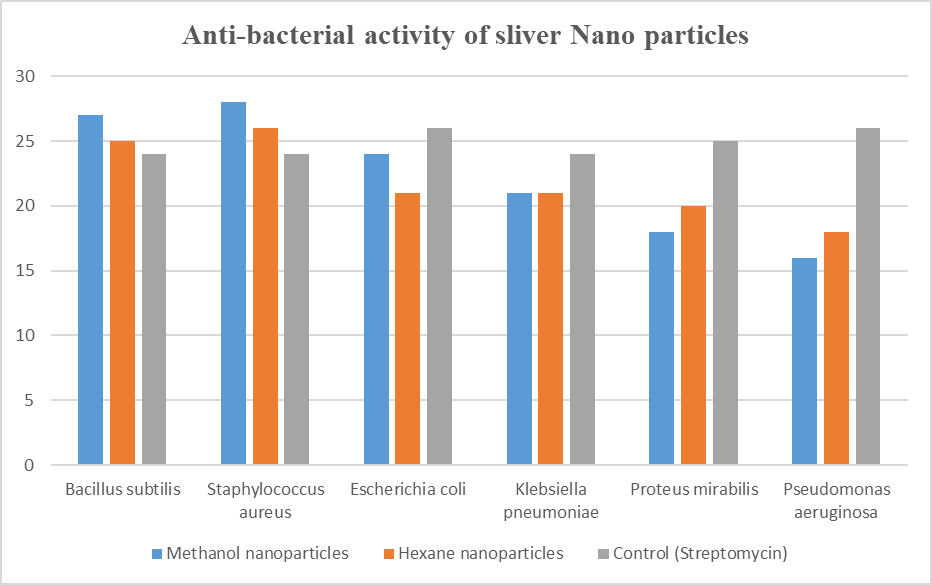
The antibacterial activity of synthesized and structurally characterized sliver Nano particles (AgNPs) were evaluated, and identified that the sliver nanoparticles were effective in inhibiting wide range of bacteria which include both gram-positive and gram-negative. In the present study it is revealed that sliver nanoparticles inhibited more gram-positive bacteria (Bacillus subtilis and Staphylococus aureus) with a zone off inhibition of 27 mm than Gram–negative bacteria which zone of inhibition ranged from21-24mm. The inhibition was more when compared with the crude extracts inhibition.
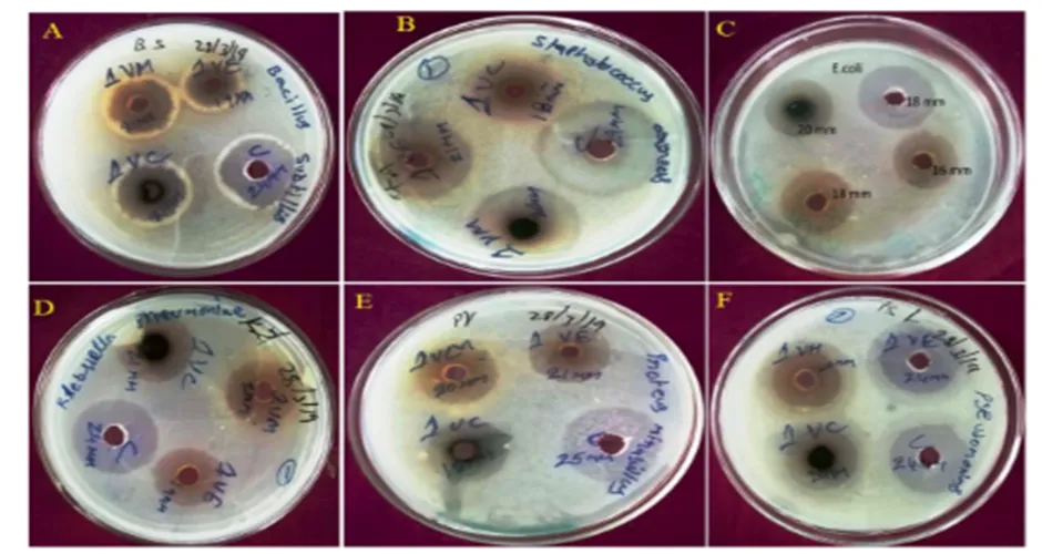
Image 01 Anti-bacterial Activity of the plant extract of C. zeylanicum bark
Image 1: The plates corroborates the antibacterial activity of plant extract as exhibited as zone of inhibition. (A) is associated with plates of Bacillus subtilis, (B) corresponds to the Staphylococcus aureus, (C) corresponds to E. coli, (D) represents the klebsiella pneumonia plate, (E) Represents Proteus mirabilis (F) shows the Pseudomonas aeruginosa plate.
Graph-01 Antibacterial Activity with plant extract of C. zeylanicum.
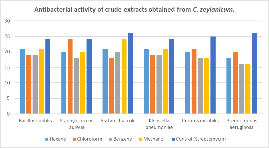
Image -02 Anti-bacterial activity of plant extract for the C. Zeylanicum

Image 2: The plates corroborates the antibacterial activity of Nanoparticles as exhibited as zone of inhibition of the B. subtilis, S. aureus, E. coli, k. pneumonia, P. mirabilis and P. aeruginosa plate.
Graph -02 Antibacterial activity C. Zeylanicum with sliver Nanoparticles
3.4 FTIR ANALYSIS OF C. ZEYLANICUM BARK EXTRACT-MEDIATED SILVER NANOPARTICLES
The FTIR spectra of C. zeylanicum bark extract-mediated silver nanoparticles (AgNPs) synthesized using four solvent extracts are presented in Figures 1–4 and the corresponding functional groups identified are summarized in Tables 1–4. The spectra revealed the presence of various bioactive functional groups, indicating the involvement of phyto-constituents in the reduction, capping, and stabilization of silver nanoparticles. The FTIR spectrum presented in Table 1 showed a broad absorption band at 3318.90 cm⁻¹, which was assigned to O–H stretching vibrations of alcohols and phenolic compounds within the range of 3200–3600 cm⁻¹. The peak observed at 2108.24 cm⁻¹ corresponded to C≡C stretching vibrations, while the absorption band at 1634.04 cm⁻¹ was attributed to C=C stretching vibrations characteristic of alkene groups. In addition, the absorption bands between 552.07 and 456.60 cm⁻¹ were associated with C–I and S–S stretching vibrations, indicating the presence of aliphatic iodol compounds and sulfur-containing functional groups.
The FTIR spectrum summarized in Table 4 exhibited a prominent absorption peak at 3292.16 cm⁻¹ corresponding to O–H stretching vibrations of alcohols and phenols. The peak at 2107.96 cm⁻¹ was assigned to C≡C stretching vibrations, whereas the band observed at 1637.80 cm⁻¹ indicated C=C stretching of alkene groups. Furthermore, the absorption peaks at 1207.96 cm⁻¹ and 1153.96 cm⁻¹ were attributed to C–O stretching vibrations of alcohols, carboxylic acids, esters, and ethers. The peak at 1054.20 cm⁻¹ corresponded to C–N stretching vibrations of aromatic amines. As shown in Table 3, a broad absorption band at 3270.18 cm⁻¹ was attributed to O–H stretching vibrations of alcohols and phenolic compounds, while the peak at 3105.15 cm⁻¹ represented N–H stretching vibrations of aliphatic amines. The peaks at 3007.97 cm⁻¹ and 2943.07 cm⁻¹ were assigned to O–H and C–H stretching vibrations, respectively. Several absorption bands recorded at 1689.96, 1642.96, 1581.57, 1546.97, 1516.64, 1452.22, and 1430.38 cm⁻¹ were attributed to C=C stretching vibrations of alkene groups. In addition, the peaks at 1356.32, 1282.44, 1269.37, 1188.82, 1176.97, 1152.82, and 1108.58 cm⁻¹ corresponded to C–O and C–N stretching vibrations, indicating the presence of alcohols, carboxylic acids, esters, ethers, and amine-containing compounds. Similarly, the FTIR spectrum presented in Table 6 exhibited a broad absorption band at 3257.80 cm⁻¹ corresponding to O–H stretching vibrations of alcohols and phenols. The peak observed at 3008.83 cm⁻¹ was assigned to N–H stretching vibrations of aliphatic amines, while the band at 2942.96 cm⁻¹ indicated C–H stretching vibrations of alkane groups. The absorption peaks at 1669.76 cm⁻¹ and 1643.56 cm⁻¹ were attributed to carbonyl-associated stretching vibrations. Furthermore, the bands observed at 1582.01, 1550.48, and 1453.56 cm⁻¹ represented C=C stretching vibrations of alkene groups. The peaks recorded at 1358.80, 1297.87, 1227.83, 1176.64, 1154.27, and 1106.49 cm⁻¹ corresponded to C–O and C–N stretching vibrations associated with alcohols, esters, ethers, and amine-containing compounds. Generally, the FTIR analysis demonstrated the presence of hydroxyl, amine, alkene, carbonyl, and ether functional groups in the synthesized AgNPs. These findings suggest that the phytochemical constituents present in C. zeylanicum bark extracts play a significant role in the bioreduction of silver ions and subsequently act as capping and stabilizing agents during nanoparticle synthesis.
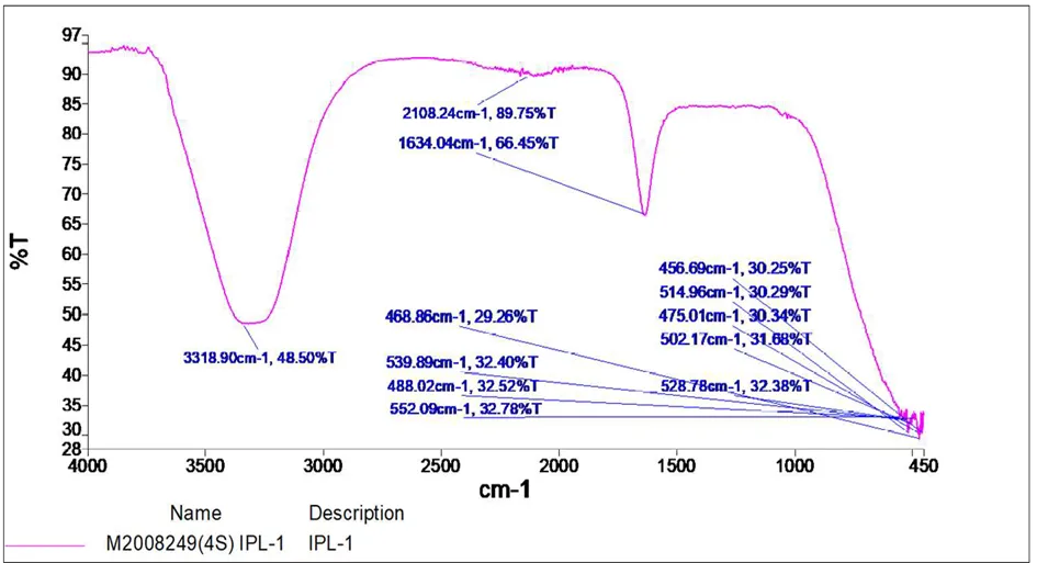
Figure 1: FTIR analysis of C. Zeylanicum Methanol extract of silver nanoparticles (AgNPs)
Table 1: FTIR Peak Values and Corresponding Functional Groups in C. Zeylanicum Methanol extract silver nanoparticles (AgNPs).
|
S. No |
Peak values(cm-1) |
Bond |
Functional groups assigned |
|
1 |
3318.90 |
O-H stretching |
Alcohol/ phenol |
|
2 |
2108.24 |
CC stretching vibration |
Silicon Compounds. |
|
3 |
1634.04 |
C=C stretching |
Alkenes |
|
4 |
552.07 |
C-I, S-S |
Aliphatic iodo |
|
5 |
468.86 |
C-I |
Halogen compound |
|
6 |
539.89 |
C-CL |
Halogen compound |
|
7 |
456.69 |
C-C stretching |
Alkyl halides |
|
8 |
514.96 |
C-CL |
Halogen compound |
|
9 |
475.01 |
C-C stretching |
Alkyl halides |
|
10 |
502.17 |
C-I |
Halogen compound |
|
11 |
528.78 |
C-CL |
Halogen compound |
|
12 |
488.02 |
C-C stretching |
Alkyl halides |
|
13 |
514.96 |
C-CL |
Halogen compound |
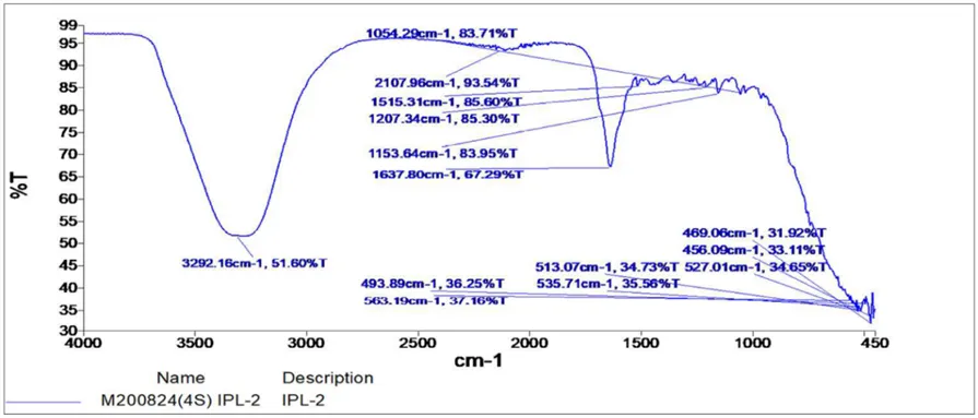
Figure 2: FTIR analysis of C. Zeylanicum extract silver nanoparticles (AgNPs) of Haxane
Table 2: FTIR Peak Values and Assigned Functional Groups for C. Zeylanicum extract silver nanoparticles (AgNPs) of Haxane.
|
S. No |
Peak values (cm-1) |
Bond |
Functional Groups Assigned |
|
1 |
3292.16 |
O-H stretching |
Alcohol/ phenol |
|
2 |
2107.96 |
CC stretching |
Silicon compounds |
|
3 |
1637.80 |
-C=C stretching |
Alkenes |
|
4 |
1207.96 |
C-O Stretching |
Alcohol, Carboxylic Acid, Ester and Either |
|
5 |
1153.96 |
C-O Stretching |
Aliphatic Amines |
|
6 |
1054.20 |
C-N Stretching |
Aromatic Amines |
|
7 |
1515.31 |
C=C |
Aromatic ring in lignin |
|
8 |
493.89 |
C-I |
Halogen compound, Iodo compound |
|
9 |
563.19 |
C-H, |
Alkene |
|
10 |
513.07 |
C-CL |
Halogen compound |
|
11 |
535.71 |
C-OH |
Carboxyl compound |
|
12 |
469.06 |
C-I |
Halogen compound, Iodo compound |
|
13 |
456.09 |
C-OH |
Carboxyl compound |
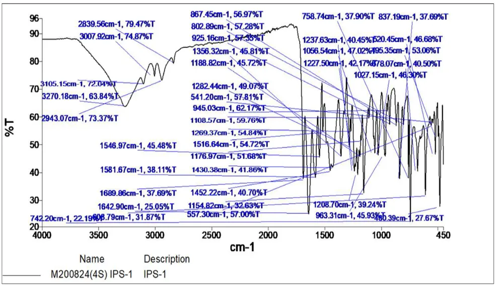
Figure 3. FTIR analysis of C. Zeylanicum extract silver nanoparticles (AgNPs) of Ethyl acetate
Table 3: FTIR Spectral Peaks and Corresponding Functional Groups for C. Zeylanicum stem methanol Extract
|
S. No |
Peak values(cm-1) |
Bond |
Functional Groups Assigned |
|
1 |
3270.18 |
O-H Stretching |
Alcohol/ Phenol |
|
2 |
3105.15 |
N-H stretching |
Aliphatic Amine |
|
3 |
3007.97 |
O-H Stretching |
Alcohol/ Phenol |
|
4 |
2943.07 |
C-H Stretching |
Alkanes |
|
5 |
2839.56 |
C-H Stretching |
Alkanes |
|
6 |
1689.96 |
C-H Stretching |
Carbonyl |
|
7 |
1642.96 |
-C=C- stretching |
Alkenes |
|
8 |
1581.57 |
-C=C- stretching |
Alkenes |
|
9 |
1546.97 |
-C=C- stretching |
Alkenes |
|
10 |
1516.64 |
-C=C- stretching |
Alkenes |
|
11 |
1452.22 |
-C=C- stretching |
Alkenes |
|
12 |
1430.38 |
-C=C- stretching |
Alkenes |
|
13 |
1356.32 |
C-H Bending |
Alkenes |
|
14 |
1282.44 |
C-O Stretching |
Alcohol, Carboxylic Acid, |
|
15 |
1269.37 |
C-O Stretching |
Alcohol, Carboxylic Acid, |
|
16 |
1188.82 |
C-O Stretching |
Alcohol, Carboxylic Acid, Ester and Either |
|
17 |
1176.97 |
C-O Stretching |
Alcohol, Carboxylic Acid, Ester and Either |
|
18 |
1152.82 |
C-O Stretching |
Aliphatic Amines |
|
19 |
1108.58 |
C-N Stretching |
Aromatic Amines |
|
20 |
963.18 |
C-O Stretching |
Aliphatic Amines |
|
21 |
608.79 |
C-Br |
Alkyl halides |
|
22 |
742.2 |
C-C |
Aromatic amine |
|
23 |
867.45 |
Ar-C |
Aromatic group |
|
24 |
802.89 |
C-C |
Aromatic |
|
25 |
925.16 |
C-O |
Ether |
|
26 |
945.03 |
C-O |
Ether |
|
27 |
541.2 |
C-C |
Nitriles |
|
28 |
1516.64 |
N-H |
Primary amine |
|
29 |
557.3 |
C-C |
Nitriles |
|
30 |
758.74 |
C-C |
Aromatic mono substituted |
|
31 |
1237. 63 |
C-O |
Carboxylic group |
|
32 |
1056.54 |
C-O |
Primary alcohol |
|
33 |
1227.5 |
C-O |
Carboxylic group |
|
34 |
837.19 |
N-H |
Secondary amine |
|
35 |
1520.45 |
N-H |
Amine primary |
|
36 |
1195.35 |
C-N |
Aromatic amine |
|
37 |
1078.07 |
C-O |
Carboxylic group |
|
38 |
1027.15 |
C-O |
Ether |
|
39 |
1208.7 |
C-O |
Carboxylic group |
|
40 |
480.39 |
C-C |
Cyclo alkane |
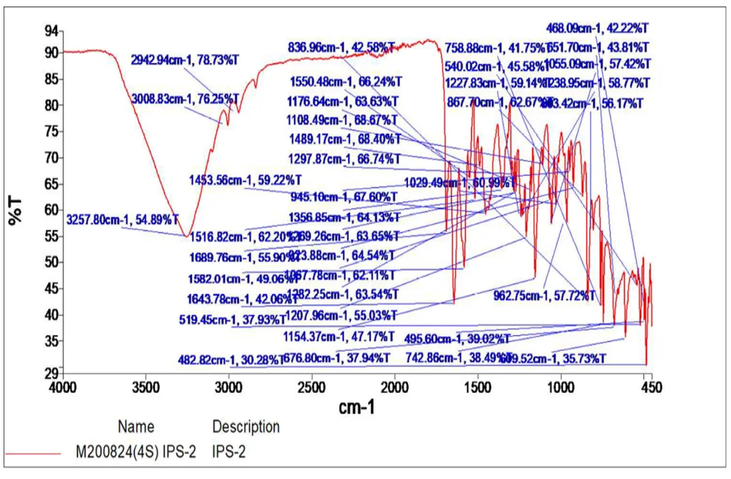
Figure 4. FTIR analysis of C. Zeylanicum extract silver nanoparticles (AgNPs) of Benzene
Table 4: FTIR Spectral Data and Functional Group Assignments for C. Zeylanicum stem methanol-AgNps (IPS-AgNps) Extracts
|
S. No |
Peak values(cm-1) |
Bond |
Functional Groups Assigned |
|
1 |
3257.80 |
O-H Stretching |
Alcohol/ Phenol |
|
2 |
3008.83 |
N-H stretching |
Aliphatic Amine |
|
3 |
2942.96 |
C-H Stretching |
Alkanes |
|
4 |
1669.76 |
C-H Stretching |
Carbonyl |
|
5 |
1582.01 |
-C=C- stretching |
Alkenes |
|
6 |
1643.56 |
C-H Stretching |
Carbonyl |
|
7 |
1453.56 |
-C=C- stretching |
Alkenes |
|
8 |
1550.48 |
-C=C- stretching |
Alkenes |
|
9 |
1176.64 |
C-O Stretching |
Alcohol, Carboxylic Acid, Ester and Either |
|
10 |
1106.49 |
C-O Stretching |
Aliphatic Amines |
|
11 |
1489.17 |
-C=C- stretching |
Alkenes |
|
12 |
1297.87 |
C-O Stretching |
Alcohol, Carboxylic Acid, |
|
13 |
1358.80 |
C-O Stretching |
Alcohol, Carboxylic Acid, |
|
14 |
1154.27 |
C-O Stretching |
Alcohol, Carboxylic Acid, |
|
15 |
1227.83 |
C-O Stretching |
Alcohol, Carboxylic Acid, |
|
16 |
1643.78 |
C=C |
Alkanes |
|
17 |
519.45 |
C-C |
Nitriles |
|
18 |
482.82 |
C-C |
Cycloalkanes |
|
19 |
836.96 |
N-H |
Secondary amines |
|
20 |
1108.49 |
C-H STRECHING |
Aromatic group |
|
21 |
945.10 |
N-H |
Primary amine |
|
22 |
1269.26 |
C-O Stretching |
Alcohol, Carboxylic Acid, |
|
23 |
973.88 |
C=C |
Alkanes |
|
24 |
1067.78 |
C-O |
Primary alcohol |
|
25 |
1282.25 |
C-O |
Carboxylic Acid, |
|
26 |
1207 |
C-O |
Carboxylic Acid, |
|
27 |
676.80 |
C-CL |
Alkyl halide |
|
28 |
758.88 |
C-C |
Aromatic mono substituted |
|
29 |
540.02 |
C-Br |
Alkyl halides |
|
30 |
867.70 |
Ar-C |
Aromatic group |
|
31 |
1029.49 |
C-O |
Ether |
|
32 |
495.60 |
C-C |
Alkyl halides |
|
33 |
742.86 |
C-C |
Aromatic mono substituted |
|
34 |
468.09 |
C-C |
Cyclo alkanes |
|
35 |
651.70 |
C-H |
Alkynes |
|
36 |
1055.09 |
C-O |
Primary alcohol |
|
37 |
1238.95 |
C-O |
Carboxylic acid |
|
38 |
1203.42 |
C-O |
Carboxylic acid |
|
39 |
962.75 |
C-O, C-C |
Amorphous |
|
40 |
609.52 |
C-H |
Alkynes |
3.5 ANTI-CANCER ACTIVITY OF C. ZEYLANICUM EXTRACT SILVER NANOPARTICLES (AgNPs) OF FOUR SOLVENTS AGAINST CANCER CELL LINES
To evaluate the anticancer activity of C. Zeylanicum extract silver nanoparticles (AgNPs) of methanol and Hexane bark extracts, we treated the cervical (Hela) and lung carcinoma cancer (A-549) cell lines with increasing concentrations of AgNPS extracts at various viz., 6.25, 12.5, 25, 50 and 100 µg/ml for 24 hr. The standard drug Cisplatin (HeLa and A-549) were used as control. C. Zeylanicum extract silver nanoparticles (AgNPs) of methanol and Hexane bark extracts were higher in percent of cell viability as compared to the untreated cell lines. It suggests that growth inhibitory principles are present in all organic fractions and we rank them as Cisplatin>AgNPS> untreated cell lines. It is evident from the calculated dose of 50% inhibition of cell viability (IC50) (Graph-3 to 6). The C. Zeylanicum extract silver nanoparticles (AgNPs) of methanol bark extracts showed lower IC50 value for both Hela (44.90 and 43.45µg/ml) and A-549 (69.42 and70.12µg/ml) cell lines. C. Zeylanicum extract silver nanoparticles (AgNPs) of Hexane bark extracts showed the IC50 value for both Hela (63.18 and 64.32µg/ml) andA-549(73.94 and 78.64µg/ml) (Image 3- 6).
The samples established a significant cytotoxicity against cervical and lung carcinoma epithelial cell lines. Based on the literature, we have tested all these extracts for anticancer activity by measuring the growth of actively proliferating cancer cells using Hela and A-549 cell lines. These AgNPS of both plants declined the cell growth in both the cell lines. The magnitude of the inhibition of cell growth is varied for both the extracts and was higher for AgNPS extract as comparison, and is dose dependent (Tables 5-8). The cell viability evaluate detects the reduction of MTT by mitochondrial dehydrogenase to blue formazan product, which infers the normal function of mitochondrial and cell viability (Merlin., et al., 2010). C. Zeylanicum AgNPS showed a more suppression of Hela and A-549 growth and also C. Zeylanicum AgNPS showed low cytotoxic activity to A-549 than Hela cell lines as compared to C. Zeylanicum AgNPS. To visualize the cancer cell viability under experimental conditions.
Table 5: Effect of Different doses of C. Zeylanicum extract silver nanoparticles (AgNPs) of methanol on HeLa cell line by MTT assay (% of Cell Viability)
|
Concentration (µg) |
Absorbance at 570nm |
% Inhibition |
% Viability |
IC50 (µg) |
|
6.5 |
0.716 |
7.73 |
92.27 |
64.32±1.672 |
|
12.5 |
0.643 |
17.13 |
82.87 |
|
|
25 |
0.512 |
34.02 |
65.98 |
|
|
50 |
0.392 |
49.48 |
50.52 |
|
|
100 |
0.267 |
65.59 |
34.41 |
|
|
untreated |
0.776 |
0 |
100 |
Graph 03: Cytotoxic activity of C. Zeylanicum extract silver nanoparticles (AgNPs) of methanol bark extracts on HELA cells
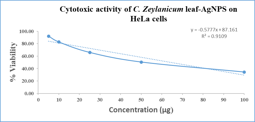
Image 3: Cytotoxic activity of C. Zeylanicum extract silver nanoparticles (AgNPs) of methanol bark extracts on HeLa cells
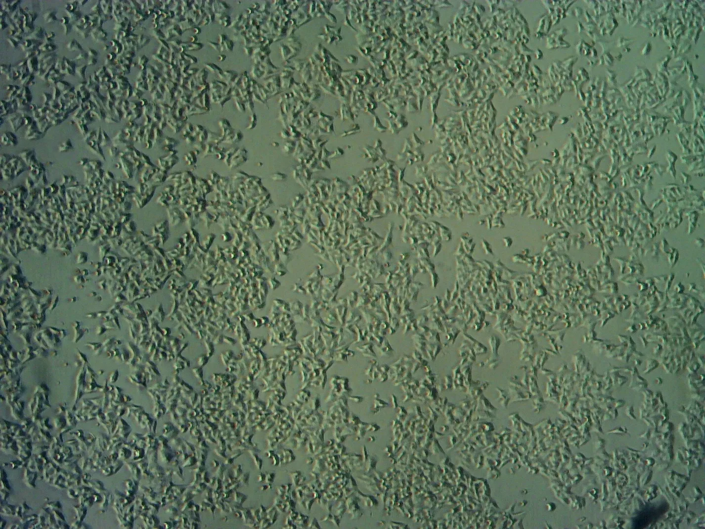
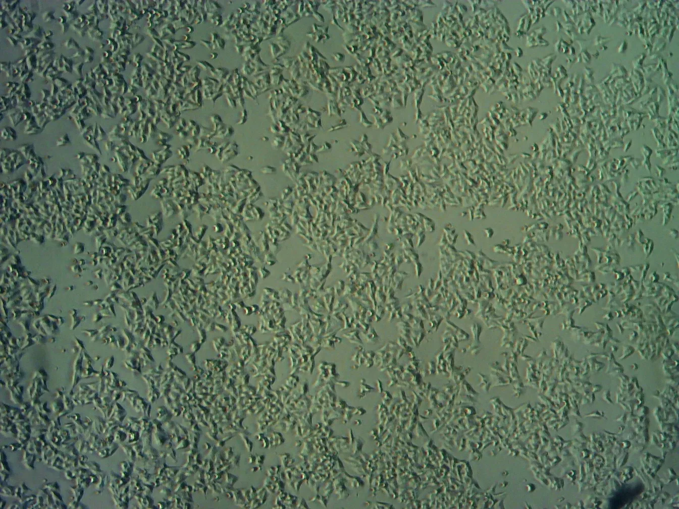
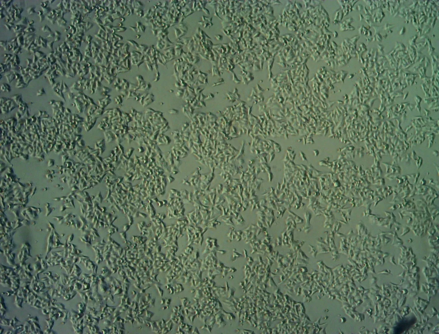
6.5µg 25 µg 100 µg
Table 6: Effect of Different doses of C. Zeylanicum extract silver nanoparticles (AgNPs) of Hexane bark extracts on HeLa cell line by MTT assay (% of Cell Viability)
|
Concentration (µg) |
Absorbance at 570nm |
% Inhibition |
% Viability |
IC50 (µg) |
|
6.5 |
0.668 |
13.91 |
86.09 |
43.45±1.159 |
|
12.5 |
0.584 |
24.74 |
75.26 |
|
|
25 |
0.413 |
46.77 |
53.23 |
|
|
50 |
0.259 |
66.62 |
33.38 |
|
|
100 |
0.156 |
79.89 |
20.11 |
|
|
untreated |
0.776 |
0 |
100 |
Graph 4: Cytotoxic activity of C. Zeylanicum extract silver nanoparticles (AgNPs) of Hexane bark extracts AgNPS on HeLa cells
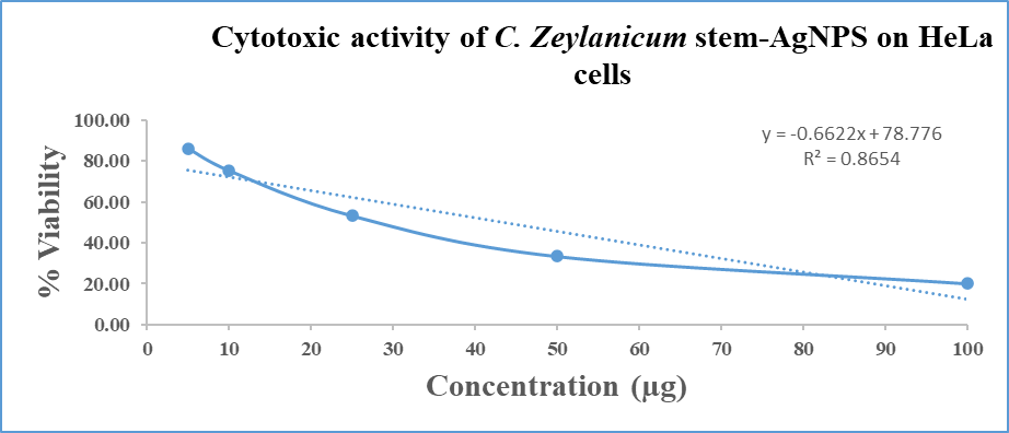
Image 4: Cytotoxic activity of C. Zeylanicum extract silver nanoparticles (AgNPs) of Hexane bark extracts AgNPS on HeLa cells
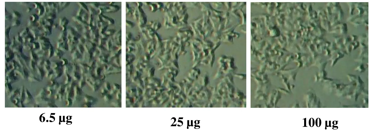
Table 7: Effect of Different doses of C. Zeylanicum extract silver nanoparticles (AgNPs) of methanol on A-549 cell line by MTT assay (% of Cell Viability)
|
Concentration (µg) |
Absorbance at 570nm |
% Inhibition |
% Viability |
IC50 (µg) |
|
6.5 |
0.534 |
4.81 |
95.19 |
78.67±2.146 |
|
12.5 |
0.506 |
9.80 |
90.20 |
|
|
25 |
0.448 |
20.14 |
79.86 |
|
|
50 |
0.347 |
38.14 |
61.86 |
|
|
100 |
0.225 |
59.89 |
40.11 |
|
|
untreated |
0.561 |
0 |
100 |
Graph 5: Cytotoxic activity of C. Zeylanicum extract silver nanoparticles (AgNPs) of methanol bark extracts on A-549
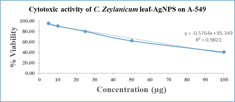
Image 5: Cytotoxic activity of C. Zeylanicum extract silver nanoparticles (AgNPs) of methanol bark extracts on A-549
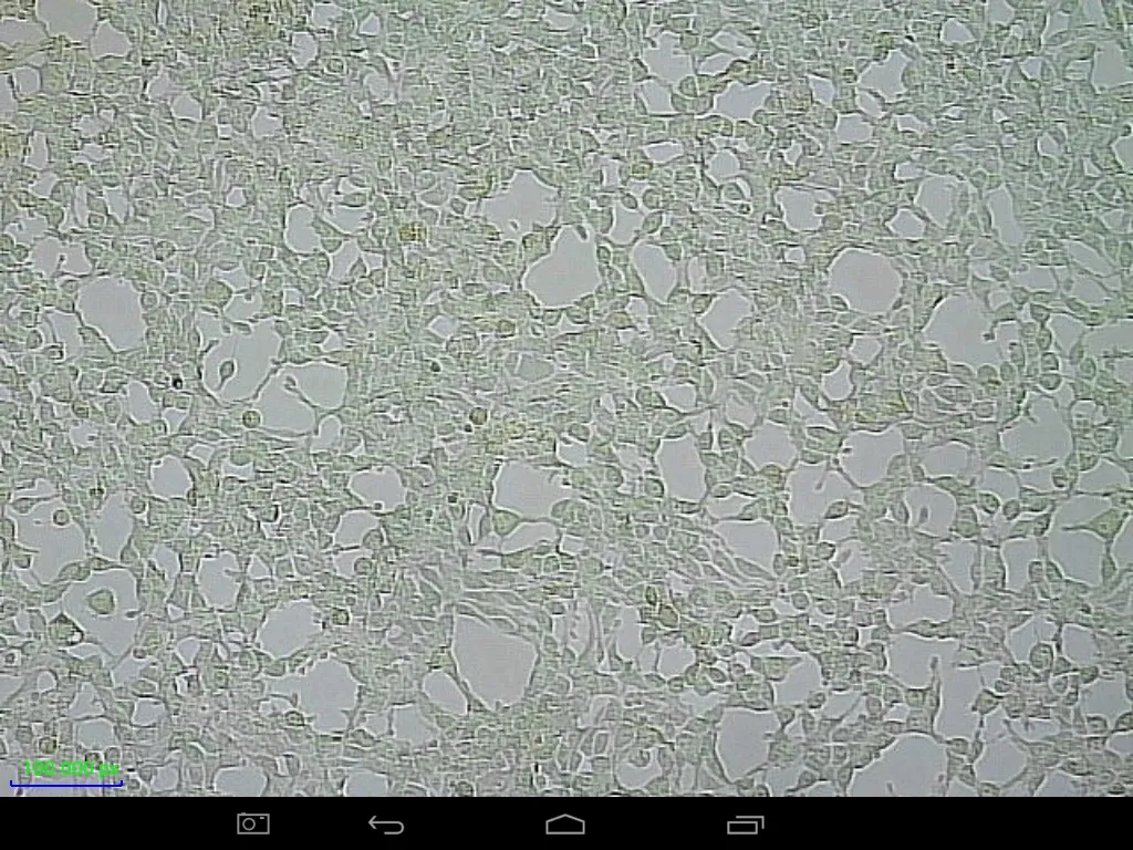
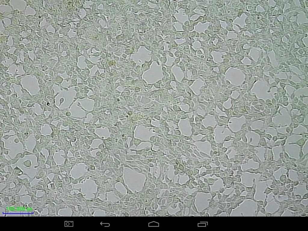
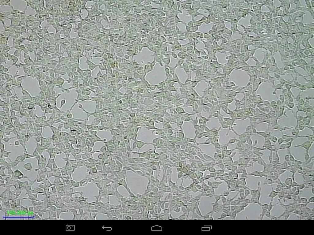
6.5µg 25 µg 100 µg
Table 8: Effect of Different doses of C. Zeylanicum extract silver nanoparticles (AgNPs) of Hexane bark extracts on A-549 cell line by MTT assay (% of Cell Viability)
|
Concentration (µg) |
Absorbance at 570nm |
% Inhibition |
% Viability |
IC50 (µg) |
|
6.5 |
0.519 |
7.48 |
92.52 |
70.12±1.823 |
|
12.5 |
0.492 |
12.29 |
87.71 |
|
|
25 |
0.421 |
24.95 |
75.05 |
|
|
50 |
0.316 |
43.67 |
56.33 |
|
|
100 |
0.197 |
64.88 |
35.12 |
|
|
untreated |
0.561 |
0 |
100 |
Graph 6: Cytotoxic activity of C. Zeylanicum extract silver nanoparticles (AgNPs) of methanol bark extracts on A-549
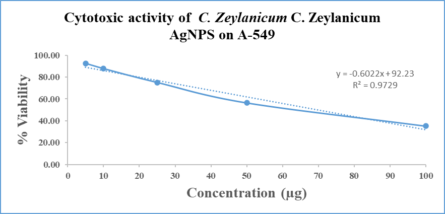
Image 6: Cytotoxic activity of C. Zeylanicum extract silver nanoparticles (AgNPs) of Hexane bark extracts on A-549
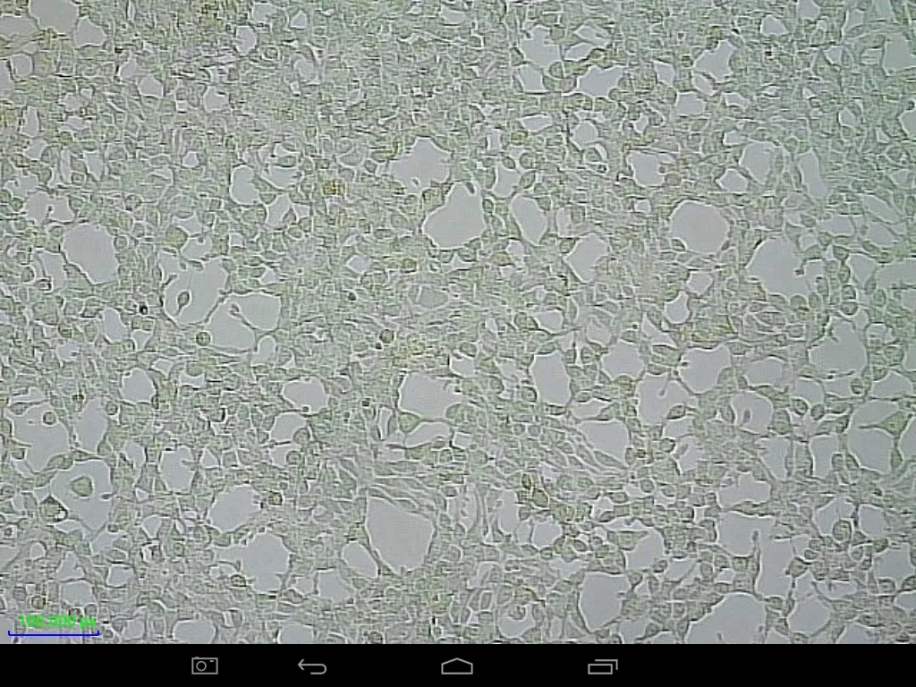
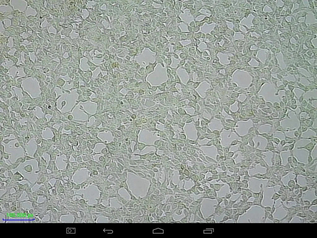
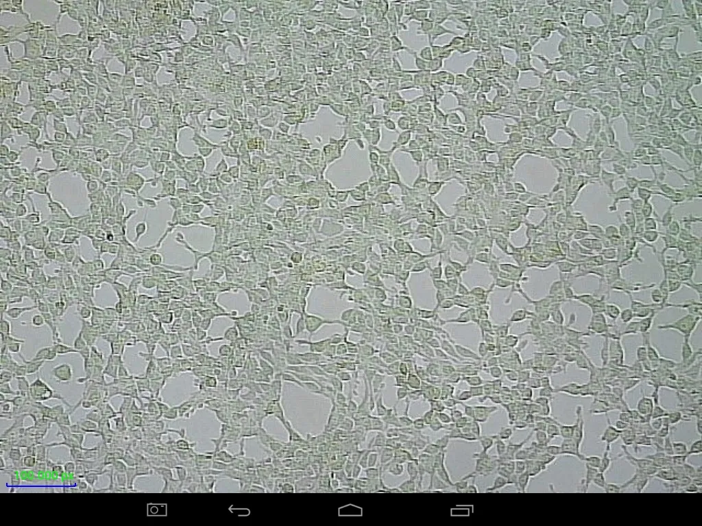
6.5µg 25 µg 100 µg
DISCUSSION
The present study demonstrated that C. zeylanicum bark extracts and their biosynthesized silver nanoparticles possess significant antibacterial and anticancer activities. The results revealed that all solvent extracts exhibited inhibitory effects against both Gram-positive and Gram-negative bacterial pathogens, although the magnitude of inhibition varied among the extracts and bacterial species tested. The methanolic extract showed the highest activity against Escherichia coli (24 mm), while hexane extract exhibited pronounced inhibition against Staphylococcus aureus (24 mm) and Proteus mirabilis (21 mm). These findings suggest that the antibacterial activity of C. zeylanicum is strongly influenced by the solvent used for extraction, which determines the recovery of bioactive phytochemicals.
The observed antibacterial activity can be attributed to the presence of cinnamaldehyde, eugenol, phenolic compounds, flavonoids, and terpenoids that are naturally abundant in C. zeylanicum bark. Recent studies have reported that cinnamon-derived phytochemicals disrupt bacterial cell membranes, interfere with cellular respiration, and inhibit biofilm formation, thereby exerting broad-spectrum antimicrobial effects (Niu et al., 2020; Elshopakey et al., 2022). Similar antibacterial effects of cinnamon extracts against both Gram-positive and Gram-negative bacteria have also been reported by Kwon et al. (2021) and Sharma et al. (2023), supporting the findings of the present investigation.
A notable outcome of the present study was the enhanced antibacterial activity exhibited by silver nanoparticles compared with the corresponding crude extracts. Methanol-derived AgNPs produced inhibition zones of 27 mm against Bacillus subtilis and 28 mm against Staphylococcus aureus, exceeding the activity of crude extracts and approaching or surpassing the activity of streptomycin in some cases. The superior antibacterial efficacy of AgNPs may be attributed to their nanoscale dimensions, increased surface area, and ability to interact directly with bacterial cell walls. Silver nanoparticles are known to induce membrane damage, generate reactive oxygen species (ROS), alter protein function, and interfere with DNA replication, ultimately leading to bacterial cell death. Similar observations have been reported in recent nanoparticle-based antimicrobial studies by Singh et al. (2021), Ahmed et al. (2022), and Alghuthaymi et al. (2024).
The FTIR analysis confirmed the presence of several functional groups, including hydroxyl (O–H), amine (N–H), alkene (C=C), carbonyl (C=O), and ether (C–O) groups in the synthesized AgNPs. These functional groups indicate the presence of phenolics, alcohols, proteins, flavonoids, and other phytoconstituents that participate in the reduction of Ag⁺ ions and stabilization of the synthesized nanoparticles. Similar FTIR profiles have been reported for green-synthesized silver nanoparticles derived from medicinal plants, where hydroxyl and carbonyl-containing compounds serve as reducing and capping agents (Kumar et al., 2021; Rajeshkumar and Bharath, 2022; Hassan et al., 2025). The presence of these bioactive functional groups further supports the role of C. zeylanicum phytochemicals in nanoparticle synthesis and stabilization.
The anticancer evaluation demonstrated that C. zeylanicum-mediated AgNPs exhibited dose-dependent cytotoxicity against HeLa and A549 cancer cell lines. The methanol-derived AgNPs displayed greater cytotoxic effects than hexane-derived nanoparticles, with lower IC₅₀ values observed against HeLa cells (43.45–64.32 µg/mL) compared with A549 cells (70.12–78.67 µg/mL). These findings indicate that cervical cancer cells were more sensitive to AgNP treatment than lung carcinoma cells. The enhanced anticancer activity may be associated with nanoparticle-mediated oxidative stress, mitochondrial dysfunction, DNA damage, and activation of apoptotic pathways. Recent investigations have shown that plant-mediated AgNPs effectively suppress cancer cell proliferation through ROS generation and apoptosis induction (Suresh et al., 2021; Almatroodi et al., 2023; El-Naggar et al., 2025).
Furthermore, the stronger activity observed in methanol-derived nanoparticles may be due to the extraction of higher concentrations of polar phytochemicals such as phenolics and flavonoids, which possess established antioxidant and anticancer properties. These compounds may act synergistically with silver nanoparticles, enhancing their biological activity. Similar synergistic interactions between phytochemicals and AgNPs have been reported in studies involving medicinal plants and nanoparticle-assisted cancer therapy (Abdel-Aziz et al., 2022; Rahman et al., 2024). Overall, the findings of the present study demonstrate that C. zeylanicum bark is a promising source of bioactive compounds for the green synthesis of silver nanoparticles with potent antibacterial and anticancer activities. The enhanced biological performance of AgNPs compared with crude extracts highlights the advantages of nanotechnology in improving the therapeutic potential of medicinal plants. Nevertheless, the present investigation was limited to in vitro experiments. Therefore, further in vivo studies, toxicity assessments, pharmacokinetic evaluations, and molecular investigations are required to validate the safety, efficacy, and clinical applicability of C. zeylanicum-mediated silver nanoparticles as potential antimicrobial and anticancer agents.
CONCLUSION
The present study successfully demonstrated the extraction of bioactive compounds from C.zeylanicum bark using different solvent systems and their application in the green synthesis of silver nanoparticles (AgNPs). The synthesized nanoparticles were characterized by FTIR analysis, which confirmed the presence of hydroxyl, amine, carbonyl, alkene, and ether functional groups. These phytochemical constituents were found to play a crucial role in the reduction, capping, and stabilization of silver nanoparticles. The antibacterial evaluation revealed that both crude extracts and biosynthesized AgNPs exhibited significant inhibitory activity against selected Gram-positive and Gram-negative bacterial pathogens. Among the tested samples, silver nanoparticles demonstrated superior antibacterial activity compared with the corresponding crude extracts, indicating the enhancement of antimicrobial efficacy through nanoparticle synthesis. The highest antibacterial activity was observed against Bacillus subtilis and Staphylococcus aureus, highlighting the potential of C. zeylanicum-mediated AgNPs as effective antimicrobial agents. The anticancer studies further demonstrated that the synthesized AgNPs possess considerable cytotoxic activity against HeLa and A549 cancer cell lines in a dose-dependent manner. Methanolic extract-mediated AgNPs exhibited greater anticancer potential than hexane-derived nanoparticles, as evidenced by lower IC₅₀ values and reduced cell viability. The observed cytotoxic effects may be attributed to the synergistic action of phytochemicals and silver nanoparticles, which induce cellular damage and inhibit cancer cell proliferation. General, the findings of this study establish C. zeylanicum bark as an excellent natural source for the green synthesis of biologically active silver nanoparticles. The enhanced antibacterial and anticancer activities exhibited by the synthesized AgNPs suggest their potential application in pharmaceutical, biomedical, and therapeutic fields. However, further investigations involving in vivo studies, toxicity assessments, molecular mechanism studies, and clinical evaluations are necessary to validate their safety and efficacy before potential therapeutic application. In conclusion, Cinnamomum zeylanicum-mediated silver nanoparticles represent a promising eco-friendly nanomaterial with significant antimicrobial and anticancer properties, providing a valuable foundation for the development of novel plant-based nanotherapeutic agents.
REFERENCES

Luay kadhim Hanoon, Characterization and Biological Evaluation of Cinnamomum zeylanicum Bark Extracts and Their Silver Nanoparticles for Biomedical Applications, Int. J. of Pharm. Sci., 2026, Vol 4, Issue 7, 4396-4417, https://doi.org/10.5281/zenodo.21484877
 10.5281/zenodo.21484877