

We use cookies to ensure our website works properly and to personalise your experience. Cookies policy
Department of Surgery (Ilmul Jarahat), National Institute of Unani Medicine, Ghaziabad 201002, Uttar Pradesh, India
Introduction: Nephrolithiasis (Hisat-e-Kulyah) is a common urinary tract disorder with a substantial risk of recurrence. Unani medicine describes renal calculi under Hisat-e-Kulyah and employs dietary, regimenal and pharmacological approaches, including medicines traditionally described as Mudirr-e-Baul and Mufattit-e-Hisat. Methods: A 35-year-old woman with recurrent bilateral nephrolithiasis presented with bilateral flank pain, increased urinary frequency, burning micturition and nausea. Baseline ultrasonography dated 18 November 2024 demonstrated bilateral renal calculi. The patient received Majoon Hajrul Yahud, Sharbat Bazoori Motadil and Jawarish Zarooni for five months, together with increased fluid intake and dietary modification. Clinical symptoms were followed during treatment and ultrasonography was repeated after five months. Results: Baseline ultrasonography showed a 4.7-mm calculus in the upper polar calyx of the right kidney with two fine renal concretions (<3.5 mm), and calculi measuring 5.8 mm in the interpolar calyx and 3.5 mm in the lower polar calyx of the left kidney; no hydronephrosis or hydroureter was reported. Symptoms progressively improved, with complete resolution by the fifth month according to the documented symptom timeline. Follow-up ultrasonography dated 15 April 2025 reported no obvious renal calculi and no hydronephrosis or hydroureter. No adverse effects were reported in the available case record. Discussion: The observed temporal association between the Unani regimen, dietary modification and hydration advice and the improvement in symptoms and radiological findings is noteworthy. However, causality cannot be established because three medicines and non-pharmacological measures were used concurrently, and spontaneous stone passage or natural variation cannot be excluded. The case therefore supports further clinical investigation rather than establishing treatment efficacy.
Urinary stone disease has plagued humans for millennia, with Egyptian mummies from 4800 BC and North American Indian graves from 1500-1000 BC showing evidence of stones [1, 2]. In the 4th century BC, Hippocrates described kidney stones and renal abscesses and famously stated in the Hippocratic Oath, “I will not cut the stone” [3]. The word “urolithiasis” (Hisat-e-Bauliyah) is derived from three Greek words: “ouron” (urine), “oros” (flow), and “lithos” (stone). This describes the formation of stones in the urinary system, including nephrolithiasis (Hisat-e-Kulyah), ureterolithiasis (Hisat-e-Halib), and cystolithiasis (Hisat-e-Masanah) [4].
Nephrolithiasis is one of the most common health problems worldwide, with a prevalence rate of about 15% worldwide and 2.3% in the Indian population [5, 6]. The prevalence of nephrolithiasis is as high as 7.6% in the Satpura region of Maharashtra, Gujarat, Madhya Pradesh, and parts of Andhra Pradesh. Pendse et al. have reported a high and increasing incidence of nephrolithiasis in Udaipur and other parts of western Rajasthan, India [7, 8]. In India, the “stones belt” covers parts of Maharashtra, Gujarat, Punjab, Haryana, Delhi, and Rajasthan. In these regions, kidney stone disease is so common that most family members will develop kidney stones at some point in their lives [9]. Nephrolithiasis is more common in men (12%) than in women (6%), and the peak incidence of both sexes is between the ages of 20 and 40 [10]. Vesicle calculi are more common in undernourished children [6]. Once the condition recurs, the risk for further relapses is increased, and the interval between recurrences is shorter [11].
1.1 Unani Concept of Nephrolithiasis (Hisat-E-Kulyah):
Ibn-e-Sina (980-1037 AD) states that Quavo-e-Fayelah (active power), which is an increased temperature exceeding the normal level of the kidney, and Maaddat-ul-Hisat (lithic matter), a viscous and sticky substance that may be phlegm, thick blood, or pus, leads to the formation of kidney stones. When the expulsive power of the kidney is impaired from altered temperament or inflammatory or ulcerous conditions, they are lodged in the calices of the kidney, notwithstanding the effort to cast them out. Then the Quvat-e-Fayelah dries the lithic matter, and crystals are formed, which gradually develop into stones [12].
Ibn-e-Zohr (1091-1162 AD) proposed that when the kidney is weak and cannot throw out thick humors, these humors will accumulate in the kidney and gradually crystallize layer by layer to form stones [13].
Ali Ibn-e-Abbas Majoosi (930-994 AD) stated that stones form in the kidneys when the calyces of the kidney are coated by thick fluids and concentrated humors. These substances become dry under high, intense heat, forming crystals that over time develop into stones [14].
Jaleenoos (Galen) believes that the main cause of nephrolithiasis is kidney ulcers. If the pus does not drain out, it solidifies and forms a stone [15].
Zakaria Razi (850-923 AD) attributes this disease to the imbalance of humors. Then the body expels these humors in the form of a thick fluid that goes to the kidneys and forms crests that lead to the formation of stones. Stones are often recurrent [15].
1.2 Types of Stone
Calcium oxalate stones (Hisat-e-Tootiyah) and calcium phosphate stones (Hisat-e-Qaimooliyavi) constitute about 80% of stones. In particular, calcium oxalate monohydrate is 40-60%, calcium oxalate dihydrate is 40-60%, calcium hydrogen phosphate is 2-4%, and calcium orthophosphate is less than 1%. Uric acid and urate stones (Hisat-e-Bauliyah) are around 10% of stones and are made up of pure uric acid or ammonium/sodium urate [16, 17, 18].
Struvite (magnesium ammonium phosphate) makes up 1% of stones in the setting of infection with urease-producing bacteria. Another 1% are cystine stones (Hisat-e-Zubaniyah), which are made of sulfur [16, 17, 18]. Xanthine stones (Hisat-e-Layyinah) are also very rare, accounting for around 1% [9, 10]. Mixed stones are common, 50-60%, including mixed calcium oxalate-phosphate stones (35-40%) and mixed uric acid-calcium oxalate stones (5%) [19].
1.3 Pathogenesis
Urinary stone formation, or urolithiasis, is a multifactorial phenomenon that commences with the supersaturation of urinary salts, then nucleation, growth, agglomeration, and eventually retention within renal tubules [20]. The supersaturation takes place due to an elevated level of salt-forming substances within urine [21]. There are many promoters of crystal growth in the body, which include calcium, sodium, oxalate, urate, acidic urine, Tamm-Horsfall protein, and decreased urine volume. On the other hand, the inhibitory factors include inorganic citrate, magnesium, pyrophosphate, and organic substances such as Tamm-Horsfall protein (glycoprotein), prothrombin fragment 1, renal lithostathine, glycosaminoglycan, osteopontin (uropontin), nephrocalcin, and increased urine volume [20, 22]. The Tamm-Horsfall protein acts as a promoter of nucleation and growth but inhibits aggregation [20]. The formation of stones is due to an imbalance of promoters and inhibitors in the kidney [23].
Hyperoxaluria is the most frequent factor responsible for the formation of oxalate stones. Diets with too little calcium intake may lead to hyperoxaluria [24, 25]. Chronic diarrhea and renal tubular acidosis and a high intake of animal protein decrease the level of citrate in urine [26]. Hyperuricosuria is linked to both uric acid and calcium stones. Uric acid reduces the solubility of calcium oxalate, which helps in stone formation [25]. The high intake of purines leads to hyperuricosuria. Hypercalciuria may be caused by hyperparathyroidism [25]. However, a low intake of calcium from the diet is not necessary since it will increase calcium stones [27].
1.4 Etiology
In classical Unani literature, the pathogenesis of nephrolithiasis is multifactorial. The creation of kidney stones is mainly related to the presence of humors, such as those of a thick and concentrated nature, and deficiencies, either structural or functional. These include weakness of the kidney itself, alteration in the renal temperaments known as Su-e-Mizaj Kulyah, nephritis or “Warm-e-Kulyah,” ulcers of the kidney or Qurooh-e-Kulyah, and a decrease in expulsive power of Quwwat-e-Dafey'ah due to increased virulence of temperature [12-15, 28]. However, based on modern concepts, the primary risk factors involved in causing nephrolithiasis include poor urine outflow; infection; high consumption of oxalate and calcium in food; vitamin deficiency, such as deficiency of vitamin A and excess intake of vitamin D; metabolic disorders such as hyperparathyroidism, cystinuria, gout, and intestinal issues; and environments with high heat and less moisture [29]. Anatomic problems in the urinary system may cause urinary stasis and infections that increase a person's risk of developing nephrolithiasis [30]. Important anatomic problems that lead to stone formation are ureteropelvic junction obstruction, horseshoe kidney, medullary sponge kidney, bifid pelvis, and ureteral duplication [31]. In addition to these factors, metabolic disorders such as obesity and hypertension, which have become common worldwide, contribute to the high incidence of urolithiasis [2]. The final important factor that leads to stone development is infections caused by urease-producing bacteria such as Proteus, Pseudomonas, and Klebsiella [6].
2. PATIENT INFORMATION
A 35-year-old married woman, a housewife, presented to the Surgery outpatient department of the National Institute of Unani Medicine with intermittent bilateral flank pain radiating to the back, increased urinary frequency, burning micturition and nausea. She reported a history of recurrent stone formation. According to the Unani assessment documented in the case record, her Mizaj was Balghami.
Table 1: Patient information
|
Age and sex |
35-year-old female |
|
Occupation |
Housewife |
|
Marital status |
Married |
|
Mizaj (temperament) |
Balghami |
|
Dietary habit |
Mixed (mostly non-vegetarian food) |
|
Water intake |
1 liter per day |
|
Appetite |
Normal |
|
Salty foods |
Prefer moderately |
|
Spicy foods |
Highly |
|
Bowel habits |
Constipation |
|
Urine |
Day – 6 to 8 times. Night – 2 to 3 times |
|
Sleep |
8-10 hours |
|
Any addiction |
None |
2.1 Past History of Illness
According to the patient's history, she had recurrent stone formation. There was no reported history of diabetes mellitus, hypertension, thyroid disease, tuberculosis or other major systemic illness.
No previous surgical intervention was reported.
2.2 Dietary Habits
The patient reported a mixed diet with frequent intake of red meat, chicken and packed fruit juice. Her baseline water intake was approximately 1 L/day.
The patient was advised to increase fluid intake during treatment.
2.3 Family History
Her father had hypertension and was receiving treatment. Her brother had a history of kidney stones.
No relevant medical history was reported for her mother.
3. CLINICAL FINDINGS
3.1 General Examination
On initial examination, the patient appeared anxious and distressed because of pain. No pallor, icterus, edema, cyanosis or lymphadenopathy was reported.
Table 2: General examination
|
General Appearance |
Anxious, Distress |
|
Built and Posture |
Obese |
|
Anemia |
Not reported |
|
Icterus |
Not reported |
|
Cyanosis |
Not reported |
|
Pigmentation |
Not reported |
|
Dyspnea |
Not reported |
|
Oedema: Local/General |
Not reported |
|
BP |
120/70 mmhg |
|
Pulse |
84 bpm |
|
Respiratory Rate |
16 /min |
|
Spo2 |
98% |
|
Temperature |
98.6°F |
3.2 Systemic Examination
Cardiovascular System: S1-S2 audible
Respiratory System: Air entry bilateral and equal
Nervous System: Conscious and well-oriented
Rectal System: NAD
Genitourinary System: NAD
3.3 Local Examination
Abdomen:
Inspection: Abdomen was described as distended, with a centrally inverted umbilicus and no pigmentation or surgical scar.
Palpation: Abdomen was soft with tenderness in the right and left lumbar regions; no organomegaly was reported.
Percussion: A dull note was reported over the abdomen.
Auscultation: Bowel sounds were reported in the right iliac fossa.
4. TIMELINE
|
Symptoms |
Baseline (18 Nov 2024) |
2nd month |
3rd month |
4th month |
5th month |
|
B/L flank pain |
++++ |
+++ |
++ |
+ |
- |
|
Frequency of urine |
++++ |
+++ |
++ |
+ |
- |
|
Burning micturition |
++++ |
+++ |
+ |
- |
- |
|
Nausea |
++++ |
+++ |
+ |
+ |
- |
5. DIAGNOSTIC ASSESSMENT
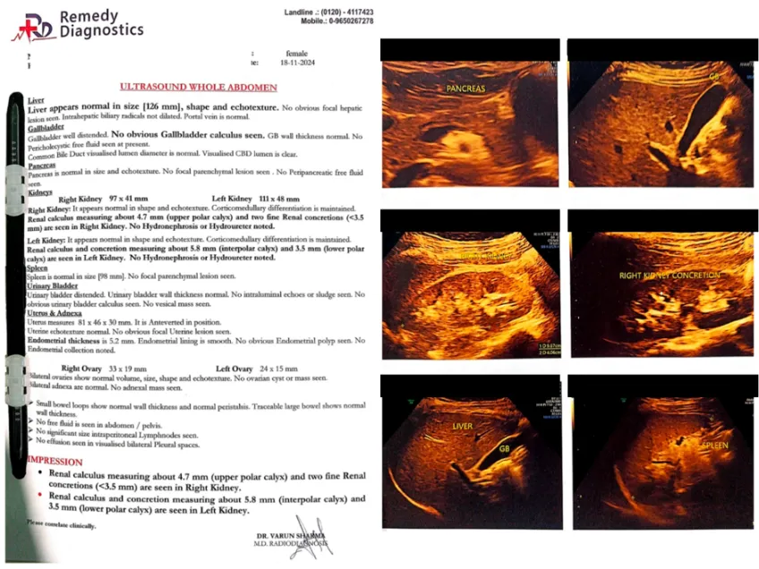
Ultrasonography (USG) of the whole abdomen was performed at baseline and after completion of the five-month treatment period. The baseline USG was dated 18 November 2024.
Pre-treatment USG (18 November 2024): The right kidney measured 97 × 41 mm and showed a renal calculus measuring approximately 4.7 mm in the upper polar calyx and two fine renal concretions measuring <3.5 mm. The left kidney measured 111 × 48 mm and showed a renal calculus measuring approximately 5.8 mm in the interpolar calyx and a 3.5-mm concretion in the lower polar calyx. No hydronephrosis or hydroureter was reported in either kidney.
6. THERAPEUTIC INTERVENTION
The patient received the documented Unani regimen for five months, from the baseline assessment in November 2024 to follow-up in April 2025, together with dietary and hydration advice. The treatment was continued because of the patient's history of recurrent renal stone formation.
6.1 Unani Medicine
Majoon Hajrul Yahud: ½ tablespoon twice daily.
Sharbat Bazoori Motadil: 20 mL twice daily.
Jawarish Zarooni: ½ tablespoon twice daily.
6.2 Dietary Advice
The patient was advised to increase fluid intake to 2.5–3 L/day, subject to clinical suitability, and to moderate dietary intake of animal protein and foods considered high in oxalate. Dietary counselling included limiting foods such as spinach, tomatoes and chips, and reducing meat and chicken intake.
7. FOLLOW-UP AND OUTCOMES
Post-treatment USG (15 April 2025): The right kidney measured 109 × 37 mm and the left kidney 110 × 47 mm. No obvious renal calculus was seen in either kidney. No hydronephrosis or hydroureter was reported. The impression stated that no significant abnormality was seen on the whole-abdomen ultrasound examination.
The documented symptoms progressively improved over the five-month follow-up period. Bilateral flank pain, urinary frequency, burning micturition and nausea decreased from the baseline assessment to complete resolution by the fifth month according to the symptom timeline. No adverse effects were reported in the available case record.
8. DISCUSSION
The five-month conservative course described here should not be interpreted as a substitute for urgent urological management when nephrolithiasis is complicated by obstruction, infection, renal impairment, uncontrolled pain or other indications for intervention. The principal limitation of this report is its single-patient design and the use of multiple concurrent interventions. Further prospective studies with standardized interventions, objective imaging outcomes, laboratory or metabolic assessment and longer follow-up are warranted.
No stone analysis or detailed metabolic evaluation is documented in the available case record, and long-term follow-up beyond the five-month treatment period is not available. Therefore, a reduction in future recurrence risk cannot be concluded from this case.
The observed temporal association between treatment and improvement is noteworthy; however, the clinical outcome cannot establish that any individual formulation caused stone clearance. The patient received three medicines simultaneously as well as increased hydration and dietary modification, so the contribution of each component cannot be separated. Spontaneous passage or natural variation in the course of nephrolithiasis also cannot be excluded.
From the Unani perspective, renal calculi are described as Hisat-e-Kulyah, and therapeutic approaches include Ilaj Bil-Ghiza, Ilaj Bit-Tadbeer and Ilaj Bid-Dawa. The medicines used in this case were selected as part of the documented Unani management approach and are traditionally described in terms of Mudirr-e-Baul and Mufattit-e-Hisat actions.
This case describes the clinical course of a patient with recurrent bilateral nephrolithiasis who received a combination of three Unani formulations together with increased fluid intake and dietary modification. At baseline, ultrasonography demonstrated bilateral renal calculi, including a largest reported calculus of 5.8 mm in the left kidney. During five months of follow-up, the patient's reported symptoms progressively improved, and follow-up ultrasonography demonstrated no obvious residual renal calculus or hydronephrosis or hydroureter.
9. CONCLUSION
This case describes progressive symptomatic improvement and radiological clearance of bilateral renal calculi following a five-month Unani regimen comprising Majoon Hajrul Yahud, Sharbat Bazoori Motadil and Jawarish Zarooni, administered with hydration and dietary advice. Although the temporal association between treatment and improvement is noteworthy, causality and treatment efficacy cannot be established from a single case with concurrent interventions. Further controlled clinical studies are required to evaluate the safety and effectiveness of this therapeutic approach in recurrent nephrolithiasis.
10. PATIENT PERSPECTIVE
The patient reported gradual improvement in flank pain, urinary frequency, burning micturition, and nausea during treatment. She expressed satisfaction with the improvement in her symptoms and follow-up ultrasonography and was pleased to avoid surgical intervention.
11. INFORMED CONSENT
Written informed consent was obtained from the patient for publication of this case report and the accompanying clinical and radiological information.
12. ETHICAL CONSIDERATIONS
Ethical approval/exemption for publication of this single case report was obtained from the competent institutional authority, as applicable.
13. FUNDING
No specific funding was received for this case report.
14. CONFLICT OF INTEREST
The authors declare that they have no conflict of interest.
15. AUTHOR CONTRIBUTIONS
Choudhary Mohd Wasim: Conceptualization, clinical management, data collection, investigation, manuscript preparation and revision.
Mohd Asif Khan: Supervision, clinical oversight, interpretation of findings, manuscript review and revision.
Both authors approved the final version of the manuscript and agreed to be accountable for all aspects of the work.
16. DATA AVAILABILITY
The data supporting the findings of this case report are available from the corresponding author on reasonable request, subject to patient confidentiality and institutional requirements.
REFERENCES

Choudhary Mohd Wasim, Mohammad Asif Khan, Clinical Response to Unani Regimen in a Patient with Recurrent Bilateral Nephrolithiasis: A Case Report, Int. J. of Pharm. Sci., 2026, Vol 4, Issue 8, 4206-4214. https://doi.org/10.5281/zenodo.22104830
 10.5281/zenodo.22104830