

We use cookies to ensure our website works properly and to personalise your experience. Cookies policy
SJVPM's Rasiklal M Dhariwal Institute of Pharmaceutical Education and Research, Pune.
The transdermal drug delivery system involves the use of skin patches to deliver medication through the skin into the bloodstream. Advantages of this system include improved bioavailability, longer duration of action, reduced side effects, and avoidance of first-pass metabolism. Transdermal patches have been successful in delivering various medicines such as scopolamine, nicotine, estrogen, nitroglycerin, lidocaine, and more. There are different generations of transdermal drug delivery systems, each with its advancements and challenges. The third generation focuses on targeting the stratum corneum for more effective drug delivery while protecting deeper tissues. Approaches in the development of transdermal therapeutic systems include micro-reservoir type systems, adhesive dispersion-controlled systems, membrane permeation-controlled systems, and matrix diffusion-controlled systems. The anatomy of human skin is crucial for understanding the penetration of drugs through the epidermis, dermis, and hypodermis layers. Despite the advantages of transdermal drug delivery, there are also disadvantages such as the development of contact dermatitis and the need for potent drugs that can penetrate the skin efficiently. Overall, transdermal drug delivery systems offer a promising alternative to traditional oral medication administration.
A transdermal patch, or skin patch, is an adhesive patch designed to deliver medication through the skin into the bloodstream at a controlled rate. It uses a special membrane to regulate drug release, and certain drugs require permeation enhancers, such as alcohol, to improve absorption. Common medications delivered via transdermal patches include scopolamine for motion sickness, nicotine for smoking cessation, estrogen for menopause and osteoporosis prevention, nitroglycerin for angina, and lidocaine for pain relief from shingles. However, larger molecules like insulin are not suitable for transdermal delivery.
Developed in the 1970s, the first FDA-approved transdermal patch (scopolamine) was introduced in 1979, followed by nitroglycerin patches in 1981. Over time, additional patches have been developed for medications such as clonidine, fentanyl, estradiol, oxybutynin, and testosterone, along with combination contraceptive and hormone therapy patches. Depending on the medication, patches can last from one to seven days. The key advantages of transdermal drug delivery include improved bioavailability, consistent drug levels, prolonged therapeutic effects, reduced dosing frequency, and minimized side effects compared to oral formulations. Additionally, transdermal patches bypass first-pass metabolism, which can reduce drug degradation and liver-related complications. Beyond therapeutic applications, transdermal patches also exist for non-medical purposes, including aromatherapy, weight loss, thermal and cooling effects, nutrient supplementation, skincare, and UV exposure monitoring. These patches continue to evolve, offering innovative solutions for both medical and cosmetic applications.[1]
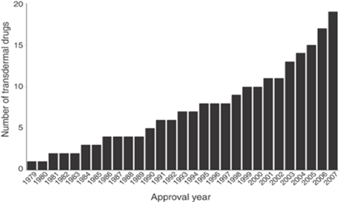
Figure No 1: Cuulative number of transdermal drugs approved by the FDA since the first approval in 1979.[63]
MECHANISM OF TRANSDERMAL DRUG DELIVERY SYSTEM:
Given the diverse nature of the skin as a membrane, it is quite remarkable. The process of percutaneous absorption can be described using the basic principles of simple diffusion laws. Transdermal delivery entails the extended use of a device applied to the skin. Over a given time frame, it is commonly taken for granted that steady-state conditions have been achieved. It is important to note that the ultimate law of diffusion to consider is Fick's first law. The second law explains non-steady state diffusion and can be applied to analyze the release rates from matrix-type transdermal patches, helping to assess the lag time. To explore the period leading up to the establishment of stable conditions and to depict the evolving concentration patterns across the skin towards linearity. The classic rendition of Fick's first law of diffusion that garners the most citations pertains to situations of equilibrium. The process of molecules moving across a membrane through diffusion.[1]
J = KD/h*(CO-Ci)
Where,
IMPORTANCE:
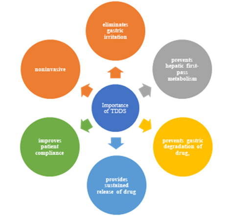
Figure 2: Importance of Transdermal Drug Delivery System[1]
HISTORY:
Table 1: Transdermal drugs approved by us FDA[62]
|
Approval Year |
Drug/ Product Name |
Indication |
Marketing Company |
|
1979 |
Scopolamine/ Transderm-Scop |
Motion sickness |
Novartis Consumer Health (Parsippany, NJ, USA) |
|
1981 |
Nitroglycerin/ Transderm-Nitro |
Angina pectoris |
Novartis (East Hannover, NJ, USA) |
|
1984 |
Clonidine/ Catapres-TTS |
Hypertension |
Boehringer Ingelheim (Ridgefield, CT, USA) |
|
1986 |
Estradiol/ Estraderm |
Menopausal symptoms |
Novartis |
|
1990
|
Fentanyl/ Duragesic |
Chronic pain |
Janssen Pharmaceutica (Titusville, NJ, USA |
|
1991 |
Nicotine/ Nicoderm, Habitrol, ProStep |
Smoking cessation |
GlaxoSmithKline (Philadelphia), Novartis Consumer Health, Elan (Gainesville, GA, USA) |
|
1993 |
Testosterone/ Testoderm |
Testosterone deficiency |
Alza (Mountain iew, CA, USA) |
|
1995 |
Lidocaine with epinephrine (iontophoresis)/ Iontocaine |
Local dermal analgesia |
Iomed (Salt Lake City, UT, USA) |
|
1998 |
Estradiol with norethidrone/ Combipatch |
Menopausal symptoms |
Novartis |
|
1999 |
Lidocaine/ Lidoderm Post-herpetic |
neuralgia pain |
Endo Pharmaceuticals (Chadds Ford, PA, USA) |
|
2001 |
Ethinyl estradiol with norelgestromin/ Ortho |
Contraception |
Ortho-McNeil Pharmaceutical (Raritan, NJ |
|
2003 |
2003 Estradiol with levonorgestrel/ Climara |
Pro Menopausal symptoms |
Bayer Healthcare Pharmaceuticals (ayne, NJ, USA) |
|
2003 |
Oxybutynin/ Oxytrol |
Overactive bladder |
atson Pharma (Corona, CA, USA) |
|
2004 |
Lidocaine (ultrasound)/ SonoPrep |
Local dermal anesthesia |
Echo Therapeutics (Franklin, MA, USA) |
|
2005 |
Lidocaine with tetracaine/ Synera |
Local dermal analgesia |
Endo Pharmaceuticals |
|
2006 |
Fentanyl HCl ( |
(iontophoresis)/ Ionsys Acute postoperative pain |
Alza |
|
2006 |
Methylphenidate/ Daytrana |
Attention deficit hyperactivity disorder |
Shire (ayne, PA, USA) |
|
2006 |
Selegiline/ Emsam |
Major depressive disorder |
Bristol-Myers Squibb (Princeton, NJ, USA) |
|
2007 |
Rotigotine/ Neupro |
Parkinson’s disease |
Schwarz Pharma (Mequon, I, USA |
|
2007 |
Rivastigmine/ Exelon |
Dementia |
Novartis |
TYPES OF TRANSDERMAL DRUG DELIVERY SYSTEMS
First-generation transdermal drug delivery system:
First-generation transdermal delivery systems, responsible for most clinical use, have seen significant advances in technology and public acceptance. These systems must be low-molecular weight, lipophilic, and efficient at low boluses, with a focus on low oral bioavailability and low frequent dosing.[1]
Second-generation transdermal drug delivery system:
The second generation of transdermal delivery systems, including chemical enhancers, iontophoresis, and ultrasound, aims to enhance skin permeability for transdermal drug delivery. However, these methods struggle to balance increased delivery across the stratum corneum and protection of deeper tissues. Despite this, these systems have improved small-molecule delivery for localized, dermatological, cosmetic, and systemic applications.[13][14][15]

Figure 3: Properties of ideal enhancer[1]
Third-generation transdermal drug delivery system:
The third generation of transdermal delivery systems is expected to significantly impact medicine delivery by targeting the stratum corneum, enabling stronger dislocation of the stratum corneum hedge and more effective transdermal delivery.
New chemical enhancers, electroporation, cavitational ultrasound, microneedles, thermal ablation, and microdermabrasion have been shown to deliver macromolecules, including remedial proteins and vaccines, across the skin in clinical trials.[1]
ADVANTAGES AND DISADVANTAGES:
Advantages:
i) an avoid gastrointestinal medicine immersion difficulties covered by gastrointestinal pH, enzymatic exertion and medicine commerce with food, drink and other orally administration medicine.
ii) They can substitute for oral administration of drugs when the route is infelicitous, as with vomiting and diarrhea.
iii) To avoid the first pass effect, e.g., Transdermal Nitroglycerin. It's fleetly metabolized by the liver when taken orally.
iv) They're noninvasive, avoiding the vexation of parenteral remedy.
vi) The exertion of medicines having a launch half-life is extended through the force of medicine in the remedial delivery system and its controlled release.
vii) medicine remedy may be terminated fleetly by junking of the operation from the face of the skin.[10][11]
Disadvantages:
i) cases develop contact dermatitis at the point of operation from one or further of the system Factors, challenging termination.
ii) Only potent medicines are suitable campaigners for transdermal patches because of the natural limits of medicine entry assessed by the skin's imperability.
iii) Some medicines, e.g., scopolamine transdermal patch placed behind the observance, it's uncomfortable.
iv) Long time cleave is delicate.[11][12]
APPROACHES IN THE DEVELOPMENT OF TRANSDERMAL THERAPEUTIC SYSTEM:
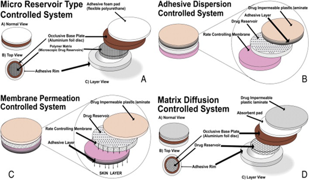
Figure 4: Approaches in the development of transdermal Therapeutic System: A.micro reservoir type system, B.adhesive controlled system dispersion, C.membrane permeation controlled system,D matrix diffusion controlled system.[1]
A. Micro Reservoir Type Controlled System:
The drug delivery method combines reservoir and matrix dispersion, suspending the drug in a liquid polymer solution and uniformly distributing it throughout lipophilic polymers using a high-energy dispersion technique. This technology allows for nitro disc release based on drug solubility and polymer matrix control. [43][44][45]
B. Adhesive Dispersion Controlled System:
A medication delivery system consists of an impermeable backing membrane and a resilient polymer medication force. A nonmedicated polymer is spread on top to control prolixity and release lines. Examples include transdermal remedies for angina pectoris and valsartan as angiotensin II type 1, which are used to treat various conditions.[1]
C. Membrane Permeation Controlled System:
The device's drug reservoir is enclosed within a rate-regulating polymeric membrane and a backing laminate. Drug molecules pass through holes, either dissolved in a solvent or uniformly distributed in a solid polymeric matrix. The rate-controlling membrane can be microporous or non-porous, with a small medication layer on its upper surface.[1]
D. Matrix Diffusion Controlled System:
This method arranges medicated stores by scattering sedate particles in a polymer lattice, creating a sedated circle with a defined surface and thickness. The polymer disc is placed on a base plate, and cement polymer is spread to create a strip along the disc. This matrix dispersion transdermal framework, like the nitroglycerin discharging therapeutic framework, prevents measurement dumping and ensures non-cracking of the polymer.[1]
ANATOMY OF HUMAN SKIN:
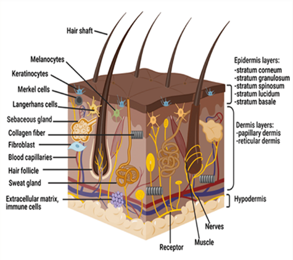
Figure 5: Anatomy of skin[67]
Table No 2:Structure and Role of Skin Layers in Drug Penetration[67]
|
Layer |
Description
|
Key Features |
Role in Drug Penetration |
|
Epidermis |
The outermost layer, stratified, avascular |
- Stratum corneum (outermost, acts as a barrier) |
- Main barrier for drug penetration |
|
Dermis |
Thick connective tissue layer |
- 3–5 mm thick |
- Capillaries absorb drugs into systemic circulation |
|
Hypodermis (Subcutaneous tissue) |
Deepest skin layer, fat storage |
- Supports the dermis & epidermis |
- Drugs must reach this layer for systemic absorption |
CASE STUDY ON ANIMAL MODELS
RODENT [Rat And Mice]
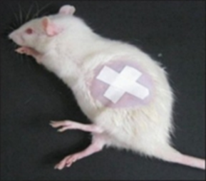
Figure 6: Application of patch to rat skin[63]
Transdermal medicine delivery offers numerous advantages, such as easy penetration, protection from gastric enzymes, and avoidance of hepatic damage. Studies have focused on improving penetration using penetration enhancers like hydrogenated soybean phospholipids, ethanol, alcohols, n-octanol, cyclic monoterpenes, nonionic surfactants, propylene glycol, and isopropyl myristate. Membranes from various animals and synthetic membranes have been used for these studies. Rat skin is a commonly used cover for in vitro saturation studies, which can help manipulate the design of transdermal remedial system (TTS) patches to achieve desired drug saturation across mortal skin. Microemulsions containing oil painting, waterless phase, surfactant, and cosurfactant have gained attention for their potential to enhance transdermal immersion. A hydrogel base was prepared and studied for its saturation potential. A nonsteroidal antirheumatic agent, DS, has potent anti-inflammatory effects but struggles to reach effective attention after transdermal operation. This study aimed to suggest new lozenge forms for enhancing topical penetration of DS and compare the study's phrasings with marketable expressions, assessing penetration and transdermal immersion.[63]
Materials and Methods:
Preparation of topical formulation:
Table 2. Contents (% wt/wt) of the Microemulsion and Gel Formulations[63]
|
Microemulsion |
Gel |
||
|
oil |
32.5 |
Carbopol |
940 0.3 |
|
water |
6.3 |
Ethanol |
25.0 |
|
Brij 58 |
5.1 |
Triethanolamine |
0.4 |
|
Span 80 |
45.9 |
Distilled water |
74.3 |
|
cosurfactant |
10.2 |
|
|
Assay of diclofenac sodium:
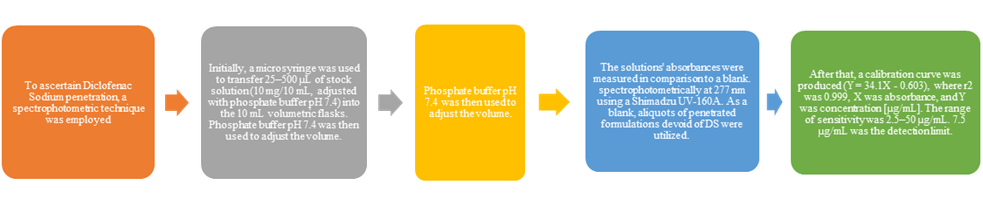
Figure 7: Flowchart of assay of diclofenac sodium assay[63]
In vitro permeation studies
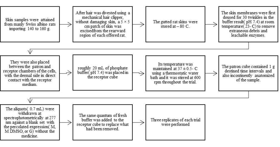
Figure 8: Flow chart of in vitro permeation studies[63]
Study on Diclofenac Sodium Permeability
Each value represents the mean ± SD( n = 3).[63]
Table 3. Means of Permeated Amounts of Diclofenac Sodium(DS) and Standard Deviations for All Formulations*[63]
|
Permeated Amount of DS (µg/cm2 ) (± SD) |
||||
|
Time |
Microemulsion |
Microemulsion + dimethyl sulfoxide |
Gel
|
Commercial formulation |
|
1 |
154.100 ± 2.0224 |
168.800 ± 3.0512 |
132.300 ± 4.2790 |
119.500 ± 2.3812 |
|
2 |
182.700 ± 2.9206 |
262.300 ± 2.8054 |
158.100 ± 3.3181 |
154.700 ± 4.3486 |
|
3 |
250.700 ± 2.6907 |
351.900 ± 4.6872 |
214.300 ± 5.1468 |
173.900 ± 4.8135 |
|
4 |
307.200 ± 3.9611 |
417.700 ± 3.5086 |
267.300 ± 6.3906 |
214.700 ± 7.4344 |
|
5 |
358.200 ± 6.1733 |
468.700 ± 2.6230 |
328.100 ± 6.4630 |
246.700 ± 6.4467 |
|
6 |
414.700 ± 4.6487 |
507.100 ± 2.7495 |
369.200 ± 5.6507 |
278.100 ± 3.2696 |
|
7 |
438.900 ± 4.9729 |
542.400 ± 6.0233 |
395.200 ± 5.5073 |
5.373 ± 2.4880 |
|
8 |
477.300 ± 5.9908 |
601.400 ± 9.8473 |
420.000 ± 5.3731 |
315.500 ± 4.1797 |
Table 4. Differences Among Formulations as to the Permeated Amount of DS for Each Hour*[63]
|
Formulations |
M+DMSO |
M |
Commercial Formulation |
|
M + DMSO |
|
|
|
|
M |
1, 2, 3, 4, 5, 6, 7, 8 |
||
|
Commercial formulation |
1, 2, 3, 4, 5, 6, 7, 8 |
1, 2, 3, 4, 5, 6, 7, 8 |
|
|
Gel |
1, 2, 3, 4, 5, 6, 7, 8 |
1, 2, 3, 4, 5, 6, 7, 8 |
1, 3, 4, 5, 6, 7, 8 |
*Numbers show significant difference (P G .05) at hours between 2 formulations crossed. M indicates microemulsion; DMSO, dimethyl sulfoxide. † At all hours, there was a significant difference between the commercial formulation and the gel, except at hour 2.[63]
Result and Discussion:
The study examined the stability of drug-loaded microemulsions (M) after adding a drug, DS. The viscosities of G and M were determined, and the mean droplet diameter was 9.19 ± 0.1 nm. DS's solubility in distilled water was 19.1 mg/mL, and its partition coefficient in n-octanol–distilled water and soybean oil–distilled water was 5.75 and 1.8, respectively. The permeation parameters of the Fick's law equation were calculated, showing a significant interaction between Factor 1 and Factor 2 and significant differences in permeability rates among the formulations studied.[63]
PIG
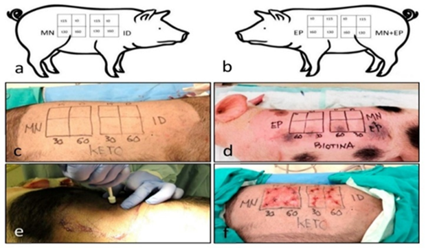
Figure 9: Design of the experimental procedure in the pigs. The working diagram of (a) the right side and (b) the left side) shows the different techniques and time intervals. The diagrams were applied to each of the differently treated animals. The images show the ketoprofen-treated pig, labeled as KETO (c), and the biotin-treated animal, labeled as BIOTINA (d). Punch biopsies were performed according to the diagram (e) and then sutured (f). Legend for quadrants: microneedle (MN), intradermal injection (ID), electroporation (EP), and the combination MN + EP; subquadrants depict the time after drug administration in minutes (0, 15, 30, and 60 min).[67]
The skin has been a focus of cosmetic and therapeutic techniques for centuries due to its simplicity in topical presentations. As the largest organ in the human body, the skin serves various purposes related to touch protection and hydration upkeep. However, small-size lipophilic drugs may still achieve therapeutic levels with topical administration, indicating that the barrier is selective. The skin comprises several barriers that function in concert, such as the stratum corneum, blood vessel endothelia, basement membrane, and epidermal tight junctions. These barriers are present in both the epidermis and the skin adnexa. Iontophoresis, a combination of electrotherapy and iontophoresis, improves the transdermal delivery of charged substances through low-intensity currents, generating a collateral water flow (electroosmosis) that can transport neutrally charged molecules across the stratum corneum. This combined approach has been established in enhancing skin permeation and has promising prospects for application in oncological treatments and vaccine administration. The stratum corneum is the most external barrier, where cells have completed the keratinization process initiated in the basal epidermal layer, losing their nuclei and acquiring lipophilic properties in a cycle of up to approximately 1 month. This lipophilic property is associated with dehydration in the stratum corneum, where water constitutes approximately 15-20% of the total mass. Epidermal tight junctions form a bidirectional barrier active for most molecules, including water. The superficial vascular plexus is the deepest skin barrier, encompassing superficial blood and lymphatic vessels. The transdermal delivery of drugs leads to an easy, quick, and efficient pharmacological action, and applied dosage does not decrease after washing. To accomplish topical permeation of pharmacological agents, permeating agents or enhancers have been developed for use on one side and epidermal disruptors on the other side. Epidermal disruptors include intradermal injections (ID), iontophoresis, electroporation (EP), and microporation employing different microneedle arrays (MN).ID or mesotherapy directly introduces medication through a needle in the dermal tissue, with performance varying with the drug employed and the depth of injection. EP generates small nanometric orifices in the stratum corneum under an electric field, creating brief disruptions in the epidermal lipophilic barrier. Microneedle (MN) technology utilizes microneedles to create micropores in the epidermis, allowing for the application of topical pharmaceutical solutions that can penetrate through these pores.[67]
Methods and materials
Animal Model and Experimental Procedure:
Microporation: The microneedle used in the study had a basculation head with six needles of specific length. The oscillation speed was 150 rpm, and the Minnesota time was limited to a 10-minute window. The quadrant was 10 by 10 centimeters, and 5 mL of irrigation was applied for each substance. The procedure was effective, but redness and small spots were observed.[67]
Electroporation-Iontophoresis: The equipment used by Mesoestetic Medical Devices in Barcelona, Spain, was designed to operate with the Spanish electrical system at 50 Hz AC power. The application process took 15 minutes and involved introducing the formulation in a plastic container connected to a steel roll-on device connected to the electroporation system. The substances were applied in a gel form, considering substance polarity and pH to improve skin condition.[67]
Table 5. Composition of the different gelified formulations of the study[67]
|
Active ingredients |
Methocel E4M |
Kathon CG |
Water P.CSP 100 |
Sodium Benzoate |
pH |
|
Clorhydrate procaine |
2% |
1.60% |
0.10% |
96.30% |
5.4 (5.1–5.8) |
|
Ketoprofen |
5% |
1.60% |
0.10% |
93.30% |
6.6 (6.2–7.0) |
|
Biotin |
0.5% |
1.60% |
0.10% |
97.80% |
6.7 (6.3–7.1) |
|
Anhydrous caffeine |
3% |
1.60% |
0.10% |
3% |
7.4 (7.1–7.7) |
Intradermal injection: The method was followed by Chos guidelines, with the ideal injection depth being half the skin fold thickness. A digital caliper was used for accurate intradermal delivery. The amount of substance was determined by dividing it in milliliters. The depth of injection was indicated by a factor of 20, as prescribed by the American Association for Laboratory Animal Science. A 4 mL dose was evenly distributed across the 10cm x 10 cm quadrant.[67]
High-pressure liquid chromatography:
The study involved homogenizing frozen cutaneous samples in a 0.9% physiological saline solution and mixing it in Ultra-Turrax T8 equipment. The material was then analyzed using a UPLC Dionex Ultimate 3000® instrument and an Impact II mass spectrophotometer. Detection was made for biotin and procaine in a positive range from 100 to 800 Da. The electrospray source was set at 4500 V, 35 psi, and 250 ?C. The samples were then centrifuged at 10,000 rotations per minute for 5 minutes.[67]
Table 6: Chromatography parameters of the different substances.[67]
|
Drug |
Procain |
Ketoprofen |
Biotin |
Caffeine |
|
Active ingredient |
50 µL |
50 µL |
50 µL |
50 µL |
|
Water |
- |
950 µL |
950 µL |
425 µL |
|
2.5 pH CH2O2 water |
950 µL |
- |
- |
- |
|
33% Ammonia |
- |
20 µL |
20 µL |
- |
|
Methanol |
- |
- |
- |
425 µL |
|
Dichloromethane |
300 µL |
300 µL |
300 µL |
- |
|
Posterior dilution |
1:1 |
1:10 |
1:10 |
1:20 |
|
Calibration parameters |
5–100ng/mL |
5–5000 ng/mL |
5–2000 ng/mL |
1–100ng/mL |
Histological assessment: The top 12 representative samples were chosen using Performance Liquid Chromatography (HPLC) methods and materials. The samples were embedded in paraffin blocks, thinly sliced into 10-micrometer-thick sections, and treated with hematoxylin-eosin and Masson trichrome stains. The representative samples took time for histological events to develop fully, requiring prompt changes.[67]
Indirect Immunohistochemistry: The study used paraffin tissue and primary antibodies to detect S100 protein, Vimentin, and Collagen type IV. The EnVision® kit by Dako included peroxidase and antiperoxidase components, as well as secondary antibodies. Immunohistochemistry was performed manually using an Olympus BX23 microscope, observing diaminobenzidine under a 40x objective.[67]
Qualitative analysis of skin: The study used paraffin tissue, sliced into 10-micrometer-thick sections, and primary antibodies targeting S100 protein, Vimentin, and Collagen type IV. Immunohistochemistry was performed manually using an Olympus BX23 microscope. The density of blood vessels, fibroblasts, melanocytes, and dendritic cells was examined in five different sections per experiment. The tissues were digitized using an SCN400F scanner from Leica Biosystems™ and a digital grid measuring 4 square millimeters. Two observers, J. A. V., independently tallied the items, and the results are presented as the average. Blood vessel and dermal fibroblast measurements were assessed by counting circular structures identified as capillaries within 1 mm below the dermo-epidermal junction. The samples were stained with hematoxylin-eosin (H&E) and immunostained for Vimentin. Melanocyte and dendritic cell density were assessed through measurement. The specimens were immunostained to detect S100 protein and Vimentin in the scanned images. Melanocyte count was determined by utilizing the number of S100+ cells, while dendritic cell count was determined by subtracting the count of S100+ cells expressing Vimentin.[67]
Result: Discoveries in tissue examination.
Skin samples from pigs' backs showed high histological quality, similar to other mammalian species. Human skin has subtle anatomical variances, especially in skin adnexa. Eccrine glands are the main sweat glands in pigs' skin, while apocrine glands are absent. Dermal capillaries are likely the ultimate structure for transdermal delivery, with a superficial dermal plexus identified at the junction between the papillary and reticular layers.[67]
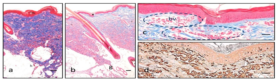
Figure 10: Features of pigskin. Masson trichrome (a–c) illustrates the three main layers in different colors with low magnification (epidermis in red, e; dermis in blue, d; hypodermis in white, h), specifically coloring collagen fibers in blue (a). Hair follicles (f) are not distinguishable from human, but closely, there are some apocrine glands (g) that help in this distinction (b). A superficial vascular plexus is readily identifiable in red with Masson trichrome (blood vessels, bv, and dashed circles), highlighted amongst the bluish collagen background of the dermis (c). Vimentin intermediate filaments are demonstrated in brown due to the diaminobenzidine and are typically present in epidermal dendritic cells and dermal fibroblasts, and endothelial cells of the blood vessels (d). Scale bar 200 µm (a,b), 80 µm (c,d).[67]
GUINEA PIGS
Studies have shown the effectiveness of the transfollicular route in absorbing medications applied topically. Hair follicles, which make up approximately 10% of the skin's surface, offer a spacious surface area for potential absorption. This makes it easier for an increased amount of medication to be absorbed through this specific route. Microparticulate systems, specifically microemulsions (ME), have been used for administering drugs directly to hair follicles. These nanocarriers provide extended-term stability and simplicity and have been found to effectively dissolve hydrophilic substances and medications attracted to fats. Adapalene, a synthetic derivative of naphthoic acid, can be used topically to treat acne vulgaris. However, the drug's percutaneous absorption rate is slow due to its insoluble nature in water. The study aimed to develop and characterize microemulsions containing adapalene and examine their penetration through the transfollicular route. Adapalene gel has been shown to have positive effects on treating various skin conditions with different underlying causes. Patients are recommended to continue using the product for up to four weeks following application, and the medication should be taken for a minimum of six months.[68]
Materials And Methods
Animal studies: The study involved mature male guinea pigs aged 20-22 weeks, weighing between 450-650 grams. Ethical approval was obtained from the Ahvaz Jundishapur University of Medical Sciences' ethical committee, who ensured the animals underwent a gentle acclimation period of 5 days before handling and application.[68]
Solubility of adapalene: The solubility of Adapalene in various oils, surfactants, and cosurfactants was evaluated. Excess Adapalene was dissolved in 5 milliliters of each oil, surfactant, and cosurfactant. Samples were mechanically agitated for 48 hours at a controlled temperature, then centrifuged at 1006 g for 30 minutes. The transparent liquids were then passed through a polytetrafluoroethylene membrane filter and analyzed using UV spectrophotometry at a wavelength of 279 nm.[68]
Adapalene Microemulsion preparation:
TABLE 7Composition of eight ME formulations of adapalene[68]
|
Formulation |
Factorial |
S/C |
% Oil |
% (S/C) |
% Water |
|
MEA-1 |
+++ |
3:1 |
50 |
40 |
10 |
|
MEA-2 |
++- |
3:1 |
50 |
45 |
5 |
|
MEA-3 |
+-+ |
3:1 |
5 |
85 |
10 |
|
MEA-4 |
+-- |
3:1 |
5 |
90 |
5 |
|
MEA-5 |
--+ |
1:1 |
5 |
85 |
10 |
|
MEA-6 |
--- |
1:1 |
5 |
90 |
5 |
|
MEA-7 |
-+- |
1:1 |
50 |
45 |
5 |
|
MEA-8 |
-++ |
1:1 |
50 |
40 |
10 |
Abbreviations: +: High level; −: low level; S/C: surfactant/cosurfactant ratio, ME: microemulsion.
Physical Stability and Drug Release Evaluation of an Adapalene-Loaded Microemulsion:
The study focuses on the physical stability of a drug-emulsion (ME) formulation, which was tested using various methods. Droplet size was measured at a temperature of 25°C, and viscosity was assessed using a Brookfield viscometer. Physical stability was assessed through stress tests and temperature stability experiments. The samples were stored according to ICH guidelines for six months, considering various temperatures and time-related physicochemical characteristics. The substance was also examined for phase segregation and physical instability.
The study also examines drug release in a laboratory setting using Franz diffusion cells equipped with a cellulose membrane. The membrane was soaked in distilled water and placed between the giver and recipient chambers. Adapalene-loaded microemulsion was placed onto the membrane, and the receptor medium was loaded with DMSO and phosphate buffer solution. The receptor fluid was stirred in motion, and the receptor medium was removed and replaced with a smoother one. An assessment was conducted to determine the drug content at a wavelength of 279 nm. The presentation featured the progressive release of medication over time, revealing a distinct pattern. Three distinct kinetic models were considered, with the highest possible amount reaching the most likely mechanism.[68]
Result: Adapalene solubility is being studied, with various excipients and microemulsions (MEs) being examined. The Middle Eastern framework integrates findings. Adapalene MEs have viscosity, mean droplet size, and polydispersity index. The formulation ME-ADP7 has the smallest particle size. The drug is gradually introduced over 24 hours, with varying effects on different skin types. In vitro permeation studies show differences in permeability among ME formulations.[68]
RABBIT
Bupranolol (BP) is a beta-adrenoceptor blocker with a chlorine substituent. After oral ingestion, healthy volunteers showed a maximum plasma concentration within 1.2 hours. However, the level is nearing the dissociation constant (k1) value at beta1. BP is susceptible to extensive metabolism and has a half-life of 2.0 hours. Transdermal delivery offers consistent drug delivery, but the quantity can be limited. Studies have shown consistent plasma levels and blood pressure impact through the daily application of a transdermal delivery system. This study explores percutaneous penetration of BP through rabbit skins using both in vivo and in vitro methods.[69]
Methods and Materials
Kaken Pharmaceutical Materials and Experiment
Preparation of Gel Ointments and Their Transdermal Systems:
Table8:Composition of BP Gel Ointments[69]
|
Components |
Rp1 |
Rp2 |
Rp3 |
|
Bupranolol |
3.0 |
3.0 |
3.0 |
|
Lauric acid |
2.21 |
2.21 |
- |
|
Carbopol 934 |
3.0 |
3.0 |
3.0 |
|
EtOH/ propylene glycol (2: 1, v/v) |
50.0 |
50.0 |
50.0 |
|
5% NH4OH |
q.s |
q.s |
q.s |
|
d-Limonene |
- |
5.0 |
- |
|
Isopropyl myristate |
- |
- |
5.0 |
|
N-Methyl-2-pyrrolidone |
- |
- |
5.0 |
|
Gentamicin solution (ml) |
1.0 |
1.0 |
1.0 |
|
Purified water |
100.0 |
100.0 |
100.0 |
Intravenous Administration, Percutaneous Penetration, and BP Determination: A Comprehensive Pharmacokinetic Evaluation
The study investigated the administration of BP hydrochloride intravenously in rabbits using a blend of dimethylformamide and ethanol. Blood samples were collected from the ear vein over 10 hours. In vitro percutaneous penetration experiments involved euthanizing rabbits, administering chloroform on the epidermis, and applying a gel ointment on the stratum corneum. In vivo experiments involved removing hair, applying a transdermal therapeutic system, and collecting blood samples. The plasma from the blood samples was isolated and frozen for testing. The study showed promising results for the transdermal therapeutic system. The determination of BP in plasma was conducted using Winkler and Lemmer's method, with a sensitivity of 5 ng/ml in plasma and 500 ng/ml in the sample solution.[69]
Result: The study investigates the relationship between plasma concentration and time following the intravenous administration of beta-blockers (BP) in rabbits. The removal of BP follows a biexponential pattern, similar to other beta-blockers like PL14 and penbutolol. In vitro penetration of BP through the skin was observed, with the rate of permeation (Js) being small without enhancers. The addition of IPM and NMP to the ointment resulted in notable penetration of BP. The lipids of the stratum corneum are influenced by NMP, which impacts the polar pathways. Collaboration between IPM and NMP is essential for effective skin care. The steady-state permeation rate corresponds directly to its solubility. IPM and NMP are agents commonly used in the stratum corneum for drug delivery. In vivo percutaneous absorption was found to be affected by plasma concentrations. One possible explanation for the significant enhancing effect seen in vivo with d-limonene could be attributed to its capability to modify the function of the enhancer. Intercellular lipids fluidize and remove lipids, boosting the diffusion of lipophilic substances. BP is absorbed quickly in the top layer of the skin, known as the stratum corneum. However, within an in vitro setting, the enhancer was unable to optimize the drug's absorption into the skin.
The plasma levels are primarily influenced by the significant metabolism of BP in the systemic circulation following percutaneous absorption. The effects are influenced by the dosage and could potentially lessen as the doses increase. In cases like percutaneous absorption, a significant systemic clearance would be evident. Transdermal systems hold promise in becoming a highly effective method of delivering drugs. A delivery system has been developed for the treatment of angina pectoris. In conclusion, it can be inferred that BP can readily permeate rabbit skin when certain enhancers are present. The drug's absorption through the skin was improved when applied in vivo.[69]
Table 9. Comparison of in Vitro Penetration Data with in Vivo Percutaneous Absorption Parameters of BP[69]
|
formulation |
In vivo |
In vitro |
Predicted Css (b) Ng/ml |
|||
|
Js (mg/h · cm2 ) |
Ratio (a) |
AUC0—∞ (ng · h/ml) |
Ratio (b) |
Cmax (ng/ml) |
||
|
1 |
12.48 |
1.00 |
210.57 |
1.0 |
12.81 |
102.21 |
|
2 |
18.38 |
1.43 |
626.96 |
2.98 |
21.78 |
146.31 |
|
3 |
45.73 |
3.56 |
471.92 |
2.24 |
16.68 |
364.02 |
The value for formulation 2 or 3/the value for formulation 1. a) Js: steady state flux b) Css: steady state concentration
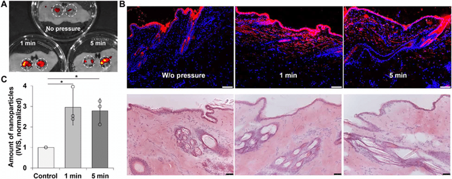
Figure 11: Transdermal delivery of NPs in rabbit skin with the temporal pressure treatment. (A) IVIS fluorescence imaging of excised rabbit skin after pressure treatment and application of NPs. Circled areas represent the area of interest. (B) Fluorescence imaging (blue, DAPI; red, NPs) and H&E staining of rabbit skin after the treatment. (C) Quantification of NP fluorescence signal in (A). Scale bars, 100 ?m. n = 3, all data are means ± SD, *P < 0.05. Photo credits: Daniel Chin Shiuan Lio, School of Chemical and Biomedical Engineering, Nanyang Technological University[69]
IN VITRO STUDY:
Two features of a dosage form that are essential for characterizing the drug dissolution profile of a controlled release dosage form and, therefore, its in vivo efficacy are the mechanism and kinetics of drug release. The kinetics of drug dissolution in controlled-release drug delivery systems has been described by several mathematical models, and the drug release rate from TDDS can be determined using a variety of methods.
The Paddle over Disc: This technique mirrors the USP paddle dissolution instrument, with the only difference being that the transdermal system is connected to a disc or cell positioned at the base of the container filled with a medium at a temperature of 32 ±5°C.[1]
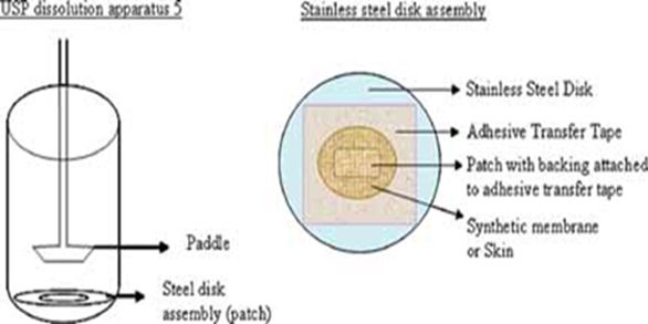
Figure 12:USP dissolution apparatus 5 [1]
The Cylinder Modified USP Basket:
This approach closely resembles the USP basket-type dissolution apparatus, with the only difference being that the setup is affixed to a hollow cylinder positioned within a medium kept at a temperature of 32 ±5°C. The absorption of drugs into the body is significantly influenced by the release of drugs from transdermal films made of polymers. Once the drug reaches the skin's surface, it is transferred to the dermal microcirculation by penetrating through the cells of the epidermis or the skin appendages.[1]
Preparation of skin for permeation studies:
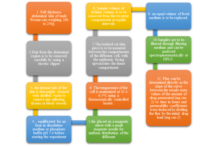
Figure 13: Flowchart of the process of preparation of skin for permeation study[1]
IN VIVO HUMAN STUDY
SKIN BARRIERS:
Biosynthetic Activities and Barrier Homeostasis
Lipid Metabolism: Epidermal differentiation involves a directional flow of lipid composition, shifting from phospholipids to ceramides, cholesterol, and free fatty acids. The outer layer of the skin, specifically the stratum corneum, contains components 11 and 13. Lipid synthesis operates with high activity and independence from systemic influences. Changes in the permeability barrier's status can trigger a recovery sequence, typically restoring functionality within 72 hours. Youthful skin undergoes a cutaneous stress test, revealing elevated cholesterol, free fatty acid, and ceramide production. Enhancement of mRNA levels and enzyme activity/mass is observed for each component. Three essential lipids are necessary for maintaining barrier homeostasis.[72]
Lamellar body secretion: The stratum corneum has a dual-compartment structure, triggered by the release of lipids from lamellar bodies. Enzymes are located between the stratum granulosum and the stratum corneum interface. Lamellar bodies are secreted gradually to maintain barrier integrity. However, sudden disruptions cause calcium loss, resulting in a significant decrease. The stratum granulosum layer controls lamellar body secretion, with calcium playing a crucial role in regulation.[72]
Epidermal differentiation: Lamellar bodies release polar lipids at the stratum, which transform into hydrophobic compounds and form mature membranes. The extracellular processing of glucosylceramides, phospholipids, and cholesterol sulfate accumulates, leading to the presence of ceramides, free fatty acids, and cholesterol in the outer skin layer. Barrier homeostasis is crucial, and inhibitors like Bromphenacylbromide or MJ33 can maintain barrier balance. Acidic sphingomyelinase can disrupt sphingomyelin breakdown, leading to skin disorders like ichthyosis and dermatosis. Cholesterol sulfate and steroid sulfatase play significant roles in epidermal differentiation but can cause barrier defects and abnormalities.[72]
Acidification: The stratum corneum, with its acidic external pH level, is crucial for maintaining the skin barrier. It is enriched with sodium-proton antiporters that create a pH gradient. Enzymes like sPLA2, β-glucocerebrosidase, and acidic sphingomyelinase regulate the breakdown of phospholipids and conversion of histidine to urocanic acid. Maintaining the acid mantle promotes homeostasis and barrier recovery. Gentle exfoliation is necessary to prevent delays.[72]
PARAMETERS AFFECTING SKIN PERMEABILITY
Transdermal drug delivery is a passive process controlled by Fick's law, which determines the absorption rate or flux of a substance. The concentration difference on either side of a barrier directly affects the rate of substance movement. Factors such as partition coefficient Km, diffusion coefficient D, and diffusion distance L govern percutaneous drug absorption kinetics, with Cv and Km being particularly significant dependent on the vehicle used. The vehicle used in drug delivery is crucial for the effectiveness of the treatment, as the structure of the vehicle can impact the speed and efficiency of drug absorption, known as bioavailability. Advancements in formulations have led to the development of various effective vehicles such as gels, solutions, and foams.
Drug concentration plays a crucial role in their effectiveness, especially in terms of percutaneous absorption. Higher concentrations are often believed to be more powerful, but some drugs may be most effective at specific concentrations. The partition coefficient, represented by K m = C sc / C v, indicates how effectively a drug can transition from the vehicle to the stratum corneum. Topically applied medications have low absorption rates and often remain on the skin's surface, making them vulnerable to being lost through exfoliation, perspiration, or other factors. Factors like skin hydration, barrier structure modification, and certain additives can enhance drug absorption significantlyVariation in different regions, such as nail, palm/sole, trunk/extremities, face/scalp, and scrotum, also influences absorption.[72]
DRUG PERMIATION:
Contaminated and chemically disinfected waters often contact with human skin, allowing for potential dermal absorption of contaminants. Accurate estimates of chemical absorption are crucial for establishing contamination thresholds and assessing transdermal drug administration feasibility. The permeability coefficient of the stratum corneum is a key parameter used to estimate absorption. However, measurements are currently unavailable, so predictive equations rely on these measurements. Alternative compounds can be used as a replacement. Numerous equations have been developed, but not all offer sufficient estimates for every chemical under scrutiny.[70]
FUTURE SCOPE ADVANCED DEVELOPMENT IN TRANSDERMAL DRUG DELIVERY SYSTEM:
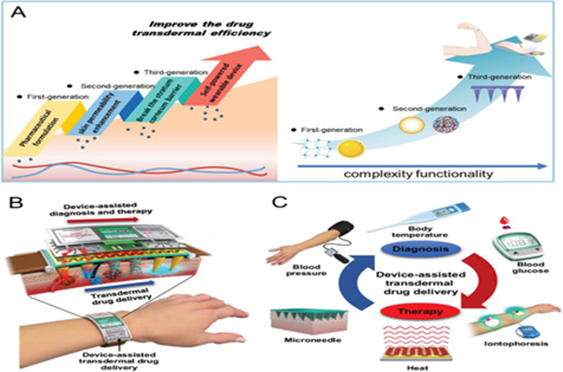
Figure 14: A) The research on transdermal drug delivery systems is gradually becoming intelligent and functionally complex, further improving the therapeutic efficacy. B) Advances in physical principles of transdermal drug delivery and research on wearable devices have promoted the development of convenient medicine. C) Wearable devices are widely used in portable medicine.[72]
Adhesive technology is currently the recommended technique for passive transdermal medication administration two The focus of formulation research is on adhesives and excipients. Modifying the adhesive to improve skin adhesion over time, reduce lag time, increase delivery rate, and improve drug stability and solubility is the aim of adhesive research. Since no one adhesive is effective for all drug and formulation chemistries, the transdermal formulator can optimize the performance of the transdermal patch polymer by tailoring the adhesive chemistry. The next generation of drug delivery systems, known as TDDS, has practical real-world uses.[59][60]
CONCLUSION
This article gives profitable information regarding the transdermal sedate conveyance frameworks and its assessment prepare all elements as a prepared reference for the inquire about researcher who are included in TDDS. The foregoing appears that TDDS have awesome potentials, being able to utilize for both hydrophobic and hydrophilic dynamic substance into promising deliverable drugs. To optimize this, a medicate delivery system, more prominent understanding of the different mechanisms of organic intuitive, and polymer are required. TDDS is a reasonable, viable application as another era of medicate conveyance framework[70][72]
REFERENCES

Ritika Sakale, Saniya Pinjari, Riya Ranadive, Pranav Raut, Shubham Waghmare, Vishnu Neharkar, Dr. Sanjay Walode, Comparative Analysis of In Vitro and In Vivo Models for Evaluating Transdermal Drug Delivery Systems, Int. J. of Pharm. Sci., 2026, Vol 4, Issue 4, 2340-2363. https://doi.org/10.5281/zenodo.19593228
 10.5281/zenodo.19593228