View Article

  • Development And Evaluation of a Polyherbal Ointment Containing Musa Paradisiaca Peel, Carica Papaya Peel And Psidium Guajava Leaf Extracts For Antimicrobial Activity

  • 1*Department of Pharmaceutics, Maharashtra Institute of Pharmacy (B. Pharm), Betala, Bramhapuri - 441206

    3Associate Professor, Dipartment of Pharmaceutics, Maharashtra Institute of Pharmacy (B. Pharm), Betala, Bramhapuri - 441206

    2Principal, Maharashtra Institute of Pharmacy (B. Pharm), Betala, Bramhapuri - 441206

Abstract

The present study was undertaken to develop and evaluate a polyherbal ointment containing extracts of Musa paradisiaca peel, Carica papaya peel, and Psidium guajava leaves for potential antimicrobial activity. The selected plant materials were subjected to extraction and incorporated into topical ointment formulations at different concentrations. Three formulations (F1, F2, and F3) were prepared and evaluated for various physicochemical and antimicrobial parameters. The formulations were assessed for appearance, homogeneity, pH, spreadability, irritancy, stability, and antimicrobial activity against selected bacterial and fungal strains. Among the prepared formulations, F2 demonstrated the most satisfactory overall physicochemical characteristics, with a pH of 6.5 and spreadability of 16.91. F2 exhibited antimicrobial activity against Staphylococcus aureus, Escherichia coli, and Candida albicans, producing zones of inhibition of 11.7, 11.3, and 11.2 mm, respectively. No significant skin irritation was observed with F2 during the evaluation period. Stability assessment further indicated that the optimized formulation remained satisfactory during the study period. The findings suggest that the combined plant extracts possess potential antimicrobial properties and that their incorporation into a topical ointment may provide a promising herbal approach for topical antimicrobial applications. Further studies are required to establish the active constituents, mechanism of action, and clinical efficacy of the formulation.

Keywords

Polyherbal ointment; Musa paradisiaca; Carica papaya; Psidium guajava; Herbal formulation; Antimicrobial activity; Topical formulation; Plant extracts.

Introduction

× Popup Image

The skin is the largest organ of the human body and serves as an essential protective barrier between the external environment and internal tissues. It provides mechanical and chemical protection, limits excessive water loss, contributes to thermoregulation and participates in immune defence. The integrity of the skin barrier is therefore important for maintaining normal physiological function and protecting the body from environmental and microbial challenges [1,2].

Skin disorders and infections represent an important health concern and may result from inflammatory, infectious, environmental and other factors. The global burden of skin diseases is substantial, with infectious and inflammatory conditions contributing considerably to morbidity and affecting quality of life [3,4]. Bacterial and fungal infections can compromise the skin barrier and may complicate wounds or other skin lesions. In particular, microorganisms associated with skin and soft-tissue infections remain clinically relevant because their management can be complicated by antimicrobial resistance and limitations associated with conventional antimicrobial therapy [5].

Wound healing is a complex biological process involving overlapping phases of inflammation, tissue formation and remodelling. Successful healing requires restoration of tissue integrity and control of factors that can delay repair, including persistent inflammation and microbial contamination [6–8]. Consequently, topical preparations capable of providing suitable local delivery and antimicrobial protection may have potential applications in the management of superficial skin conditions.

Topical drug delivery provides several advantages for the localized administration of therapeutic agents to the skin. Ointments are semisolid dosage forms that can provide prolonged contact with the application site and may facilitate localized delivery of active constituents. The selection of an appropriate base and optimization of formulation characteristics such as consistency, spreadability, viscosity, pH and extrudability are important for the performance and patient acceptability of topical preparations [9–11].

Medicinal plants have traditionally been used as sources of therapeutic agents, and herbal medicines continue to receive considerable attention because of their diverse phytochemical constituents. However, appropriate quality control, standardization and evaluation are necessary to ensure the reproducibility and safety of herbal preparations [12]. Polyherbal formulations involve the combination of more than one medicinal plant and may provide a broader phytochemical profile than preparations containing a single plant material. The concept of polyherbal therapy is well recognized in traditional systems of medicine and has also been investigated from a pharmacological and pharmaceutical perspective [13].

The biological effects of plant-derived preparations are associated with their complex mixtures of phytoconstituents. Phytochemical screening is therefore useful for the preliminary detection of major classes of constituents and for establishing the phytochemical profile of plant extracts. Commonly investigated groups include alkaloids, flavonoids, tannins, saponins, glycosides and phenolic compounds [14–17]. Evaluation of herbal materials and their extracts using appropriate quality-control approaches is essential for the development of reproducible herbal formulations [15,18].

In the present study, a polyherbal topical ointment was developed using extracts of Musa paradisiaca peel, Carica papaya peel and Psidium guajava leaves. The study focused on extraction, preliminary phytochemical screening, formulation of different ointment batches and evaluation of their physicochemical and pharmaceutical characteristics. The formulations were further investigated for antimicrobial activity against selected bacterial and fungal microorganisms, skin compatibility and short-term stability. The study also explores the pharmaceutical potential of combining these plant materials in a topical dosage form while providing a value-added approach to plant materials such as fruit peels.

2. Materials and Methods

2.1 Plant Materials and Authentication

Fresh banana (Musa paradisiaca) peels, papaya (Carica papaya) peels, and guava (Psidium guajava) leaves were selected as herbal materials. Fresh banana and papaya fruits were obtained from local markets, while guava leaves were collected from healthy plants in the local area. The collected materials were free from visible disease, insect infestation, and physical damage. The plant materials were authenticated by a qualified botanist from the Department of Botany, and voucher specimens were prepared and preserved for reference.

2.2 Preparation of Plant Materials

The collected plant materials were thoroughly washed with running tap water followed by distilled water to remove adhering dust and foreign matter. The cleaned materials were shade-dried at room temperature for 10–15 days, avoiding direct sunlight. The dried materials were separately pulverized using a mechanical grinder, passed through sieve No. 40, and stored in airtight containers until extraction.

2.3 Extraction of Plant Materials

Each powdered plant material (100 g) was separately subjected to Soxhlet extraction using ethanol. Extraction was continued for 6–8 h until exhaustion. The extracts were filtered through Whatman filter paper, concentrated using a rotary vacuum evaporator, dried, and stored at 4°C until further use. The percentage extractive yield was calculated as the weight of extract obtained relative to the weight of plant material used, multiplied by 100.

2.4 Preliminary Phytochemical Screening

The ethanolic extracts of banana peel, papaya peel, and guava leaves were subjected to preliminary qualitative phytochemical screening for major secondary metabolites. Standard chemical tests were performed for alkaloids, flavonoids, tannins, glycosides, saponins, and phenolic compounds using established phytochemical screening procedures.

2.5 Formulation of Polyherbal Ointment

Three polyherbal ointment formulations (F1–F3) were prepared with different concentrations of the three plant extracts. F1 contained 2% each of banana peel, papaya peel, and guava leaf extracts; F2 contained 3% each; and F3 contained 4% each. White soft paraffin was used as the base, with beeswax (5%), liquid paraffin (10%), methyl paraben (0.20%), and propyl paraben (0.02%) incorporated into each formulation.

Table 1- Formulation of Polyherbal Ointment

Ingredient (% w/w)

F1

F2

F3

Musa paradisiaca peel extract

2

3

4

Carica papaya peel extract

2

3

4

Psidium guajava leaf extract

2

3

4

White soft paraffin

q.s. to 100

q.s. to 100

q.s. to 100

Beeswax

5

5

5

Liquid paraffin

10

10

10

Methyl paraben

0.20

0.20

0.20

Propyl paraben

0.02

0.02

0.02

2.6 Preparation of Ointment

The formulations were prepared by the fusion method. Beeswax and white soft paraffin were accurately weighed and heated together on a water bath at approximately 70°C until completely melted. Liquid paraffin was then incorporated with continuous stirring to obtain a uniform base. The herbal extracts were separately triturated and incorporated into a small quantity of the molten base to form a smooth paste. This paste was gradually incorporated into the molten base with continuous stirring. The preservatives were incorporated as described in the formulation procedure, followed by continuous mixing until a smooth and homogeneous ointment was obtained. The formulations were subsequently cooled gradually at room temperature with stirring and filled into suitable containers.

2.7 Evaluation of Ointment Formulations

The prepared formulations were evaluated for appearance, colour, odour, consistency, homogeneity, pH, spreadability, viscosity, extrudability, washability, and drug content. These parameters were used to assess the physical quality, uniformity, application characteristics, and overall acceptability of the formulations.

For drug-content determination, 1 g of ointment was accurately weighed, dissolved in a suitable solvent, filtered to remove insoluble base material, and analysed using a UV–Visible spectrophotometer at the selected wavelength. The extract concentration was determined from the calibration curve and expressed as percentage drug content.

2.8 Antimicrobial Activity

The antimicrobial activity of F1, F2, and F3 was evaluated by the cup-plate (agar well diffusion) method against Staphylococcus aureus, Escherichia coli, and Candida albicans. Nutrient agar was used for bacterial evaluation and Sabouraud dextrose agar for fungal evaluation. The media were sterilized by autoclaving at 121°C for 15 min. Approximately 20–25 mL of molten medium was poured into sterile Petri plates and allowed to solidify.

Fresh microbial cultures were uniformly inoculated onto the respective agar surfaces using sterile cotton swabs. Wells of approximately 6 mm diameter were prepared using a sterile cork borer. The test ointment formulations were introduced into separate wells, while an appropriate standard antimicrobial preparation and ointment base without herbal extracts were used as positive and negative controls, respectively. The plates were allowed to stand at room temperature for approximately 30–60 min to facilitate diffusion. Bacterial plates were incubated at approximately 37°C for 24 h, whereas C. albicans plates were incubated at approximately 28°C for 48 h. Zones of inhibition were measured in millimetres, and each experiment was performed in triplicate.

2.9 Skin Irritation Study

Skin compatibility of the prepared ointments was evaluated in healthy human volunteers after obtaining informed consent. A small quantity of the formulation was applied to a marked area on the inner forearm and left undisturbed for 24 h under normal environmental conditions. The treated area was subsequently examined for visible signs of redness, swelling, or irritation.

2.10 Stability Study

The prepared formulations were subjected to stability evaluation for three months under room-temperature conditions. During the study period, the formulations were examined for changes in physical appearance, pH, consistency, and phase separation. The optimized formulation was selected based on its overall physical, physicochemical, antimicrobial, skin-compatibility, and stability characteristics.

2.11 Statistical Analysis

Experimental observations were compiled and subjected to statistical evaluation. Antimicrobial experiments were performed in triplicate, and the results were expressed as mean values.

3. Results and Discussion

3.1 Authentication and Extractive Yield

The selected plant materials were authenticated as Musa paradisiaca Linn. (banana peel), Carica papaya Linn. (papaya peel), and Psidium guajava Linn. (guava leaves). The percentage extractive yields obtained by ethanolic Soxhlet extraction were 12.8% for banana peel, 10.6% for papaya peel, and 15.4% for guava leaves. Guava leaves showed the highest extractive yield among the three plant materials.

Table 2- Results and Discussion

Plant material

Weight taken (g)

Extract obtained (g)

Yield (%)

Banana peel

100

12.8

12.8

Papaya peel

100

10.6

10.6

Guava leaves

100

15.4

15.4

3.2 Preliminary Phytochemical Screening

Phytochemical screening demonstrated the presence of several secondary metabolites in all three extracts. Alkaloids and glycosides were detected in all extracts, while flavonoids and phenolics were particularly abundant in guava leaf extract. Tannins were moderately present in banana peel and abundantly present in guava leaves, whereas saponins were moderately present in papaya peel. The presence of these phytoconstituents provides a chemical basis for the observed biological activity of the herbal formulations.

Table 3- Preliminary Phytochemical Screening

Phytoconstituent

Banana peel

Papaya peel

Guava leaves

Alkaloids

+

+

+

Flavonoids

++

++

+++

Tannins

++

+

+++

Saponins

+

++

+

Glycosides

++

++

++

Phenolics

++

++

+++


Key: + Present; ++ Moderately present; +++ Abundantly present.

3.3 Formulation and Physical Evaluation

Three formulations containing 2%, 3%, and 4% each of banana peel, papaya peel, and guava leaf extracts were developed. All formulations exhibited smooth appearance, characteristic herbal odour, and absence of phase separation. F2 showed excellent consistency and homogeneity, whereas F3 was slightly thicker, indicating an effect of increasing extract concentration on the physical characteristics of the ointment.

Table 4 - Formulation and Physical Evaluation

Parameter

F1

F2

F3

Colour

Light green

Greenish brown

Dark green

Odour

Characteristic

Characteristic

Characteristic

Appearance

Smooth

Smooth

Smooth

Consistency

Good

Excellent

Slightly thick

Homogeneity

Good

Excellent

Good

Phase separation

Absent

Absent

Absent

3.4 Physicochemical Evaluation

The pH values of F1, F2, and F3 were 6.2, 6.5, and 6.6, respectively. Spreadability values were 14.32, 16.91, and 13.99 g·cm/s, with F2 showing the highest spreadability. The viscosity values were 18,420, 16,915, and 20,235 cP for F1, F2, and F3, respectively. F2 demonstrated excellent extrudability and easy washability, whereas F3 was moderately washable.

Table 5- Physicochemical Evaluation

Parameter

F1

F2

F3

pH

6.2

6.5

6.6

Spreadability (g·cm/s)

14.32

16.91

13.99

Viscosity (cP)

18,420

16,915

20,235

Extrudability

Good

Excellent

Good

Washability

Easily washable

Easily washable

Moderately washable

The drug-content values ranged from 96.65% to 98.28%, with F2 showing the highest reported value of 98.28%, indicating good content uniformity.

3.5 Antimicrobial Activity

The agar well diffusion study demonstrated antimicrobial activity of the developed formulations against Staphylococcus aureus, Escherichia coli, and Candida albicans. F2 produced the highest reported antibacterial zones against S. aureus (11.7 mm) and E. coli (11.3 mm). F3 produced the highest reported antifungal zone against C. albicans (11.3 mm), although its activity was reported as comparable to F2.
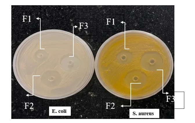


Figure No. 01: Results Against Bacterial Strains
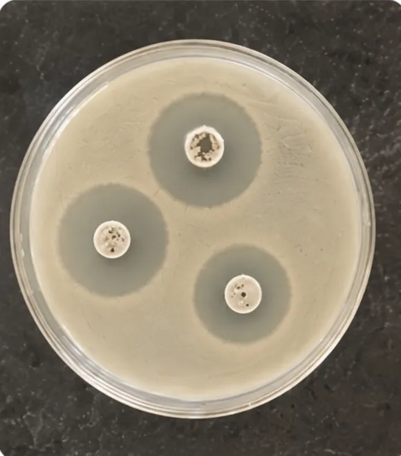


Figure No. 02: Results Against Fungal Strain (C. albicans)

The observed antimicrobial activity may be associated with the combined contribution of phytoconstituents such as flavonoids, tannins, phenolics, alkaloids, saponins, and glycosides identified in the plant extracts. However, the present study demonstrates antimicrobial activity rather than establishing pharmacological synergy mechanistically.

3.6 Skin Compatibility and Stability

Skin irritation evaluation showed that F1 and F2 were non-irritant, with no observed redness, swelling, or irritation, whereas F3 produced mild irritation. Stability evaluation over three months under room-temperature conditions showed no significant changes in pH, consistency, or phase separation. F1 and F2 retained their original appearance, while F3 showed a slight colour change. Overall, F2 exhibited the most favourable stability profile.

3.7 Selection of Optimized Formulation

Considering the combined results of physical characteristics, pH, spreadability, viscosity, extrudability, drug content, antimicrobial activity, skin compatibility, and stability, F2 was selected as the optimized formulation. F2 contained 3% each of banana peel, papaya peel, and guava leaf extracts and demonstrated a pH of 6.5, maximum spreadability of 16.91 g·cm/s, highest reported drug content of 98.28%, excellent extrudability, antimicrobial activity, absence of skin irritation, and satisfactory stability.

4. CONCLUSION

The present study successfully developed and evaluated a polyherbal topical ointment containing Musa paradisiaca peel, Carica papaya peel, and Psidium guajava leaf extracts. Preliminary phytochemical screening indicated the presence of important secondary metabolites, including alkaloids, flavonoids, tannins, saponins, glycosides, and phenolic compounds. Three formulations (F1, F2, and F3) containing 2%, 3%, and 4% of each herbal extract, respectively, were prepared by the fusion method and evaluated for their physicochemical, antimicrobial, skin-compatibility, and stability characteristics.

Among the formulations, F2 demonstrated the most favorable overall characteristics, including a pH of 6.5, spreadability of 16.91 g·cm/sec, viscosity of 16,915 cP, and drug content of 98.28%. The formulation exhibited good homogeneity, consistency, extrudability, and washability, with no phase separation. F2 also demonstrated notable antimicrobial activity against Staphylococcus aureus and Escherichia coli, with inhibition zones of 11.7 and 11.3 mm, respectively. Antifungal activity against Candida albicans was also observed. The formulations remained physically stable during the three-month stability study under long-term and accelerated conditions, although slight colour change was observed in F3. F1 and F2 showed no irritation, whereas F3 produced mild irritation.

Overall, F2 was selected as the optimized formulation based on its balanced physicochemical properties, antimicrobial performance, skin compatibility, and stability. The findings indicate that these plant extracts can be successfully incorporated into a topical ointment and provide a potential approach for utilizing plant-derived materials, including agro-waste such as fruit peels, in herbal topical formulations.

5. Future Scope

Further studies are required to establish the therapeutic potential of the optimized polyherbal ointment. Future investigations should include comprehensive in-vivo wound-healing studies, histopathological evaluation, assessment of wound contraction and epithelialization, and evaluation of relevant biochemical and inflammatory markers. Additional studies on mechanism of antimicrobial action, formulation optimization, long-term stability, and clinical safety may further support the development of the formulation for potential topical applications.

REFERENCES
  1. Proksch E, Brandner JM, Jensen JM. The skin: an indispensable barrier. Exp Dermatol. 2008;17(12):1063-1072.
  2. Tortora GJ, Derrickson BH. Principles of Anatomy and Physiology. 16th ed. Hoboken, NJ: John Wiley & Sons; 2021. p.146-180.
  3. Hay RJ, Johns NE, Williams HC, Bolliger IW, Dellavalle RP, Margolis DJ, et al. The global burden of skin disease in 2010. J Invest Dermatol. 2014;134(6):1527-1534.
  4. Bickers DR, Lim HW, Margolis D, Weinstock MA, Goodman C, Faulkner E, et al. The burden of skin diseases. J Am Acad Dermatol. 2006;55(3):490-500.
  5. Dryden M. Skin and soft tissue infection: microbiology and epidemiology. Int J Antimicrob Agents. 2009;34(Suppl 1):S2-S7.
  6. Guo S, DiPietro LA. Factors affecting wound healing. J Dent Res. 2010;89(3):219-226.
  7. Gurtner GC, Werner S, Barrandon Y, Longaker MT. Wound repair and regeneration. Nature. 2008;453(7193):314-321.
  8. Velnar T, Bailey T, Smrkolj V. The wound healing process: an overview of the cellular and molecular mechanisms. J Int Med Res. 2009;37(5):1528-1542.
  9. Aulton ME, Taylor KMG. Aulton's Pharmaceutics: The Design and Manufacture of Medicines. 6th ed. London: Elsevier; 2022. p.565-592.
  10. Benson HAE, Watkinson AC. Topical and Transdermal Drug Delivery: Principles and Practice. Hoboken: Wiley; 2012. p.1-25.
  11. Allen LV. Ansel's Pharmaceutical Dosage Forms and Drug Delivery Systems. 12th ed. Philadelphia: Wolters Kluwer; 2022. p.385-410.
  12. Ekor M. The growing use of herbal medicines: issues relating to adverse reactions and challenges in monitoring safety. Front Pharmacol. 2014;4:177.
  13. Parasuraman S, Thing GS, Dhanaraj SA. Polyherbal formulation: concept of Ayurveda. Pharmacogn Rev. 2014;8(16):73-80.
  14. Williamson EM. Synergy and other interactions in phytomedicines. Phytomedicine. 2001;8(5):401-409.
  15. Mukherjee PK. Quality Control and Evaluation of Herbal Drugs. 2nd ed. New Delhi: Business Horizons; 2019. p.35-60.
  16. Harborne JB. Phytochemical Methods: A Guide to Modern Techniques of Plant Analysis. 3rd ed. London: Chapman and Hall; 1998. p.1-40.
  17. Tiwari P, Kumar B, Kaur M, Kaur G, Kaur H. Phytochemical screening and extraction: a review. Int Pharm Sci. 2011;1(1):98-106.
  18. World Health Organization. Quality Control Methods for Herbal Materials. Geneva: World Health Organization; 2011. p.8-45.
  19. Tortora GJ, Derrickson BH. Principles of Anatomy and Physiology. 16th ed. Hoboken, NJ: John Wiley & Sons; 2021. p. 146-180.
  20. Proksch E, Brandner JM, Jensen JM. The skin: an indispensable barrier. Exp Dermatol. 2008;17(12):1063-1072.
  21. Mescher AL. Junqueira's Basic Histology: Text and Atlas. 16th ed. New York: McGraw-Hill Education; 2021. p. 349-378.
  22. Ross MH, Pawlina W. Histology: A Text and Atlas. 8th ed. Philadelphia: Wolters Kluwer; 2020. p. 453-478.
  23. McGrath JA, Uitto J. Anatomy and organization of human skin. In: Kang S, Amagai M, Bruckner AL, Enk AH, Margolis DJ, McMichael AJ, et al., editors. Fitzpatrick's Dermatology. 9th ed. New York: McGraw-Hill; 2019. p. 1-18.
  24. Elias PM. Skin barrier function. Curr Allergy Asthma Rep. 2008;8(4):299-305.
  25. Young B, O'Dowd G, Woodford P. Wheater's Functional Histology. 6th ed. Philadelphia: Elsevier; 2018. p. 343-360.
  26. Kolarsick PAJ, Kolarsick MA, Goodwin C. Anatomy and physiology of the skin. J Dermatol Nurses Assoc. 2011;3(4):203-213.
  27. Saladin KS. Anatomy and Physiology: The Unity of Form and Function. 9th ed. New York: McGraw-Hill Education; 2023. p. 159-181.
  28. Hall JE. Guyton and Hall Textbook of Medical Physiology. 15th ed. Philadelphia: Elsevier; 2021. p. 897-904.
  29. Tortora GJ, Nielsen MT. Principles of Human Anatomy. 15th ed. Hoboken: Wiley; 2017. p. 130-152.
  30. Hay RJ, Johns NE, Williams HC, Bolliger IW, Dellavalle RP, Margolis DJ, et al. The global burden of skin disease in 2010. J Invest Dermatol. 2014;134(6):1527-1534.
  31. Bickers DR, Lim HW, Margolis D, Weinstock MA, Goodman C, Faulkner E, et al. The burden of skin diseases. J Am Acad Dermatol. 2006;55(3):490-500.
  32. Leung DYM, Bieber T. Atopic dermatitis. Lancet. 2003;361(9352):151-160.
  33. Boehncke WH, Schön MP. Psoriasis. Lancet. 2015;386(9997):983-994.
  34. Zaenglein AL, Pathy AL, Schlosser BJ, Alikhan A, Baldwin HE, Berson DS, et al. Guidelines of care for the management of acne vulgaris. J Am Acad Dermatol. 2016;74(5):945-973.
  35. Dryden M. Skin and soft tissue infection: microbiology and epidemiology. Int J Antimicrob Agents. 2009;34(Suppl 1):S2-S7.
  36. Boateng J, Catanzano O. Advanced therapeutic dressings for effective wound healing. J Pharm Sci. 2015;104(11):3653-3680.
  37. Guo S, DiPietro LA. Factors affecting wound healing. J Dent Res. 2010;89(3):219-229.
  38. Gurtner GC, Werner S, Barrandon Y, Longaker MT. Wound repair and regeneration. Nature. 2008;453(7193):314-321.
  39. Velnar T, Bailey T, Smrkolj V. The wound healing process: an overview. J Int Med Res. 2009;37(5):1528-1542.
  40. Eming SA, Krieg T, Davidson JM. Inflammation in wound repair. J Invest Dermatol. 2007;127(3):514-525.
  41. Singer AJ, Clark RAF. Cutaneous wound healing. N Engl J Med. 1999;341(10):738-746.
  42. Gonzalez ACDO, Costa TF, Andrade ZA, Medrado ARAP. Wound healing: a literature review. An Bras Dermatol. 2016;91(5):614-620.
  43. Demidova-Rice TN, Hamblin MR, Herman IM. Acute and impaired wound healing. Adv Skin Wound Care. 2012;25(7):304-314.
  44. Aulton ME, Taylor KMG. Aulton's Pharmaceutics: The Design and Manufacture of Medicines. 6th ed. London: Elsevier; 2022. p. 565-592.
  45. Allen LV. Ansel's Pharmaceutical Dosage Forms and Drug Delivery Systems. 12th ed. Philadelphia: Wolters Kluwer; 2022. p. 385-410.
  46. Benson HAE, Watkinson AC. Topical and Transdermal Drug Delivery: Principles and Practice. Hoboken: Wiley; 2012. p. 1-25.
  47. Brown MB, Martin GP, Jones SA, Akomeah FK. Dermal and transdermal drug delivery systems. Drug Deliv. 2006;13(3):175-187.
  48. Barry BW. Dermatological Formulations: Percutaneous Absorption. New York: Marcel Dekker; 1983. p. 1-28.
  49. Prausnitz MR, Langer R. Transdermal drug delivery. Nat Biotechnol. 2008;26(11):1261-1268.
  50. Williams AC. Transdermal and Topical Drug Delivery. London: Pharmaceutical Press; 2003. p. 20-50.
  51. Hadgraft J. Skin, the final frontier. Int J Pharm. 2001;224(1-2):1-18.
  52. Lachman L, Lieberman HA, Kanig JL. The Theory and Practice of Industrial Pharmacy. 4th ed. Mumbai: Varghese Publishing House; 2019. p. 534-560.
  53. Banker GS, Rhodes CT. Modern Pharmaceutics. 5th ed. New York: Marcel Dekker; 2009. p. 689-716.
  54. Remington JP. Remington: The Science and Practice of Pharmacy. 23rd ed. London: Pharmaceutical Press; 2020. p. 873-910.
  55. Sinko PJ. Martin's Physical Pharmacy and Pharmaceutical Sciences. 7th ed. Philadelphia: Wolters Kluwer; 2017. p. 527-548.
  56. Indian Pharmacopoeia Commission. Indian Pharmacopoeia. Ghaziabad: IPC; 2022. Vol. I. p. 741-744.
  57. British Pharmacopoeia Commission. British Pharmacopoeia. London: The Stationery Office; 2023. Vol. IV. p. A370-A378.
  58. United States Pharmacopeial Convention. USP 47-NF 42. Rockville, MD: USP Convention; 2024. p. 1121-1134.
  59. Gennaro AR. Remington's Pharmaceutical Sciences. 22nd ed. Philadelphia: Lippincott Williams & Wilkins; 2018. p. 1673-1692.
  60. Kokate CK, Purohit AP, Gokhale SB. Pharmacognosy. 57th ed. Pune: Nirali Prakashan; 2022. p. 1-22.
  61. Evans WC. Trease and Evans Pharmacognosy. 17th ed. London: Elsevier; 2020. p. 1-30.
  62. Harborne JB. Phytochemical Methods: A Guide to Modern Techniques of Plant Analysis. 3rd ed. London: Chapman and Hall; 2018. p. 1-40.
  63. Ekor M. The growing use of herbal medicines: issues relating to adverse reactions and challenges in monitoring safety. Front Pharmacol. 2014;4:177.
  64. Parasuraman S, Thing GS, Dhanaraj SA. Polyherbal formulation: concept of Ayurveda. Pharmacogn Rev. 2014;8(16):73-80.
  65. Williamson EM. Synergy and other interactions in phytomedicines. Phytomedicine. 2001;8(5):401-409.
  66. Wagner H. Synergy research: approaching a new generation of phytopharmaceuticals. Fitoterapia. 2011;82(1):34-37.
  67. Mukherjee PK. Quality Control and Evaluation of Herbal Drugs. 2nd ed. New Delhi: Business Horizons; 2019. p. 35-60.
  68. Patwardhan B, Vaidya ADB, Chorghade M. Ayurveda and natural products drug discovery. Curr Sci. 2004;86(6):789-799.
  69. Fabricant DS, Farnsworth NR. The value of plants used in traditional medicine for drug discovery. Environ Health Perspect. 2001;109(Suppl 1):69-75.
  70. Sofowora A, Ogunbodede E, Onayade A. The role and place of medicinal plants in healthcare delivery. Afr J Tradit Complement Altern Med. 2013;10(5):210-229.
  71. Harborne JB. Phytochemical Methods: A Guide to Modern Techniques of Plant Analysis. 3rd ed. London: Chapman and Hall; 1998. p. 1–302.
  72. Trease GE, Evans WC. Trease and Evans Pharmacognosy. 16th ed. London: Saunders Elsevier; 2009. p. 135–380.
  73. Kokate CK, Purohit AP, Gokhale SB. Pharmacognosy. 56th ed. Pune: Nirali Prakashan; 2021. p. 6.1–6.35.
  74. Khandelwal KR. Practical Pharmacognosy: Techniques and Experiments. 30th ed. Pune: Nirali Prakashan; 2021. p. 149–170.
  75. Mukherjee PK. Quality Control and Evaluation of Herbal Drugs. 2nd ed. New Delhi: Elsevier India Pvt. Ltd.; 2019. p. 186–245.
  76. Sofowora A. Medicinal Plants and Traditional Medicine in Africa. 3rd ed. Ibadan: Spectrum Books Ltd.; 2008. p. 150–191.
  77. Evans WC. Pharmacognosy. 15th ed. Edinburgh: Saunders Elsevier; 2002. p. 95–135.
  78. Wallis TE. Textbook of Pharmacognosy. 5th ed. New Delhi: CBS Publishers and Distributors; 2005. p. 210–245.
  79. Brain KR, Turner TD. The Practical Evaluation of Phytopharmaceuticals. Bristol: Wright-Scientechnica; 1975. p. 81–95.
  80. Wagner H, Bladt S. Plant Drug Analysis: A Thin Layer Chromatography Atlas. 2nd ed. Berlin: Springer-Verlag; 1996. p. 1–384.
  81. Indian Pharmacopoeia Commission. Indian Pharmacopoeia. Vol. IV. Ghaziabad: Indian Pharmacopoeia Commission; 2022. p. 3120–3150.
  82. World Health Organization. Quality Control Methods for Herbal Materials. Geneva: World Health Organization; 2011. p. 8–45.
  83. Farnsworth NR. Biological and phytochemical screening of plants. J Pharm Sci. 1966;55(3):225–276.
  84. Edeoga HO, Okwu DE, Mbaebie BO. Phytochemical constituents of some Nigerian medicinal plants. Afr J Biotechnol. 2005;4(7):685–688.
  85. Tiwari P, Kumar B, Kaur M, Kaur G, Kaur H. Phytochemical screening and extraction: A review. Int Pharm Sci. 2011;1(1):98–106.
  86. Aulton ME, Taylor KMG. Aulton's Pharmaceutics: The Design and Manufacture of Medicines. 6th ed. London: Elsevier; 2022. p. 675–710.
  87. Allen LV. Ansel's Pharmaceutical Dosage Forms and Drug Delivery Systems. 12th ed. Philadelphia: Wolters Kluwer; 2022. p. 275–310.
  88. Lachman L, Lieberman HA, Kanig JL. The Theory and Practice of Industrial Pharmacy. 3rd ed. Mumbai: Varghese Publishing House; 2009. p. 534–563.
  89. Banker GS, Rhodes CT. Modern Pharmaceutics. 4th ed. New York: Marcel Dekker; 2002. p. 395–420.
  90. Indian Pharmacopoeia Commission. Indian Pharmacopoeia. Vol. II. Ghaziabad: IPC; 2022. p. 1980–1988.
  91. Remington JP. Remington: The Science and Practice of Pharmacy. 23rd ed. Philadelphia: Pharmaceutical Press; 2020. p. 879–905.
  92. Indian Pharmacopoeia Commission. Indian Pharmacopoeia. Vol. I. Ghaziabad: IPC; 2022. p. 2.7.2–2.7.8.
  93. Cappuccino JG, Welsh CT. Microbiology: A Laboratory Manual. 12th ed. New York: Pearson Education; 2020. p. 255–270.
  94. Pelczar MJ, Chan ECS, Krieg NR. Microbiology: Concepts and Applications. New York: McGraw-Hill; 2001. p. 460–485.
  95. Aneja KR. Experiments in Microbiology, Plant Pathology and Biotechnology. 4th ed. New Delhi: New Age International Publishers; 2014. p. 245–260.
  96. CLSI. Performance Standards for Antimicrobial Susceptibility Testing. 35th ed. Wayne (PA): Clinical and Laboratory Standards Institute; 2025.
  97. Khandelwal KR. Practical Pharmacognosy: Techniques and Experiments. 30th ed. Pune: Nirali Prakashan; 2021. p. 181–195

Reference

  1. Proksch E, Brandner JM, Jensen JM. The skin: an indispensable barrier. Exp Dermatol. 2008;17(12):1063-1072.
  2. Tortora GJ, Derrickson BH. Principles of Anatomy and Physiology. 16th ed. Hoboken, NJ: John Wiley & Sons; 2021. p.146-180.
  3. Hay RJ, Johns NE, Williams HC, Bolliger IW, Dellavalle RP, Margolis DJ, et al. The global burden of skin disease in 2010. J Invest Dermatol. 2014;134(6):1527-1534.
  4. Bickers DR, Lim HW, Margolis D, Weinstock MA, Goodman C, Faulkner E, et al. The burden of skin diseases. J Am Acad Dermatol. 2006;55(3):490-500.
  5. Dryden M. Skin and soft tissue infection: microbiology and epidemiology. Int J Antimicrob Agents. 2009;34(Suppl 1):S2-S7.
  6. Guo S, DiPietro LA. Factors affecting wound healing. J Dent Res. 2010;89(3):219-226.
  7. Gurtner GC, Werner S, Barrandon Y, Longaker MT. Wound repair and regeneration. Nature. 2008;453(7193):314-321.
  8. Velnar T, Bailey T, Smrkolj V. The wound healing process: an overview of the cellular and molecular mechanisms. J Int Med Res. 2009;37(5):1528-1542.
  9. Aulton ME, Taylor KMG. Aulton's Pharmaceutics: The Design and Manufacture of Medicines. 6th ed. London: Elsevier; 2022. p.565-592.
  10. Benson HAE, Watkinson AC. Topical and Transdermal Drug Delivery: Principles and Practice. Hoboken: Wiley; 2012. p.1-25.
  11. Allen LV. Ansel's Pharmaceutical Dosage Forms and Drug Delivery Systems. 12th ed. Philadelphia: Wolters Kluwer; 2022. p.385-410.
  12. Ekor M. The growing use of herbal medicines: issues relating to adverse reactions and challenges in monitoring safety. Front Pharmacol. 2014;4:177.
  13. Parasuraman S, Thing GS, Dhanaraj SA. Polyherbal formulation: concept of Ayurveda. Pharmacogn Rev. 2014;8(16):73-80.
  14. Williamson EM. Synergy and other interactions in phytomedicines. Phytomedicine. 2001;8(5):401-409.
  15. Mukherjee PK. Quality Control and Evaluation of Herbal Drugs. 2nd ed. New Delhi: Business Horizons; 2019. p.35-60.
  16. Harborne JB. Phytochemical Methods: A Guide to Modern Techniques of Plant Analysis. 3rd ed. London: Chapman and Hall; 1998. p.1-40.
  17. Tiwari P, Kumar B, Kaur M, Kaur G, Kaur H. Phytochemical screening and extraction: a review. Int Pharm Sci. 2011;1(1):98-106.
  18. World Health Organization. Quality Control Methods for Herbal Materials. Geneva: World Health Organization; 2011. p.8-45.
  19. Tortora GJ, Derrickson BH. Principles of Anatomy and Physiology. 16th ed. Hoboken, NJ: John Wiley & Sons; 2021. p. 146-180.
  20. Proksch E, Brandner JM, Jensen JM. The skin: an indispensable barrier. Exp Dermatol. 2008;17(12):1063-1072.
  21. Mescher AL. Junqueira's Basic Histology: Text and Atlas. 16th ed. New York: McGraw-Hill Education; 2021. p. 349-378.
  22. Ross MH, Pawlina W. Histology: A Text and Atlas. 8th ed. Philadelphia: Wolters Kluwer; 2020. p. 453-478.
  23. McGrath JA, Uitto J. Anatomy and organization of human skin. In: Kang S, Amagai M, Bruckner AL, Enk AH, Margolis DJ, McMichael AJ, et al., editors. Fitzpatrick's Dermatology. 9th ed. New York: McGraw-Hill; 2019. p. 1-18.
  24. Elias PM. Skin barrier function. Curr Allergy Asthma Rep. 2008;8(4):299-305.
  25. Young B, O'Dowd G, Woodford P. Wheater's Functional Histology. 6th ed. Philadelphia: Elsevier; 2018. p. 343-360.
  26. Kolarsick PAJ, Kolarsick MA, Goodwin C. Anatomy and physiology of the skin. J Dermatol Nurses Assoc. 2011;3(4):203-213.
  27. Saladin KS. Anatomy and Physiology: The Unity of Form and Function. 9th ed. New York: McGraw-Hill Education; 2023. p. 159-181.
  28. Hall JE. Guyton and Hall Textbook of Medical Physiology. 15th ed. Philadelphia: Elsevier; 2021. p. 897-904.
  29. Tortora GJ, Nielsen MT. Principles of Human Anatomy. 15th ed. Hoboken: Wiley; 2017. p. 130-152.
  30. Hay RJ, Johns NE, Williams HC, Bolliger IW, Dellavalle RP, Margolis DJ, et al. The global burden of skin disease in 2010. J Invest Dermatol. 2014;134(6):1527-1534.
  31. Bickers DR, Lim HW, Margolis D, Weinstock MA, Goodman C, Faulkner E, et al. The burden of skin diseases. J Am Acad Dermatol. 2006;55(3):490-500.
  32. Leung DYM, Bieber T. Atopic dermatitis. Lancet. 2003;361(9352):151-160.
  33. Boehncke WH, Schön MP. Psoriasis. Lancet. 2015;386(9997):983-994.
  34. Zaenglein AL, Pathy AL, Schlosser BJ, Alikhan A, Baldwin HE, Berson DS, et al. Guidelines of care for the management of acne vulgaris. J Am Acad Dermatol. 2016;74(5):945-973.
  35. Dryden M. Skin and soft tissue infection: microbiology and epidemiology. Int J Antimicrob Agents. 2009;34(Suppl 1):S2-S7.
  36. Boateng J, Catanzano O. Advanced therapeutic dressings for effective wound healing. J Pharm Sci. 2015;104(11):3653-3680.
  37. Guo S, DiPietro LA. Factors affecting wound healing. J Dent Res. 2010;89(3):219-229.
  38. Gurtner GC, Werner S, Barrandon Y, Longaker MT. Wound repair and regeneration. Nature. 2008;453(7193):314-321.
  39. Velnar T, Bailey T, Smrkolj V. The wound healing process: an overview. J Int Med Res. 2009;37(5):1528-1542.
  40. Eming SA, Krieg T, Davidson JM. Inflammation in wound repair. J Invest Dermatol. 2007;127(3):514-525.
  41. Singer AJ, Clark RAF. Cutaneous wound healing. N Engl J Med. 1999;341(10):738-746.
  42. Gonzalez ACDO, Costa TF, Andrade ZA, Medrado ARAP. Wound healing: a literature review. An Bras Dermatol. 2016;91(5):614-620.
  43. Demidova-Rice TN, Hamblin MR, Herman IM. Acute and impaired wound healing. Adv Skin Wound Care. 2012;25(7):304-314.
  44. Aulton ME, Taylor KMG. Aulton's Pharmaceutics: The Design and Manufacture of Medicines. 6th ed. London: Elsevier; 2022. p. 565-592.
  45. Allen LV. Ansel's Pharmaceutical Dosage Forms and Drug Delivery Systems. 12th ed. Philadelphia: Wolters Kluwer; 2022. p. 385-410.
  46. Benson HAE, Watkinson AC. Topical and Transdermal Drug Delivery: Principles and Practice. Hoboken: Wiley; 2012. p. 1-25.
  47. Brown MB, Martin GP, Jones SA, Akomeah FK. Dermal and transdermal drug delivery systems. Drug Deliv. 2006;13(3):175-187.
  48. Barry BW. Dermatological Formulations: Percutaneous Absorption. New York: Marcel Dekker; 1983. p. 1-28.
  49. Prausnitz MR, Langer R. Transdermal drug delivery. Nat Biotechnol. 2008;26(11):1261-1268.
  50. Williams AC. Transdermal and Topical Drug Delivery. London: Pharmaceutical Press; 2003. p. 20-50.
  51. Hadgraft J. Skin, the final frontier. Int J Pharm. 2001;224(1-2):1-18.
  52. Lachman L, Lieberman HA, Kanig JL. The Theory and Practice of Industrial Pharmacy. 4th ed. Mumbai: Varghese Publishing House; 2019. p. 534-560.
  53. Banker GS, Rhodes CT. Modern Pharmaceutics. 5th ed. New York: Marcel Dekker; 2009. p. 689-716.
  54. Remington JP. Remington: The Science and Practice of Pharmacy. 23rd ed. London: Pharmaceutical Press; 2020. p. 873-910.
  55. Sinko PJ. Martin's Physical Pharmacy and Pharmaceutical Sciences. 7th ed. Philadelphia: Wolters Kluwer; 2017. p. 527-548.
  56. Indian Pharmacopoeia Commission. Indian Pharmacopoeia. Ghaziabad: IPC; 2022. Vol. I. p. 741-744.
  57. British Pharmacopoeia Commission. British Pharmacopoeia. London: The Stationery Office; 2023. Vol. IV. p. A370-A378.
  58. United States Pharmacopeial Convention. USP 47-NF 42. Rockville, MD: USP Convention; 2024. p. 1121-1134.
  59. Gennaro AR. Remington's Pharmaceutical Sciences. 22nd ed. Philadelphia: Lippincott Williams & Wilkins; 2018. p. 1673-1692.
  60. Kokate CK, Purohit AP, Gokhale SB. Pharmacognosy. 57th ed. Pune: Nirali Prakashan; 2022. p. 1-22.
  61. Evans WC. Trease and Evans Pharmacognosy. 17th ed. London: Elsevier; 2020. p. 1-30.
  62. Harborne JB. Phytochemical Methods: A Guide to Modern Techniques of Plant Analysis. 3rd ed. London: Chapman and Hall; 2018. p. 1-40.
  63. Ekor M. The growing use of herbal medicines: issues relating to adverse reactions and challenges in monitoring safety. Front Pharmacol. 2014;4:177.
  64. Parasuraman S, Thing GS, Dhanaraj SA. Polyherbal formulation: concept of Ayurveda. Pharmacogn Rev. 2014;8(16):73-80.
  65. Williamson EM. Synergy and other interactions in phytomedicines. Phytomedicine. 2001;8(5):401-409.
  66. Wagner H. Synergy research: approaching a new generation of phytopharmaceuticals. Fitoterapia. 2011;82(1):34-37.
  67. Mukherjee PK. Quality Control and Evaluation of Herbal Drugs. 2nd ed. New Delhi: Business Horizons; 2019. p. 35-60.
  68. Patwardhan B, Vaidya ADB, Chorghade M. Ayurveda and natural products drug discovery. Curr Sci. 2004;86(6):789-799.
  69. Fabricant DS, Farnsworth NR. The value of plants used in traditional medicine for drug discovery. Environ Health Perspect. 2001;109(Suppl 1):69-75.
  70. Sofowora A, Ogunbodede E, Onayade A. The role and place of medicinal plants in healthcare delivery. Afr J Tradit Complement Altern Med. 2013;10(5):210-229.
  71. Harborne JB. Phytochemical Methods: A Guide to Modern Techniques of Plant Analysis. 3rd ed. London: Chapman and Hall; 1998. p. 1–302.
  72. Trease GE, Evans WC. Trease and Evans Pharmacognosy. 16th ed. London: Saunders Elsevier; 2009. p. 135–380.
  73. Kokate CK, Purohit AP, Gokhale SB. Pharmacognosy. 56th ed. Pune: Nirali Prakashan; 2021. p. 6.1–6.35.
  74. Khandelwal KR. Practical Pharmacognosy: Techniques and Experiments. 30th ed. Pune: Nirali Prakashan; 2021. p. 149–170.
  75. Mukherjee PK. Quality Control and Evaluation of Herbal Drugs. 2nd ed. New Delhi: Elsevier India Pvt. Ltd.; 2019. p. 186–245.
  76. Sofowora A. Medicinal Plants and Traditional Medicine in Africa. 3rd ed. Ibadan: Spectrum Books Ltd.; 2008. p. 150–191.
  77. Evans WC. Pharmacognosy. 15th ed. Edinburgh: Saunders Elsevier; 2002. p. 95–135.
  78. Wallis TE. Textbook of Pharmacognosy. 5th ed. New Delhi: CBS Publishers and Distributors; 2005. p. 210–245.
  79. Brain KR, Turner TD. The Practical Evaluation of Phytopharmaceuticals. Bristol: Wright-Scientechnica; 1975. p. 81–95.
  80. Wagner H, Bladt S. Plant Drug Analysis: A Thin Layer Chromatography Atlas. 2nd ed. Berlin: Springer-Verlag; 1996. p. 1–384.
  81. Indian Pharmacopoeia Commission. Indian Pharmacopoeia. Vol. IV. Ghaziabad: Indian Pharmacopoeia Commission; 2022. p. 3120–3150.
  82. World Health Organization. Quality Control Methods for Herbal Materials. Geneva: World Health Organization; 2011. p. 8–45.
  83. Farnsworth NR. Biological and phytochemical screening of plants. J Pharm Sci. 1966;55(3):225–276.
  84. Edeoga HO, Okwu DE, Mbaebie BO. Phytochemical constituents of some Nigerian medicinal plants. Afr J Biotechnol. 2005;4(7):685–688.
  85. Tiwari P, Kumar B, Kaur M, Kaur G, Kaur H. Phytochemical screening and extraction: A review. Int Pharm Sci. 2011;1(1):98–106.
  86. Aulton ME, Taylor KMG. Aulton's Pharmaceutics: The Design and Manufacture of Medicines. 6th ed. London: Elsevier; 2022. p. 675–710.
  87. Allen LV. Ansel's Pharmaceutical Dosage Forms and Drug Delivery Systems. 12th ed. Philadelphia: Wolters Kluwer; 2022. p. 275–310.
  88. Lachman L, Lieberman HA, Kanig JL. The Theory and Practice of Industrial Pharmacy. 3rd ed. Mumbai: Varghese Publishing House; 2009. p. 534–563.
  89. Banker GS, Rhodes CT. Modern Pharmaceutics. 4th ed. New York: Marcel Dekker; 2002. p. 395–420.
  90. Indian Pharmacopoeia Commission. Indian Pharmacopoeia. Vol. II. Ghaziabad: IPC; 2022. p. 1980–1988.
  91. Remington JP. Remington: The Science and Practice of Pharmacy. 23rd ed. Philadelphia: Pharmaceutical Press; 2020. p. 879–905.
  92. Indian Pharmacopoeia Commission. Indian Pharmacopoeia. Vol. I. Ghaziabad: IPC; 2022. p. 2.7.2–2.7.8.
  93. Cappuccino JG, Welsh CT. Microbiology: A Laboratory Manual. 12th ed. New York: Pearson Education; 2020. p. 255–270.
  94. Pelczar MJ, Chan ECS, Krieg NR. Microbiology: Concepts and Applications. New York: McGraw-Hill; 2001. p. 460–485.
  95. Aneja KR. Experiments in Microbiology, Plant Pathology and Biotechnology. 4th ed. New Delhi: New Age International Publishers; 2014. p. 245–260.
  96. CLSI. Performance Standards for Antimicrobial Susceptibility Testing. 35th ed. Wayne (PA): Clinical and Laboratory Standards Institute; 2025.
  97. Khandelwal KR. Practical Pharmacognosy: Techniques and Experiments. 30th ed. Pune: Nirali Prakashan; 2021. p. 181–195

Photo
Gauri Patankar
Corresponding author

Department of Pharmaceutics, Maharashtra Institute of Pharmacy (B. Pharm), Betala, Bramhapuri - 441206

Photo
Pruthviraj Meshram
Co-author

Associate Professor, Dipartment of Pharmaceutics, Maharashtra Institute of Pharmacy (B. Pharm), Betala, Bramhapuri - 441206

Photo
Dr. Sachin Dudhe
Co-author

Principal, Maharashtra Institute of Pharmacy (B. Pharm), Betala, Bramhapuri - 441206

Gauri Patankar, Dr. Sachin Dudhe, Pruthviraj Meshram, Development And Evaluation Of A Polyherbal Ointment Containing Musa Paradisiaca Peel, Carica Papaya Peel And Psidium Guajava Leaf Extracts For Antimicrobial Activity , Int. J. of Pharm. Sci., 2026, Vol 4, Issue 9, 3854-3865. https://doi.org/ 10.5281/zenodo.23034207

More related articles
Artificial Intelligence in Pharmacy Practice: Tran...
Dr. Mohana Priya Pamidi, Dr. K. Venkata Gopaiah...
Inflammation and Carcinogenesis: Molecular Targets...
K. Malleswari, A. Lakshmi Prasanna Sahithi, Dr. D. Rama Brahma Re...
Moringa oleifera-Based Nanogels for Topical Management of Inflammatory Disorders...
Rahul Punia, Vanshika Dabur, Pooja, Dheeraj, Manisha Sharma...
Comparative Study of Different Samples of Chandamarutha sindoora for Neuro-Regen...
Dr. Samarth Ballyaya D, Dr. Rachana C, Dr. Savitha K Bhat...
Nanofabrication of Stimuli-Responsive Nanocarriers for Targeted Overcoming of Ch...
Balakoti Erothi, Chunduru Venkata Ragha Vinay, Ummadisetty Manoj Kumar, K Eswar Kumar, Routhu Pratyu...