

We use cookies to ensure our website works properly and to personalise your experience. Cookies policy
1,2,3,5 Department of chemistry, Satavahana University, Karimnagar, Telangana, INDIA, 505001
4Bioinformatics Division, PGRRCDE, Osmania University, Hyderabad, Telangana-500007, INDIA.
Construction of triazolothione, thiadiazole, triazole-functionalized quinoline derivatives 5a-d, 6a-d and 7a-d respectively, were prepared starting from 6-fluoro-2-(trifluoromethyl)quinolin-4-ol 1 through reaction with bromoethyl acetate to afford selective O-alkylation derivative 2, followed by reaction with hydrazine hydrate resulted carbohydrazide derivatives 3 and further reacted with diverse substituted phenyl isothiocyanates to form quinoline hydrazine carbothiamide derivatives 4a-d. Each compound is independently reacted in presence of NaOH, H2SO4, and N2H4.H2O to form triazolothione, thiadiazole, triazole-functionalized quinoline derivatives 5a-d, 6a-d and 7a-d respectively. All the products 5a-d, 6a-d and 7a-d were screened against anticancer activity on four human cancer cell lines HeLa (cervical cancer, CCL-2), COLO 205 (colon cancer, CCL-222), HepG2 (liver cancer, HB-8065), and MCF7 (breast cancer, HTB-22), using the MTT assay promising compounds 7b and 7c identified. All the products were screened anti-inflammatory activity, among the compounds tested, the compounds 5a, 5b, 5d and 6a exhibited significant inhibition of IL-1b secretion as a measure of anti-inflammatory activity
Quinoline is a bicyclic aromatic heterocycle containing a single nitrogen atom. It is widely found in numerous natural products as well as many clinically used drugs, which has established its significance in drug discovery. The quinoline core is present in a variety of therapeutically important agents, including antibiotics [1], anticancer [2], antiviral [3], antimalarial [4], and antitubercular [5] drugs. Notable examples include Levofloxacin, Foretinib, Cabozantinib, Neratinib, Saquinavir, Primaquine, and Bedaquiline.
In drug discovery, the strategy of combining two or more pharmacophores into a single molecular framework to achieve enhanced or dual biological activity compared to the parent compounds is known as hybridization [6]. Hybrid molecules often help address limitations associated with conventional drugs, such as poor bioavailability, drug resistance, toxicity, and adverse side effects. Owing to these advantages, hybrid drugs have attracted significant attention in medicinal chemistry. Numerous review articles have reported on the synthesis and biological applications of quinoline derivatives [7–9], and green synthetic approaches for quinoline derivatives have also been extensively reviewed [10].
Moreover, the quinoline ring system is widely present in many natural products, particularly alkaloids, and serves as an important framework in the design of numerous synthetic compounds with diverse pharmacological activities. Several quinoline-based natural products are used directly as therapeutic agents or act as lead molecules for the development of more potent analogues. For instance, quinine (Structure 1), isolated from the bark of Cinchona trees, has long been used in the treatment of malaria. Its structural elucidation and structure–activity relationship (SAR) studies led to the development of improved antimalarial drugs such as chloroquine (Structure 2), primaquine (Structure 3), and mefloquine (Structure 4) [11]. Chimanine alkaloids and simple quinoline derivatives (Structures 5–6), obtained from the bark of Galipea longiflora (Rutaceae family), exhibit activity against Leishmania species, the causative agents of leishmaniasis [12]. Cryptolepine (Structure 7), an indoloquinoline alkaloid, is found in the West African shrub Cryptolepis sanguinolenta [13]. Additionally, dynemicin A (Structure 8) is naturally occurring quinoline-containing antitumor antibiotics [14,15,16].
Figure 1: Bioactive compounds based on quinoline
The incorporation of trifluoromethyl group/fluoro group [17-23] at specific positions within organic molecules can lead to significant enhance in their properties, such as increased lipid solubility, greater oxidative thermal stability, enhanced membrane permeability, and increased oral bioavailability.
To continue our research efforts[24-29], in our research program we designed and synthesized novel triazolothione, thiadiazole, triazole functionalized quinoline derivatives and evaluated for their anticancer and anti-inflammatory activities, and promising compounds which showed good activity have been identified and further evaluated for docking interactions.
CHEMISTRY:
Compound 6-fluoro-2-(trifluoromethyl)quinolin-4-ol 1 on reaction with bromoethyl acetate in the presence of K2CO3 and cat. amount of NaI in acetone as solvent for about 6h refluxing condition to get ethyl 2-((6-fluoro-2-(trifluoromethyl)quinolin-4-yl)oxy)acetate 2. Further compound 2 reaction with hydrazine hydrate in ethanol refluxing condition and obtained 2-((6-fluoro-2-(trifluoromethyl)quinolin-4-yl)oxy)acetohydrazide 3. Reaction between Compound 3 and diverse substituted isothiocyanatobenzene in ethanol refluxing condition to obtain 2-(2-((6-fluoro-2-(trifluoromethyl)quinolin-4-yl)oxy)acetyl)-N-phenyl hydrazine carbothioamide 4a-d. Each compound 4a-d is independently reacted in presence of NaOH in ethanol, H2SO4, and N2H4.H2O in methanol to form triazolothione, thiadiazole, triazole-functionalized quinoline derivatives 5a-d, 6a-d and 7a-d respectively. Reaction details were outlined in scheme.
5a=R=C6H5 6a=R=C6H5 7a=R=C6H5
5b=R=3-OCH3-C6H4 6b=R=3-OCH3-C6H4 7b=R=3-OCH3-C6H4
5c=R=4CH3-C6H4 6c=R=4CH3-C6H4 7c=R=4CH3-C6H4
5d=R=4Cl-C6H4 6d=R=4Cl-C6H4 7d=R=4Cl-C6H4
Scheme: Synthesis of novel triazolothione, thiadiazole, triazole functionalized quinoline derivatives 5a-d, 6a-d and 7a-d.
RESULTS AND DISCUSSION:
Anticancer activity
The synthesized compounds 5a-d, 6a-d and 7a-d were screened for their inhibitory activity against four human cancer cell lines: HeLa (cervical cancer, CCL-2), COLO 205 (colon cancer, CCL-222), HepG2 (liver cancer, HB-8065), and MCF7 (breast cancer, HTB-22), using the MTT assay [30]. 5-Fluorouracil was used as the standard control in the study. Most of the products showed promising cytotoxic activity across all the four cell lines, although some compounds displayed no significant response up to concentrations of 118.2 µM (Table 1).
Compounds 7b and 7c, which contain 4-methoxyphenyl and 4-methl phenyl substituents, demonstrated superior activity to their analogs. This finding suggests these scaffolds are promising for drug development. Anticancer activity results were tabulated in table 1
Table 1. In vitro cytotoxicity of compounds 5a-d, 6a-d and 7a-d.
|
Compound |
IC50 values (in µM) HeLa COLO205 HepG2 MCF7 |
|||
|
5a |
29.80 ± 1.52 |
25.70 ± 1.28 |
--- |
49.44 ± 3.25 |
|
5b |
31.25 ± 0.21 |
34.50 ± 1.25 |
43.60 ± 0.22 |
63.30 ± 4.41 |
|
5c |
45.10 ± 3.41 |
--- |
24.20 ± 1.23 |
21.33 ± 0.13 |
|
5d |
--- |
--- |
--- |
118.2±0.38 |
|
6a |
25.20 ± 1.68 |
42.52 ± 3.49 |
--- |
--- |
|
6b |
--- |
55.76 ± 0.26 |
--- |
96.80 ± 5.38 |
|
6c |
--- |
27.74 ± 0.12 |
--- |
--- |
|
6d |
18.20 ± 0.32 |
--- |
--- |
--- |
|
7a |
22.23 ± 0.37 |
12.3 ± 0.20 |
32.2 ± 1.37 |
|
|
7b |
18.50 ± 0.22 |
9.21 ± 0.37 |
7.10 ± 0.20 |
14.40 ± 0.22 |
|
7c |
13.26 ± 0.62 |
--- |
19.50 ± 0.42 |
10.42 ± 0.28 |
|
7d |
--- |
32.30 ± 0.26 |
--- |
71.42 ± 0.28 |
|
5-Fluorouracil (Std control) |
1.8 ± 0.09 |
1.9 ± 0.11 |
1.7±0.08 |
1.8±0.07 |
---indicates IC50 value >118.2 µg/mL, Cell lines used: HeLa - Cervical cancer (CCL-2), COLO 205- Colon cancer (CCL-222), HepG2- Liver cancer (HB-8065), MCF7 - Breast cancer (HTB-22),
Anti-inflammatory activity
All final compounds (5a–d, 6a–d, and 7a–d) were evaluated for their anti-inflammatory activity using a PMA (phorbol 13-myristate 12-acetate)-induced inflammation model in THP-1 monocyte cells. Among the tested compounds, 5a, 5b, 5d and 6a demonstrated significant inhibition of IL-1β secretion, indicating notable anti-inflammatory potential, with IC₅₀ values ranging from 5.9 to 9.6 mM (see Table 5). Under identical experimental conditions, piroxicam, a well-known COX-2 inhibitor, exhibited an IC₅₀ value of 18 mM. Furthermore, none of the compounds showed any effect on cell viability, even at a concentration of 20 mM (data not shown), confirming that their anti-inflammatory activity is not associated with cytotoxic effects. Structure–activity relationship (SAR) analysis suggested that the triazolothione quinoline derivatives observed more prominent activity. Further investigations are in progress to optimize the lead compounds. Activity results were tabulated in table 2.
Table 2:IL-1β secretion inhibition efficacy (as a measure of anti-inflammatory activity) of the synthesized compounds 5a-d, 6a-d and 7a-d.a
S. No Compounds IL-1β (IC50 mM)b
1 5a 7.2 ± 1.8
2 5b 9.6 ± 3.4
3 5c NA
4 5d 5.9 ± 2.1
5 6a 6.2 ± 2.4
6 6b 11.8 ± 1.4
7 6c NA
8 6d 20.0 ± 1.6
9 7a NA
10 7b 14.9 ± 3.2
11 7c NA
12 7d 16.4 ± 2.8
13 Piroxicamc 18.0 ± 2.7
___________________________________________________________________________
aTHP1 monocytes were pre-treated with 5, 10 and 20 mM concentrations of the above mentioned Quinoline derivatives 5a-d, 6a-d and 7a-d for 2 h before simulation with 100 nM Phorbol 13-myristate 12-acetate (PMA) to induce inflammation for a period of 48 h. At the end of the treatment, conditioned media was collected and the level of IL-1β was measured by ELISA as described in the experimental section.
bIC50 values are mean ± SD of three independent experiments, NA: indicates IC50 value >20 mM.
cPiroxicam, a known anti-inflammatory agent was used as a positive control
Anti-inflammatory activity
All final compounds (5a–d, 6a–d, and 7a–d) were evaluated for their anti-inflammatory activity using a PMA (phorbol 13-myristate 12-acetate)-induced inflammation model in THP-1 monocyte cells.[31] Among the tested compounds, 5a, 5b, 5d and 6a demonstrated significant inhibition of IL-1β secretion, indicating notable anti-inflammatory potential, with IC₅₀ values ranging from 5.9 to 9.6 mM (see Table 2). Under identical experimental conditions, piroxicam, a well-known COX-2 inhibitor, exhibited an IC₅₀ value of 18 mM. Furthermore, none of the compounds showed any effect on cell viability, even at a concentration of 20 mM (data not shown), confirming that their anti-inflammatory activity is not associated with cytotoxic effects. Structure–activity relationship (SAR) analysis suggested that the triazolothione quinoline derivatives observed more prominent activity. Further investigations are in progress to optimize the lead compounds.
MATERIALS AND METHODS:
The in-silico molecular docking study was carried out for the two synthesized compounds and one control drug i.e., Piroxicam by using the Autodock 4.2. Ligand, protein preparation, grid generation and receptor-ligand docking were carried out by using Autodock4.2 software.[32].
Selection and preparation of target protein:
The Crystal “HUMAN INTERLEUKIN-1 BETA” whose PDB ID is 9ILB[33], was retrieved from the PDB (Protein Data Bank) and used for molecular docking. It seems to have a 2.28 Å resolution and one 153 aminoacid residues of protein chain A. The molecular weight of the protein is 17376.87 kDa.[34] AutoDockTools (ADT) 1.5.6 was used to prepare the input files and removed water molecules. Added all hydrogen’s and saved it in PDBQT format.
Selection and Preparation of ligand molecules:
The molecules were sketched and energy minimized by using Gasteiger-Hückel charges in sybyl6.7 software and saved it in .mol2 format.[35] The ligands being uploaded individually in .mol2 format into the AutoDockTools, root was selected and saved it in PDBQT format.
Molecular docking Analysis:
AutoDock 4.2 was performed to validate the binding efficacy of the protein-ligand complex. Molecular docking analysis was performed using Autodock 4.2 tool. The protein 9ILB and ligand was uploaded individually and removed water molecules and added all Hydrogens. Later the protein was saved in pdbqt format. Autodock uses Genetic Algorithm. The standard docking procedure was employed for a rigid protein and a flexible ligand whose torsion angles were identified. Grid dimensions selected as 60, 60, and 60 points and the amino acids which are present in the active site were selected and centered the in x, y, and z directions was built with a grid spacing of 0.375 Å and a distance-dependent function of the dielectric constant were used for the calculation of the energetic map[36]. The default settings were used for all other parameters[37]. Lamarckian genetic algorithm method was employed for docking simulations. The default settings were used for all other parameters. At the end of docking, the best poses were analyzed for hydrogen bonding calculations using Autodock 4.2, Ligplot plus[38] and Biovia Discovery studio visualizer[39]. Estimated ΔGbinding, kcal/mol (free energy of ligand binding), the Ki (inhibition constant) for each compound was calculated.
Docking Interactions:
The docking analysis was performed for two synthesized and the control Piroxicam compounds against the Crystal Structure of HUMAN INTERLEUKIN-1 BETA, represented by the crystal structure 9ILB, at a resolution of 2.28 Å, which provides a reliable structural framework for evaluating ligand interactions.
The molecular docking results revealed notable differences in binding affinities and interaction profiles among the tested compounds, including the reference drug Piroxicam.
Compound 1 exhibited a most negative binding affinity of −5.00 kcal/mol, interacting with residues Val41, Lys63, and Lys65. Similarly, Compound 2 showed a slightly improved binding affinity of −5.53 kcal/mol, with interacting Gln39, Val41, Lys63, and Lys65. The additional interaction with Gln39 in Compound 2 may account for its marginally better binding energy compared to Compound 1, suggesting improved stabilization within the binding pocket.
In contrast, Piroxicam demonstrated a significantly stronger binding affinity of −7.63 kcal/mol, interacting with Met20, Glu37, Gln39, and Val41. The more favorable binding energy indicates a stronger and more stable ligand–protein complex. This enhanced interaction may be attributed to the involvement of key residues such as Glu37 and Met20, which are not engaged by the test compounds. These residues could play a critical role in ligand stabilization, possibly through hydrogen bonding, electrostatic interactions, or hydrophobic contacts.
A comparative analysis highlights that Val41 is a common interacting residue across all ligands, suggesting its importance in the binding pocket architecture. Additionally, Gln39 appears in both Compound 2 and Piroxicam interactions, indicating its potential contribution to improved binding affinity when involved.
Despite showing interactions with key residues like Lys63 and Lys65, Compounds 1 and 2 displayed weaker binding affinities compared to Piroxicam. This suggests that their interaction profiles may lack the optimal combination of contacts required for strong binding, particularly with residues such as Glu37 and Met20. Furthermore, the absence of interactions with these critical residues may limit their overall inhibitory potential.
Overall, while Compound 2 demonstrates slightly better binding than Compound 1, both compounds exhibit lower binding affinity compared to Piroxicam. These findings suggest that although the tested compounds can occupy the active site and interact with important residues, further structural optimization is necessary to enhance their binding strength and mimic the interaction pattern of the standard drug.
|
Compound number |
Binding energy ΔG (kcal/mol) |
Dissociation constant (kl) |
Interacting amino acids |
|
5d |
-5.00 |
215.71 µM |
Val41, Lys63, Lys65 |
|
6a |
-5.53 |
89.11 µM |
Gln39, Val41, Lys63, Lys65 |
|
Piroxicam |
-7.63 |
2.57 µM |
Met20, Glu37, Gln39, Val41 |
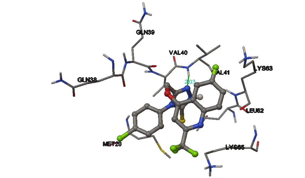
Fig. 2. 3-Dimensional representation of compound 5d against human interleukin 1beta target (9ILB)
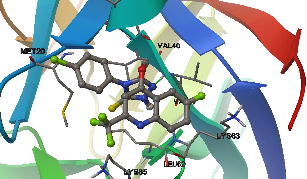
Fig.3. 3D-with secondary structure docking confirmation of compound 5d against human interleukin 1beta target (9ILB)
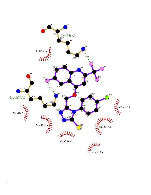
Fig.4. 2D-docking and Ligplot docking confirmation of compound 8d against human interleukin 1beta target (9ILB)
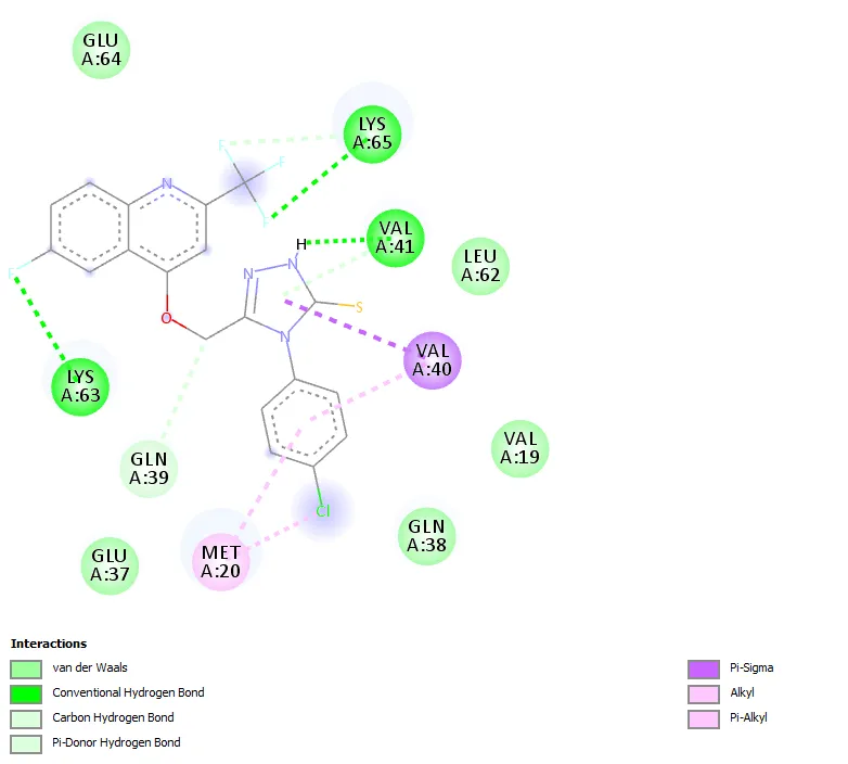
Fig.5. 2D-docking confirmation of ligand 5d against human interleukin 1beta target (9ILB)
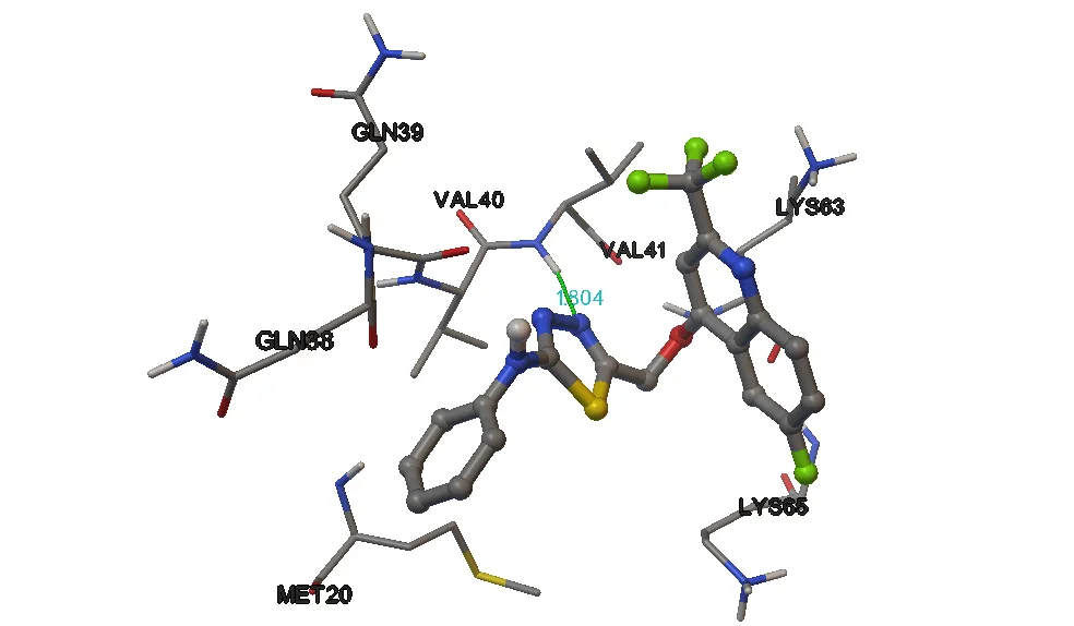
Fig.6. 3-Dimensional representation of compound 6a against human interleukin 1beta target (9ILB)
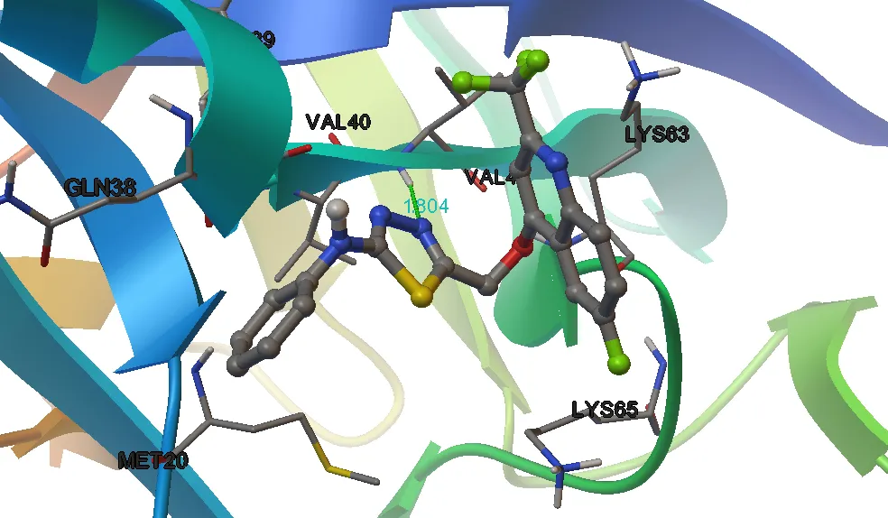
Fig.7. 3D-with secondary structure docking confirmation of compound 6a against human interleukin 1beta target (9ILB)
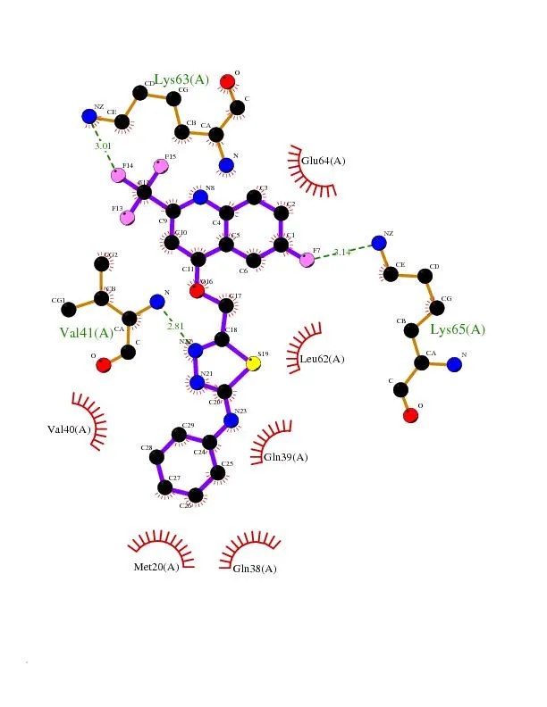
Fig.8. 2D-docking and Ligplot docking confirmation of compound 6a against human interleukin 1beta target (9ILB)
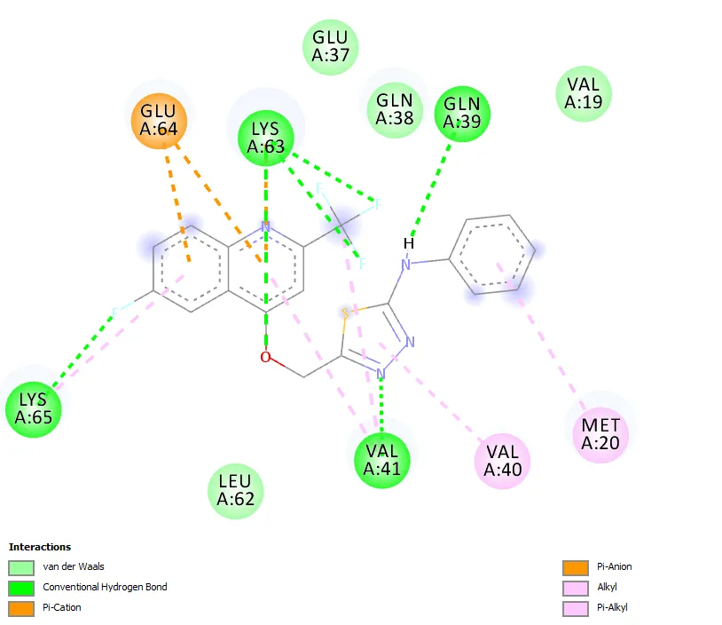
Fig.9. 2D-docking confirmation of ligand 6a against human interleukin 1beta target (9ILB)
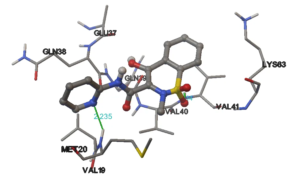
Fig.10. 3-Dimensional representation of compound Piroxicam against human interleukin 1beta target (9ILB)
.
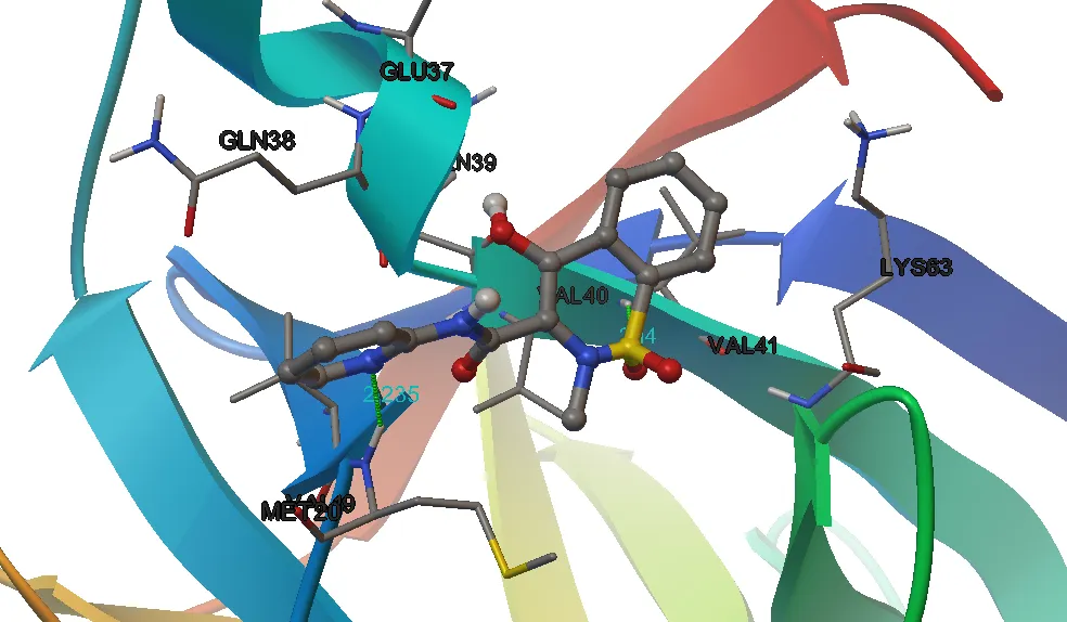
Fig.11. 3D-with secondary structure docking confirmation of compound Piroxicam against human interleukin 1beta target (9ILB)
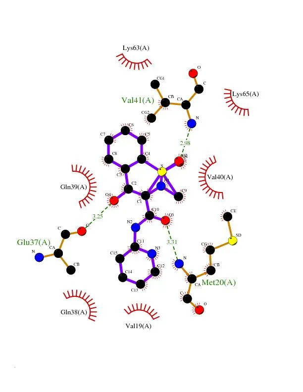
Fig.12. 2D-docking and Ligplot docking confirmation of compound Piroxicam against human interleukin 1beta target (9ILB)
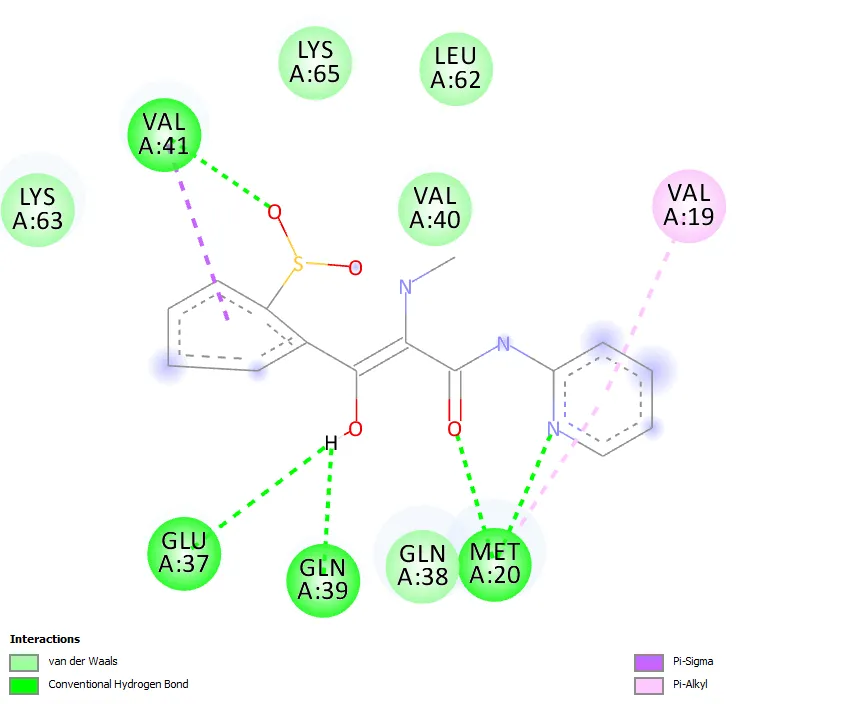
Fig.13. 2D-docking confirmation of ligand Piroxicam against human interleukin 1beta target (9ILB)
General procedure for the preparation of ethyl 2-((6-fluoro-2-(trifluoromethyl)quinolin-4-yl)oxy)acetate (2)
Charged Compound 6-fluoro-2-(trifluoromethyl)quinolin-4-ol 1, bromoethylacetate, K2CO3 and cat. amount of NaI in acetone. The reaction mixture was refluxed for 6-8 h and after cooling to room temperature the acetone was removed under vacuum. The residue was washed with n-hexane and then water was added to give a grey solid which was filtered with water and dried. Grey solid; Yield 71%; 1H NMR (CDCl3, 300 MHz): δ 1.27 t (3H, -CH3), 4.23 q (2H, -CH2), 4.68 s (2H, -OCH2), 7.10 s (1H, Ar-H), 7.44 dd (J=7.6 Hz, 1H, Ar-H), 7.62 dd (J=7.6 Hz, 1H, Ar-H), 8.05 m (1H, Ar-H); MS (ESI): m/z [(M+H)+]: Found, 318. Calcd, 317; Anal. calc. for C14H11F4NO3: C 53.00; H 3.49; N 4.42 %. Found: C 53.02; H 3.51; N 4.45%.
Preparation of 2-((6-fluoro-2-(trifluoromethyl)quinolin-4-yl)oxy)acetohydrazide (3)
Ethyl 2-((6-fluoro-2-(trifluoromethyl)quinolin-4-yl)oxy)acetate 2 on reaction hdrazine hydrate in the presence of ethanol refluxing condition for about 6-8 hours, and after confirmation of the product by TLC, reaction mixture was allowed to cool and obtained 2-((6-fluoro-2-(trifluoromethyl)quinolin-4-yl)oxy)acetohydrazide 3 by filtration. Grey solid; Yield 71%; 1H NMR (CDCl3, 300 MHz): δ 4.52 (br. s., 2H, -NH2), 4.72 s (2H, -OCH2), 7.12 (s, 1H, Ar-H), 7.49 (dd, J=7.9 Hz, 1H, Ar-H), 7.65 (d, J=7.9 Hz, 1H, Ar-H), 8.09 (m, 1H, Ar-H), 9.28 (br. s., 1H, -CONH-); MS (ESI): m/z [(M+H)+]: 304; Anal. calc. for C12H9F4N3O2: C 47.53, H 2.99, N 13.86 %. Found: C 47.55, H 2.98, N 13.88 %.
General procedure for the preparation of 2-(2-((6-fluoro-2-(trifluoromethyl)quinolin-4-yl)oxy)acetyl)-N-phenylhydrazinecarbothioamide (4a-d)
The 2-((6-fluoro-2-(trifluoromethyl)quinolin-4-yl)oxy)acetohydrazide 3 (2 mmol) was taken in 95% ethanol (20 ml) and phenylisothiocyanate (2 mmol) was added, the reaction mixture was refluxed for 5-6 hrs and after cooling to room temperature the ethanol was removed under vacuum. The residue was washed with n-hexane and it gives light yellow solid and dried to get 2-(2-((6-fluoro-2-(trifluoromethyl) quinolin-4-yl)oxy)acetyl)-N-phenyl hydrazine carbothioamide. Grey solid; Yield 81%; 3522 (-NHCO-), 3375, 3310 (-NH2), 1671 (-NHCO); 1H NMR (DMSO-d6, 300 MHz): δ 4.81 (s, 2H, -OCH2), 7.09 (s, 1H, Ar-H), 7.18 (dd, 1H, Ar-H), 7.26-7.32 (m, 3H, Ar-H), 7.41-7.45 (m, 2H, Ar-H), 7.60 (dd, 1H, Ar-H), 8.15 (m, 1H, Ar-H), 9.15 (br. s, 1H, -NH-), 9.90 (br. s, 1H, -NH-), 10.84 (br. S, 1H, -NH-); MS (ESI): m/z [(M+H)+]: 439; Anal. calc. for C19H14F4N4O2S: C 52.05, H 3.22, N 12.78 %. Found: C 52.08, H 3.24, N 12.80 %.
General procedure for the preparation of 3-(((6-fluoro-2-(trifluoromethyl)quinolin-4-yl)oxy)methyl)-4-phenyl-1H-1,2,4-triazole-5(4H)-thione derivatives (5a-d):
A solution of 2-(2-((6-fluoro-2-(trifluoromethyl)quinolin-4-yl) oxy) acetyl)-N-phenyl hydrazine carbothioamide 4 (0.01 mol) in 2N NaOH was allowed to reflux for 2h. The resulting solution was cooled to room temperature and acidified to pH 3-4 with 37% hydrochloric acid. The precipitate formed was filtered and washed with distilled water, dried and crystallized to furnish the compound 5a-d.
3-(((6-Fluoro-2-(trifluoromethyl)quinolin-4-yl)oxy)methyl)-4-phenyl-1H-1,2,4-triazole-5(4H)-thione (5a). Yellow solid; Yield 75%; M.Pt. 185-187 °C; 1H NMR (DMSO-d6, 300 MHz): δ 4.81 (s, 2H, -OCH2), 7.09 (s, 1H, Ar-H), 7.15 (dd, 1H, Ar-H), 7.26-7.32 (m, 3H, Ar-H), 7.41-7.45 (m, 2H, Ar-H), 7.67 (dd, 1H, Ar-H), 8.11 (m, 1H, Ar-H), 12.84 (br. s, 1H, -NHCS-); 13C NMR (DMSO-d6, 75 MHz): δ 78.4, 120.5, 122.4, 123.1, 124.7, 125.3, 126.5, 128.6, 129.7, 130.4, 133.5, 135.8, 136.7, 142.3, 148.3, 162.1, 165.4; MS (ESI): m/z [(M+H)+]: 421; Anal. calc. for C19H12F4N4OS: C 54.28, H 2.88, N 13.33 %. Found: C 54.30, H 2.86, N 13.35 %.
3-(((6-Fluoro-2-(trifluoromethyl)quinolin-4-yl)oxy)methyl)-4-(3-methoxyphenyl)-1H-1,2,4-triazole-5(4H)-thione (5b). Yellow solid; Yield 70 %; M.Pt. 195-197 °C; 1H NMR (DMSO-d6, 300 MHz): δ 3.82 (s, 3H, -OCH3), 4.83 (s, 2H, -OCH2), 7.10 (s, 1H, Ar-H), 7.19 (dd, 1H, Ar-H), 7.27 (s, 1H, Ar-H), 7.42-7.47 (m, 3H, Ar-H), 7.65 (dd, 1H, Ar-H), 8.08 (m, 1H, Ar-H), 12.81 (br. s, 1H, -NHCS-); 13C NMR (DMSO-d6, 75 MHz): δ 55.6, 78.2, 119.5, 121.4, 122.6, 123.5, 124.8, 125.6, 126.5, 127.4, 128.5, 129.8, 131.3, 133.6, 135.4, 136.8, 142.4, 148.3, 162.3, 165.5; MS (ESI): m/z [(M+H)+]: 451; Anal. calc. for C20H14F4N4O2S: C 53.33, H 3.13, N 12.44 %. Found: C 53.35, H 3.15, N 12.46 %.
3-(((6-fluoro-2-(trifluoromethyl)quinolin-4-yl)oxy)methyl)-4-(p-tolyl)-1H-1,2,4-triazole-5(4H)-thione (5c). Yellow solid; Yield 68 %; M.Pt. 201-203 °C; 1H NMR (DMSO-d6, 300 MHz): δ 2.34 (s, 3H, -CH3), 4.81 (s, 2H, -OCH2), 7.08 (s, 1H, Ar-H), 7.17 (dd, 1H, Ar-H), 7.29 (d, J= 8.0 Hz, 2H, Ar-H), 7.63 (dd, 1H, Ar-H), 7.74 (d, J= 8.0 Hz, 2H, Ar-H), 8.10 (m, 1H, Ar-H), 12.84 (br. s, 1H, -NHCS-); 13C NMR (DMSO-d6, 75 MHz): δ 21.6, 78.1, 120.4, 122.2, 123.6, 124.7, 125.4, 126.6, 127.3, 128.6, 129.7, 130.5, 133.4, 136.7, 142.3, 148.6, 162.4, 165.6; MS (ESI): m/z [(M+H)+]: 435; Anal. calc. for C20H14F4N4OS: C 55.30, H 3.25, N 12.90 %. Found: C 55.32, H 3.28, N 12.89 %.
4-(4-Chlorophenyl)-3-(((6-fluoro-2-(trifluoromethyl)quinolin-4-yl)oxy)methyl)-1H-1,2,4-triazole-5(4H)-thione (5d). Yellow solid; Yield 65 %; M.Pt. 178-180 °C; 1H NMR (DMSO-d6, 300 MHz): δ 4.84 (s, 2H, -OCH2), 7.06 (s, 1H, Ar-H), 7.18 (dd, 1H, Ar-H), 7.28 (d, J= 8.0 Hz, 2H, Ar-H), 7.65 (dd, 1H, Ar-H), 7.77 (d, J= 8.0 Hz, 2H, Ar-H), 8.12 (m, 1H, Ar-H), 12.82 (br. s, 1H, -NHCS-); 13C NMR (DMSO-d6, 75 MHz): δ 78.1, 120.6, 122.4, 123.5, 124.7, 125.6, 126.7, 127.6, 128.9, 129.8, 130.3, 133.5, 136.6, 142.3, 148.5, 162.2, 165.3; MS (ESI): m/z [(M+H)+]: 455; Anal. calc. for C19H11ClF4N4OS: C 50.17, H 2.44, N 12.32 %. Found: C 50.18, H 2.46, N 12.35 %.
General procedure for the preparation of 5-(((6-fluoro-2-(trifluoromethyl)quinolin-4-yl)oxy)methyl)-N-phenyl-1,3,4-thiadiazol-2-amine derivatives (6a-d)
A mixture of 0.001 mol of 2-(2-((6-fluoro-2-(trifluoromethyl)quinolin-4-yl) oxy) acetyl)-N-phenyl hydrazine carbothioamide (4a) and concentrated H2SO4 (1 mL) was stirred at room temperature for 1-2 h. Then the reaction mixture was poured over crushed ice. The precipitated solid was washed with sodium carbonate solution followed by water to afford compound .
5-(((6-Fluoro-2-(trifluoromethyl)quinolin-4-yl)oxy)methyl)-N-phenyl-1,3,4-thiadiazol-2-amine (6a). Light yellow solid; Yield 80 %; M.Pt. 155-157 °C; 1H NMR (DMSO-d6, 300 MHz): δ 4.91 (s, 2H, -OCH2), 7.09 (s, 1H, Ar-H), 7.11 (dd, 1H, Ar-H), 7.26-7.31 (m, 3H, Ar-H), 7.39-7.43 (m, 2H, Ar-H), 7.63 (dd, 1H, Ar-H), 8.16 (m, 1H, Ar-H), 10.16 (br. s, 1H, -NH-); 13C NMR (DMSO-d6, 75 MHz): δ 68.8, 121.3, 122.5, 123.3, 124.8, 125.4, 126.5, 128.9, 129.7, 130.4, 133.8, 135.3, 136.5, 142.3, 148.3, 150.2, 162.2; MS (ESI): m/z [(M+H)+]: 421; Anal. calc. for C19H12F4N4OS: C 54.28, H 2.88, N 13.33 %. Found: C 54.29, H 2.89, N 13.35 %.
5-(((6-fluoro-2-(trifluoromethyl)quinolin-4-yl)oxy)methyl)-N-(3-methoxyphenyl)-1,3,4-thiadiazol-2-amine (6b). Yellow solid; Yield 74 %; M.Pt. 163-165 °C; 1H NMR (DMSO-d6, 300 MHz): δ 3.82 (s, 3H, -OCH3), 4.89 (s, 2H, -OCH2), 7.09 (s, 1H, Ar-H), 7.18 (dd, 1H, Ar-H), 7.26 (s, 1H, Ar-H), 7.40-7.46 (m, 3H, Ar-H), 7.66 (dd, 1H, Ar-H), 8.12 (m, 1H, Ar-H), 12.85 (br. s, 1H, -NHCS-); 13C NMR (DMSO-d6, 75 MHz): δ 55.5, 69.2, 120.5, 121.6, 122.4, 123.6, 124.3, 125.7, 126.6, 127.5, 128.7, 129.7, 131.2, 133.4, 135.6, 136.9, 142.5, 148.3, 162.3, 165.7; MS (ESI): m/z [(M+H)+]: 451; Anal. calc. for C20H14F4N4O2S: C 53.33, H 3.13, N 12.44 %. Found: C 53.35, H 3.15, N 12.45 %.
5-(((6-fluoro-2-(trifluoromethyl)quinolin-4-yl)oxy)methyl)-N-(p-tolyl)-1,3,4-thiadiazol-2-amine (6c). Yellow solid; Yield 60 %; M.Pt. 181-183 °C; 1H NMR (DMSO-d6, 300 MHz): δ 2.35 (s, 3H, -CH3), 4.92 (s, 2H, -OCH2), 7.07 (s, 1H, Ar-H), 7.16 (dd, 1H, Ar-H), 7.28 (d, J= 8.0 Hz, 2H, Ar-H), 7.63 (dd, 1H, Ar-H), 7.77 (d, J= 8.0 Hz, 2H, Ar-H), 8.12 (m, 1H, Ar-H), 12.86 (br. s, 1H, -NHCS-); 13C NMR (DMSO-d6, 75 MHz): δ 21.7, 69.1, 120.3, 122.5, 123.7, 124.8, 125.5, 126.8, 127.4, 128.5, 129.7, 130.4, 133.5, 136.8, 142.4, 148.7, 162.6, 165.8; MS (ESI): m/z [(M+H)+]: 435; Anal. calc. for C20H14F4N4OS: C 55.30, H 3.25, N 12.90 %. Found: C 55.31, H 3.28, N 12.89 %.
N-(4-chlorophenyl)-5-(((6-fluoro-2-(trifluoromethyl)quinolin-4-yl)oxy)methyl)-1,3,4-thiadiazol-2-amine (6d). Yellow solid; Yield 62 %; M.Pt. 162-164 °C; 1H NMR (DMSO-d6, 300 MHz): δ 4.90 (s, 2H, -OCH2), 7.05 (s, 1H, Ar-H), 7.15 (dd, 1H, Ar-H), 7.26 (d, J= 8.0 Hz, 2H, Ar-H), 7.66 (dd, 1H, Ar-H), 7.77 (d, J= 8.0 Hz, 2H, Ar-H), 8.12 (m, 1H, Ar-H), 12.81 (br. s, 1H, -NHCS-); 13C NMR (DMSO-d6, 75 MHz): δ 68.9, 120.5, 122.6, 123.5, 124.7, 125.5, 126.8, 127.6, 128.9, 129.6, 130.5, 132.4, 136.7, 142.6, 148.4, 162.3, 165.4; MS (ESI): m/z [(M+H)+]: 455; Anal. calc. for C19H11ClF4N4OS: C 50.17, H 2.44, N 12.32 %. Found: C 50.19, H 2.45, N 12.34 %.
General procedure for the preparation of 5-(((6-fluoro-2-(trifluoromethyl)quinolin-4-yl)oxy)methyl)-N3-phenyl-4H-1,2,4-triazole-3,4-diamine (7a-d)
Charged compound 2-(2-((6-fluoro-2-(trifluoromethyl)quinolin-4-yl) oxy) acetyl)-N-phenyl hydrazine carbothioamide (4a) (0.01 mol) with N2H4.H2O (0.01 mol) in MeOH (1 mL). The solution was refluxed for 5–6 h. After cooling to room temperature, ice water (10 mL) was added to the reaction mixture, which was then neutralized with 3N HCl to form a precipitate. The precipitate was isolated by filtration to afford the triazole derivatives 7a-d.
5-(((6-fluoro-2-(trifluoromethyl)quinolin-4-yl)oxy)methyl)-N3-phenyl-4H-1,2,4-triazole-3,4-diamine (7a). Light cream solid; Yield 82 %; M.Pt. 179-181 °C; 1H NMR (DMSO-d6, 300 MHz): δ 4.90 (s, 2H, -OCH2), 5.19 (br. s, 2H, -NH2), 6.99 (br. s, 1H, -NH-), 7.12 (s, 1H, Ar-H), 7.25 (dd, 1H, Ar-H), 7.30-7.36 (m, 3H, Ar-H), 7.45-7.48 (m, 2H, Ar-H), 7.66 (dd, 1H, Ar-H), 8.11 (m, 1H, Ar-H); 13C NMR (DMSO-d6, 75 MHz): δ 71.2, 120.3, 121.5, 123.4, 124.7, 125.5, 126.6, 128.6, 129.8, 130.5, 133.7, 136.5, 142.3, 148.3, 151.2, 156.4, 162.1; MS (ESI): m/z [(M+H)+]: 419; Anal. calc. for C19H14F4N6O: C 54.55, H 3.37, N 20.09 %. Found: C 54.57, H 3.39, N 20.11 %.
5-(((6-fluoro-2-(trifluoromethyl)quinolin-4-yl)oxy)methyl)-N3-(3-methoxyphenyl)-4H-1,2,4-triazole-3,4-diamine (7b). Light Yellow solid; Yield 69 %; M.Pt. 181-183 °C; 1H NMR (DMSO-d6, 300 MHz): δ 3.83 (s, 3H, -OCH3), 4.91 (s, 2H, -OCH2), 5.20 (br. s, 2H, -NH2), 7.01 (br. s, 1H, -NH-), 7.15 (s, 1H, Ar-H), 7.26 (dd, 1H, Ar-H), 7.32 (s, 1H, Ar-H), 7.42-7.48 (m, 3H, Ar-H), 7.69 (dd, 1H, Ar-H), 8.13 (m, 1H, Ar-H); 13C NMR (DMSO-d6, 75 MHz): δ 55.6, 71.3, 119.8, 120.6, 122.1, 123.7, 124.6, 125.6, 126.7, 127.8, 128.9, 129.7, 131.2, 133.4, 135.6, 142.5, 148.3, 152.1, 155.6, 162.3,; MS (ESI): m/z [(M+H)+]: 449; Anal. calc. for C20H16F4N6O2: C 53.57, H 3.60, N 18.74 %. Found: C 53.59, H 3.62, N 18.76 %.
5-(((6-fluoro-2-(trifluoromethyl)quinolin-4-yl)oxy)methyl)-N3-(p-tolyl)-4H-1,2,4-triazole-3,4-diamine (7c). Yellow solid; Yield 62 %; M.Pt. 171-173 °C; 1H NMR (DMSO-d6, 300 MHz): δ 2.36 (s, 3H, -CH3), 4.92 (s, 2H, -OCH2), 5.21 (br. s, 2H, -NH2), 7.03 (br. s, 1H, -NH-), 7.15 (s, 1H, Ar-H), 7.28 (dd, 1H, Ar-H), 7.36 (d, J= 8.0 Hz, 2H, Ar-H), 7.65 (dd, 1H, Ar-H), 7.78 (d, J= 8.0 Hz, 2H, Ar-H), 8.14 (m, 1H, Ar-H); 13C NMR (DMSO-d6, 75 MHz): δ 21.5, 71.5, 120.4, 122.6, 123.8, 124.6, 125.4, 126.7, 127.5, 128.6, 129.8, 130.5, 133.6, 136.7, 140.5, 142.5, 148.8, 152.4, 155.6, 162.6; MS (ESI): m/z [(M+H)+]: 433; Anal. calc. for C20H16F4N6O: C 55.56, H 3.73, N 19.44 %. Found: C 55.58, H 3.75, N 19.46 %.
N3-(4-chlorophenyl)-5-(((6-fluoro-2-(trifluoromethyl)quinolin-4-yl)oxy)methyl)-4H-1,2,4-triazole-3,4-diamine (7d). Yellow solid; Yield 673%; M.Pt. 192-194 °C; 1H NMR (DMSO-d6, 300 MHz): δ 4.93 (s, 2H, -OCH2), 5.21 (br. s, 2H, -NH2), 7.03 (br. s, 1H, -NH-), 7.16 (s, 1H, Ar-H), 7.25 (dd, 1H, Ar-H), 7.35 (d, J= 8.0 Hz, 2H, Ar-H), 7.64 (dd, 1H, Ar-H), 7.79 (d, J= 8.0 Hz, 2H, Ar-H), 8.10 (m, 1H, Ar-H); 13C NMR (DMSO-d6, 75 MHz): δ 71.5, 120.4, 122.5, 123.7, 124.6, 125.6, 126.7, 127.8, 128.5, 129.3, 130.6, 132.5, 136.7, 142.6, 148.4, 151.4, 155.8, 162.3; MS (ESI): m/z [(M+H)+]: 453; Anal. calc. for C19H13ClF4N6O: C 50.40, H 2.89, N 18.56 %. Found: C 50.42, H 2.91, N 18.58 %.
CONCLUSION
In our research program we designed and synthesized novel triazolothione, thiadiazole, triazole functionalized quinoline derivatives. The structures of all new compounds were confirmed by comprehensive spectroscopic analysis (IR, 1H, 13C NMR, and ESI-MS). All the products were screened against anticancer activity on four human cancer cell lines ''HeLa (cervical cancer, CCL-2), COLO 205 (colon cancer, CCL-222), HepG2 (liver cancer, HB-8065), and MCF7 (breast cancer, HTB-22)'', using the MTT assay promising compounds identified. All the products were screened anti-inflammatory activity, among the compounds tested, Some of the compounds exhibited significant inhibition of IL-1b secretion as a measure of anti-inflammatory activity.
REFERENCES


Chakravarthy Bhupathi, Raghu Dasari, Kavitha Tula, Shravan Kumar Gunda, Namratha Vaddiraju, Construction of Novel Triazolothione, Thiadiazole, Triazole Functionalized Quinoline Derivatives; Their Biological Evolutions and Docking Interactions, Int. J. of Pharm. Sci., 2026, Vol 4, Issue 7, 4132-4147, https://doi.org/10.5281/zenodo.21470978
 10.5281/zenodo.21470978