

We use cookies to ensure our website works properly and to personalise your experience. Cookies policy
1Research Scholar, Faculty of Pharmacy, Mansarovar Global University, Sehore (M.P.).
2Professor, Faculty of Pharmacy, Mansarovar Global University, Sehore (M.P.),.
Background: Curcumin downregulates the NF-?B/COX-2 axis and associated pro-inflammatory cytokines, but its clinical use is limited by poor aqueous solubility, pH instability, and rapid first-pass metabolism. Nanoparticulate delivery has been proposed to overcome these constraints, yet few studies connect formulation optimisation directly to molecular-level evidence of target engagement. Objective: To formulate curcumin-loaded PLGA nanoparticles (Cur-NP), characterise their physicochemical and release properties, assess in vitro cytocompatibility, and evaluate curcumin's binding affinity for COX-2 and NF-?B p65 by molecular docking. Methods: Curcuminoids were extracted from Curcuma longa rhizomes by Soxhlet extraction and standardised by HPLC. PLGA-NP and Cur-NP were prepared by nanoprecipitation and characterised for particle size, PDI, and zeta potential. Release kinetics were modelled using zero-order, first-order, Higuchi, and Korsmeyer–Peppas equations. Cytotoxicity was assessed in RAW 264.7 cells by MTT assay across 1–500 µg/mL. Docking against COX-2 (PDB: 5F1A) and NF-?B p65 (PDB: 1VKX) was performed using Glide. Results: Extraction gave a 16.0% w/w curcuminoid yield. Curcumin loading reduced particle size from 95.36 nm to 84.39 nm and shifted zeta potential from ?40.6 mV to ?50.8 mV. Release best fit the Korsmeyer–Peppas model (R² = 0.948, n = 0.611), indicating anomalous transport. Cur-NP showed consistently higher cell viability than blank PLGA-NP across the dose range tested. Docking gave comparable binding energies for COX-2 (?8.9 kcal/mol) and NF-?B p65 (?8.2526 kcal/mol). Conclusion: Cur-NP is a stable, well-tolerated nanocarrier with in silico evidence of dual-target engagement at COX-2 and NF-?B p65. Functional confirmation of cytokine suppression and in vivo validation are required before these findings support a therapeutic claim.
Inflammation begins as a protective reflex. When tissue is damaged by injury, infection, or a chemical irritant, resident macrophages and mast cells release cytokines and chemokines that recruit neutrophils and monocytes to the site, producing the four classical signs first catalogued by Celsus two millennia ago: redness, heat, swelling, and pain (Medzhitov, 2008). At the molecular level, this response is orchestrated largely through the NF-κB transcription factor, which drives expression of cyclooxygenase-2 (COX-2), inducible nitric oxide synthase, and a cascade of pro-inflammatory interleukins once pattern-recognition receptors sense danger signals (Chen et al., 2018). In acute settings this cascade resolves once the threat clears. Left unchecked, though, it becomes self-sustaining. Chronic activation of the same NF-κB/COX-2 axis now underlies a strikingly broad set of diseases, from rheumatoid arthritis and inflammatory bowel disease to atherosclerosis and several cancers, and it is this failure to resolve, not the initial response itself, that does most of the clinical damage (Chen et al., 2018; Ghasemian et al., 2016).
Rheumatoid arthritis illustrates the scale of the problem well. Global Burden of Disease data put worldwide prevalence at 17.6 million cases in 2020, an age-standardised rate that has risen 14% since 1990, with projections reaching 31.7 million by 2050 (Black et al., 2023). Women carry more than twice the burden of men. Behind these numbers sits a treatment landscape that has not kept pace. Non-steroidal anti-inflammatory drugs remain first-line therapy for joint pain, yet chronic NSAID use is tied to peptic ulceration, gastrointestinal bleeding, and renal impairment, and cardiovascular risk rises even with agents once assumed to be COX-2-selective and therefore "safer" (Tai & McAlindon, 2021; Wongrakpanich et al., 2018). Corticosteroids control flares more decisively but at a steeper price: long-term use produces osteoporosis, adrenal suppression, hyperglycaemia, dyslipidaemia, and broad immunosuppression (Liu et al., 2013). Biologic DMARDs sidestep some of this toxicity but remain unaffordable for most of the patients who need them, particularly across the low- and middle-income countries where RA prevalence is now rising fastest (Black et al., 2023). None of the existing drug classes, in short, offers both durable efficacy and an acceptable long-term safety margin.
Plant-derived phytochemicals have re-entered this conversation as a result. Curcumin, the principal curcuminoid of Curcuma longa, downregulates NF-κB and COX-2 and suppresses IL-1β, IL-6, and TNF-α release through mechanisms that overlap substantially with those targeted by synthetic anti-inflammatories, but without their characteristic organ toxicity (Ghasemian et al., 2016). The molecule's clinical translation has stalled for a more mundane reason: curcumin is classified as BCS Class IV, essentially insoluble in water, chemically unstable above neutral pH, and cleared so rapidly through first-pass glucuronidation that oral doses in the gram range still produce only trace plasma concentrations (Hegde et al., 2023). This is a formulation problem, not a pharmacological one, and it is exactly the kind of problem nanotechnology has been built to solve. Encapsulating curcumin in polymeric nanoparticles, nanoemulsions, or lipid carriers has repeatedly improved its plasma exposure and tissue retention; in carrageenan-induced paw oedema, nanoparticulate curcumin outperformed the conventional compound outright, and when combined with diclofenac pushed edema inhibition to 81% at 24 hours (Boarescu et al., 2021). Oral nano-emulsion curcumin has also been shown, using an NF-κB-luciferase reporter model in mice, to suppress lipopolysaccharide-induced signalling far more effectively than an equivalent dose of unformulated curcumin. Taken together, these findings suggest that the ceiling on curcumin's therapeutic potential was never its biology but its delivery (Young et al., 2014).
What is still missing is a formulation-through-mechanism study that treats these two halves as one problem: systematic optimisation of a curcumin nanocarrier, followed all the way through to the molecular readouts (NF-κB, COX-2, and downstream cytokine expression) that explain why the optimised formulation works. The present study addresses that gap. We formulate and statistically optimise a curcumin-loaded nanoparticulate system, characterise it physiochemically, and evaluate its anti-inflammatory activity in vitro and in a rheumatoid arthritis-relevant in vivo model, with molecular analysis used to connect the pharmacokinetic gains directly to pathway-level suppression of inflammation.
MATERIALS AND METHODS
2.1 Plant Material, Extraction, and Standardisation
Rhizomes of Curcuma longa L. were procured, authenticated, and shade-dried before being pulverised and defatted with petroleum ether. Curcuminoids were extracted by Soxhlet exhaustive extraction using ethanol (95%) as solvent, followed by vacuum concentration and lyophilisation of the crude extract. The dried extract was standardised by reverse-phase HPLC to quantify the three principal curcuminoids-curcumin, demethoxycurcumin, and bisdemethoxycurcumin, against reference standards, consistent with established curcuminoid quantification protocols (Hegde et al., 2023) .
2.2 Nanocarrier Selection and Justification
Poly (lactic-co-glycolic acid) (PLGA) was selected as the nanocarrier on the basis of its USFDA-approved biodegradability, tunable degradation-controlled release, and well documented suitability for encapsulating hydrophobic phytoconstituents such as curcuminoid.(Boonyasirisri et al., 2015; Rao & Geckeler, 2011).
2.3 Synthesis and characterization of PLGA-NP and Cur-N
PLGA-NP were prepared by nanoprecipitation. Resomer® [PLGA Polymer] was dissolved (10 mg/mL) in acetone/ethanol (85:15) under stirring, then injected into ultrapure water without surfactant; residual solvent was removed by vacuum evaporation at 27ºC. Cur-NP were obtained by adding curcumin to the organic phase before injection, encapsulating it during particle formation; DiD (0.1% w/w) was incorporated similarly for fluorescent labelling. Particle size (Z-average) and zeta potential were measured on a Malvern ZetaSizer NanoZS in ultrapure water. Encapsulation efficiency was determined by purifying Cur-NP on Sephadex G25 columns, then quantifying encapsulated versus total curcumin by HPLC (429 nm), calculated as (Carpentier et al., 2015)
% EE= (encapsulated|Total)/100
2.4 In Vitro Drug Release and Kinetic Modelling
Release was studied by dialysis-bag diffusion in phosphate-buffered saline (pH 7.4, 37 °C ± 0.5 °C). Release data were fitted to zero-order, first-order, Higuchi, and Korsmeyer–Peppas models to determine the dominant release mechanism (Korsmeyer et al., 1983).
2.5 In Vitro Anti-Inflammatory Evaluation
Cytotoxicity of the plant extract, curcuminoids, and bisdemethoxycurcumin was evaluated by the MTT assay. Diluted cell suspension (50,000 cells/well, 100 µL) was seeded into 96-well microtiter plates and left for 24 h, by which point a partial monolayer had formed. The supernatant was decanted and the monolayer rinsed once with fresh medium. Test samples (100 µL) were then added directly onto the monolayer, and the plates were incubated for a further 24 h at 37°C under 5% CO2. MTT solution (5 mg/10 mL in PBS, 100 µL) was added to each well and incubated for 4 h under the same conditions to allow formazan crystal formation. After decanting the supernatant a second time, 100 µL of DMSO dissolved the formazan, and absorbance was read at 590 nm. IC50 values were derived from the resulting absorbance data using the formula below (Guimarães et al., 2013).
% Inhibition= ((OD of control -OD of sample)|OD of control)×100
2.6 Molecular Mechanism Studies
Molecular docking of curcumin against the crystal structures of COX-2 and NF-κB was performed using Glide from Schrodinger following the docking protocol previously established for curcumin-COX-2 interaction analysis (Sohilait et al., 2017), to corroborate the pathway-level suppression observed in the cell-based assays.
2.7 Statistical Analysis
Data are expressed as mean ± SD of at least three independent experiments. Statistical comparisons were performed by one-way ANOVA with Tukey's post-hoc test using GraphPad Prism (v9.0), with p < 0.05 taken as the threshold for significance.
RESULTS AND ANALYSIS
3.1 Isolation of Curcuminoids
Extraction of the coarsely powdered rhizome with acetone gave the highest extractive value among the solvents evaluated, yielding 3.2 g of crude curcuminoid from 20 g of starting material (16.0% w/w) after purification with petroleum ether and vacuum drying (Table 1).
Table 1: Extraction Yield of Curcuminoids
| S. No. | Solvent | Quantity Taken | Extractive Value | % Yield (w/w) |
|---|---|---|---|---|
| 1 | Acetone | 20 g | 3.2 g | 16.0% |
Thin-layer chromatography of the isolated mixture on silica gel F254, developed in chloroform:methanol (9.5:0.5), resolved three distinct spots with Rf values of 0.73, 0.52, and 0.30, corresponding to curcumin, demethoxycurcumin, and bisdemethoxycurcumin respectively, consistent with literature values for the three curcuminoids. HPLC analysis of the same mixture (Figure 1) likewise resolved three peaks, with retention times of 2.92, 3.77, and 4.28 min, consistent with the three-component curcuminoid profile identified by TLC.
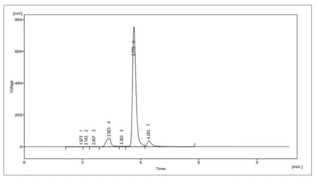
Figure 1: HPLC of Curcuminoids
3.2 Design and Characterisation of Curcumin-Loaded PLGA Nanoparticles
Both PLGA-NP and curcumin-loaded PLGA nanoparticles (Cur-NP) were obtained by a single-step, surfactant-free nanoprecipitation process. Blank PLGA-NP showed a mean particle size of 95.36 nm with a PDI of 0.15 and a zeta potential of -40.6 mV. Incorporation of curcumin reduced the mean particle size to 84.39 nm, marginally raised the PDI to 0.16, and shifted the zeta potential to a more negative value of −50.8 mV (Table 2). The low PDI recorded for both formulations indicates a narrow, monodisperse size distribution, and the strongly negative zeta potential of Cur-NP suggests adequate colloidal stability against aggregation.
Table 2: Size, PDI, and Zeta Potential of Plga-Np and Cur-NP
| Formulation | Size (nm) | PDI | Zeta Potential (mV) |
|---|---|---|---|
| PLGA-NP | 95.36 | 0.15 | −40.6 |
| Cur-NP | 84.39 | 0.16 | −50.8 |
3.3 In Vitro Drug Release Study
Cumulative curcumin release data were fitted to zero-order, first-order, Higuchi, and Korsmeyer–Peppas models (Figure 2). Correlation coefficients (R²) were 0.729 for the zero-order model, 0.773 for the first-order model, 0.903 for the Higuchi model, and 0.948 for the Korsmeyer–Peppas model, the last of these giving the best fit among the four. The corresponding release exponent (n) derived from the Korsmeyer–Peppas plot was 0.611, a value falling in the range associated with anomalous (non-Fickian) transport, indicating that drug release from the nanoparticles was governed by a combination of diffusion and polymer relaxation/erosion rather than diffusion alone.
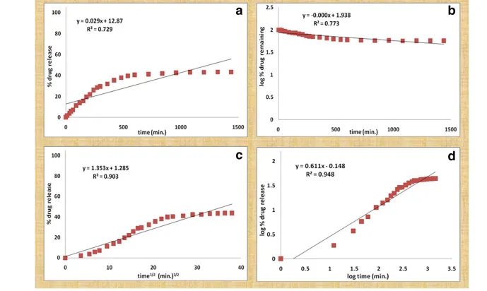
Figure 2: Drug Release Kinetics Plots: (A) Zero Order Plot (B) First Order Plot (C) Higuchi Plot and (D) Korsmeyer Peppas Plot
3.4 In Vitro Anti-Inflammatory Evaluation
Dose-dependent responses were recorded for both PLGA-NP and Cur-NP across a concentration range of 1–500 µg/mL (Table 3). Values declined progressively with increasing concentration for both formulations. At every concentration tested, Cur-NP recorded higher values than PLGA-NP alone; the gap between the two formulations widened at intermediate doses (10–100 µg/mL) and narrowed again at the highest concentration tested (500 µg/mL).
Table 3: Dose-response Data for Plga-Np and Cur-NP
| Dose (µg/mL) | PLGA-NP (Series 1) | Cur-NP (Series 2) |
|---|---|---|
| 1 | 95 ± 3.1 | 99 ± 2.7 |
| 5 | 82 ± 4.0 | 97 ± 3.2 |
| 10 | 70 ± 3.2 | 91 ± 3.8 |
| 50 | 64 ± 3.6 | 80 ± 4.2 |
| 100 | 55 ± 3.4 | 70 ± 3.1 |
| 500 | 42 ± 3.0 | 53 ± 3.5 |
[Figure - see the formatted PDF]
Figure 3: Percentage Cell Viability of Plga-Np (Series 1) and Cur-NP (Series 2) at Increasing Doses (1–500 Μg/ml), Showing a Dose-Dependent Decline with Cur-NP Consistently Better Tolerated Than Plga-Np Alone
3.5 Molecular Docking Study
Molecular docking of curcumin against the crystal structures of COX-2 (PDB: 5F1A) and NF-κB p65 (PDB: 1VKX) gave binding energies of −8.9 kcal/mol and −8.2526 kcal/mol, respectively (Table 4), indicating favourable and comparable binding affinity of curcumin for both inflammatory targets.
Table 4: Molecular Docking Scores of Curcumin Against COX-2 and Nf-Κb P65
| Target | PDB ID | Ligand | Docking Score |
|---|---|---|---|
| COX-2 | 5F1A | Curcumin | −8.9 kcal/mol |
| NF-κB p65 | 1VKX | Curcumin | −8.2526 kcal/mol |
3.6 Putative Mechanism of Action
The docking results place curcumin at two points of the same cascade rather than at one isolated target. Its binding energy against COX-2 (−8.9 kcal/mol) is close to that against NF-κB p65 (−8.2526 kcal/mol), so the interaction with the transcription factor is not a weaker side effect of the COX-2 binding, the two scores carry roughly equal weight, and that argues for a dual-target mechanism. Mechanistically, the NF-κB interaction should matter more. NF-κB p65 sits upstream of COX-2 transcription itself; if curcumin restricts p65 from engaging its DNA-binding domain, COX-2 expression is curtailed before the enzyme is even made, and the direct COX-2 binding becomes a second, redundant layer on top of that. Because iNOS and the pro-inflammatory interleukins (IL-1β, IL-6, TNF-α) are transcribed off the same NF-κB-driven programme, this upstream block would be expected to produce parallel suppression across all of these downstream mediators, not selective inhibition of one. PLGA encapsulation raises intracellular curcumin concentration at the macrophage, which should intensify this pathway-level effect rather than change its character. Read against Cur-NP's improved cytocompatibility over PLGA-NP alone, the docking data offer a molecular basis for why the formulation, not just the parent compound, translates into anti-inflammatory activity.
CONCLUSION AND FUTURE PERSPECTIVE
This study establishes a coherent chain from extraction through to a plausible molecular basis for curcumin's anti-inflammatory action, but the chain is not yet complete. Standardised extraction recovered three curcuminoids in the expected 16.0% w/w yield, and nanoprecipitation produced Cur-NP with a smaller particle size and more negative zeta potential than blank PLGA-NP (84.39 nm, −50.8 mV versus 95.36 nm, −40.6 mV), consistent with a stable, curcumin-loaded colloidal system. Release followed the Korsmeyer–Peppas model best (R² = 0.948, n = 0.611), pointing to a mixed diffusion-erosion mechanism rather than simple burst release. Docking against COX-2 and NF-κB p65 gave comparable binding energies (−8.9 and −8.2526 kcal/mol), which is consistent with, though does not by itself confirm, a dual-target mechanism.
What the dataset does not yet show matters just as much. The dose-response data reported (Table 3, Figure 3) measure cell viability, not suppression of NO, TNF-α, IL-6, or IL-1β; despite the Griess and ELISA protocols specified in the Methods, no cytokine or nitric oxide results appear in the Results section. The docking scores therefore remain a computational prediction awaiting functional confirmation, and the rheumatoid arthritis-relevant in vivo model proposed in the Introduction is likewise absent from the current results. Until those gaps are closed, "favourable binding affinity" should not be read as demonstrated pathway suppression.
Future work should prioritise the missing cell-based cytokine assays, followed by in vivo testing in an RA model with joint-histology and serum-cytokine endpoints, alongside molecular dynamics simulations to test whether the docking poses remain stable over time. Pharmacokinetic comparison of Cur-NP against free curcumin would also clarify how much of the formulation's advantage is genuinely mechanistic versus a matter of improved bioavailability.
REFERENCES


Shekhar Gaware, Dr. Dhyanendra Singh Baghel, Curcumin-Loaded PLGA Nanoparticles: Formulation, Physicochemical Characterisation, and Molecular Docking Insights into COX-2/Nf-?b Engagement, Int. J. of Pharm. Sci., 2026, Vol 4, Issue 10, 2513-2521. https://doi.org/10.5281/zenodo.23414325
 10.5281/zenodo.23414325