Fucoidan is a sulphated marine polysaccharide that comes from brown algae and has a variety of biological properties, such as immunomodulatory, antioxidant, and anticancer properties. Recent developments in nanotechnology have made it possible to create hybrid nanocarriers that combine fucoidan with polymers and herbal bio-actives to increase the effectiveness of medication delivery. The medicinal use of herbal substances like curcumin, quercetin, and Asiatic acid is limited by their low solubility and bioavailability. Hybrid nanocarriers based on fucoidan provide improved stability, controlled release, and targeted delivery. The structural characteristics of fucoidan, hybrid nanocarrier types, drug transport methods, and therapeutic uses are all summarized in this paper. The necessity of clinical translation is further highlighted by the discussion of difficulties and future prospects.
Keywords
Fucoidan, Sulphated polysaccharide, Brown algae, Hybrid nanocarriers, Drug delivery, Herbal bioactive.
Introduction
×
Fucoidan is a fucose-rich sulphated polysaccharide primarily obtained from brown seaweeds such as Fucus, Laminaria, and Sargassum. It exhibits a wide range of biological activities including anticoagulant, antiviral, anticancer, and anti-inflammatory effects. The first extraction of fucoidan was in 1913 from a species of brown algae [1] such as Laminaria digitata, Ascophyllum nodosum and Fucus vesiculosus. Fucoidan is a negatively charged and highly hygroscopic polysaccharide [2]. A high content of fucoidan is mainly found in the leaves of L. digitata, A. nodosum, Macrocystis porifera and F. vesiculosus. Fucoidan is soluble in both water and acid solutions. After the first publication took place in 1913, the number of published articles (studies) on fucoidan has increased significantly, especially in the modern era. The reason behind the increase in studies is that fucoidan has anti-tumour, anti-coagulant and anti-oxidant activities, as well as the importance in terms of regulating the metabolism of glucose and cholesterol [3]. Also, there has been an interest in fucoidan because of its potential to provide protection against liver damage and urinary system failures. It is evident that research on fucoidan is gradually flourishing, as these activities are carried out, and more of its bio-activities and health-related benefits are being discovered as studies continue to accumulate.
Sources Of Fucoidan
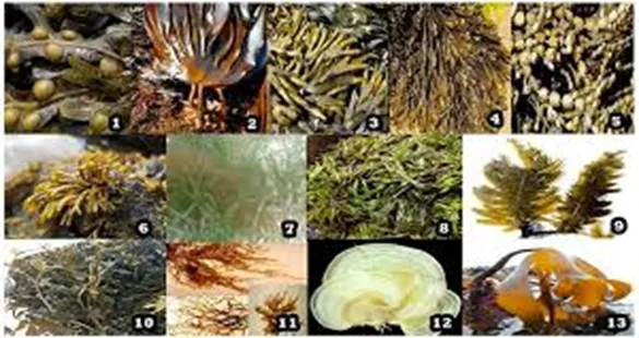
Fucoidan is a sulphated polysaccharide which can be found amongst a number of marine sources, including sea cucumbers [4] or brown algae [5]. A great number of algae and invertebrates have been ascertained for their fucoidan contents inclusive of Fucus vesiculosus, Sargassum stenophyllum, Chorda filum, Ascophyllum nodosum, Dictyota menstrual is, Fucus evanescens, Fucus serratus, Fucus distichus, Caulerpa racemosa, Hizikia fusiforme, Padina gymnospora, Kjellmaniella crassifolia, Analipus japonicus and Laminaria hyperborea exhibited in Figure 1. In these sources, different types of fucoidan can be obtained and the methods of extraction employed are different, especially when they are reported in different studies.
However, herbal bioactives (phytochemicals) such as curcumin and asiatic acid suffer from:
Low aqueous solubility
Poor bioavailability
Rapid metabolism
Nanotechnology-based delivery systems have emerged as a promising strategy to overcome these limitations. Hybrid nanocarriers combining fucoidan with polymers or lipids provide enhanced targeting and controlled release properties.
Structure And Properties Of Fucoidan
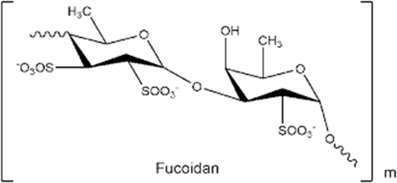
Fucoidan is known as a fucose-enriched and sulphated polysaccharide that is mainly sourced from the extracellular matrix of brown algae. Fucoidan is made up of L-fucose, sulphate groups and one or more small proportions of xylose, mannose, galactose, rhamnose, arabinose, glucose, glucuronic acid and acetyl groups in a variety of brown algae [6-8]. In a number of studies, researchers have also used galactofucan to represent a kind of fucoidan. Galactofucan is known as a monosaccharide, and the composition of the monosaccharide is galactose accompanied by fucose, similar to rhamnofucan (rhamnose and fucose) and rhamnoalactofucan (rhamnose, galactose and fucose). In addition to the structure of fucoidan, there is also a variation amongst different seaweed types. Nevertheless, fucoidan normally has two types of homos fucose One type (I) encompasses repeated (1→3)-L-fucopyranose, and the other type (II) encompasses alternating and repeated (1→3)- and (1→4)-L-fucopyranose [9]. Reports based on structures of fucoidan, sourced from different species of brown algae, brought about an improved categorization in terms of structures. By a way of illustration, most of the fucoidans sourced from species belonging to the Fucales have an alternating linkage of (1→3)-α-L-fucose and (1→4)-α-L-fucose [10-14]. Structures of Ascophyllum nodosum fucoidan [15] and F. vesiculosus fucoidan show a resemblance of one another, the difference is only significant based on sulfate patterns and the presence of glucuronic acid. A number of Fucales species, such as Fucus serratus, Fucus distichus and Pelvetia canaliculate, present similar fucoidan backbone, but show more diversity in the branching and the presence of different monosaccharides [13,14,16]. However, exceptions do exist, for instance, fucoidans from the Bifurcaria bifucardia and Himanthalia elongate do not follow or ascribe to such a structural feature [17]. Hence, identifying the structure of fucoidan based on the specie.
Fig. 2. Chemical Structure of Fucoidan
Another important fact that deserves mentioning is that the structure of fucoidan is also highly dependent on the harvest season. This is based on the Undaria pinnafida fucoidan, which exhibited distinct characteristics and bioactivity, especially when harvested during different seasons [18,19]. In addition, it should be indicated that the purification method also plays a critical role in the structure of fucoidan. To such an extent that new purification methods have led to the revelation that the fucoidan structure is comprised of multiple fractions [20]. An investigation reported that the structure of crude fucoidan sourced from A. nodosum showed a predominant repetition of [→(3)-α-L-Fuc(2SO3−) − (1→4)-α-Fuc(2,3diSO3−)-(1)]n [21]. However, from the same species, a purified fraction comprised of primarily α-(1→3)-fructosyl residues with a sparse linkage of α-(1→4) and was found to be highly branched [22]. Therefore, the employment of different extraction methods results in distinct structures. For example, a report states that one species produced two distinct fucoidan structures, particularly galactofucan and uronofucoidans [23]. Hence, it should be emphasized that purification techniques are one of the determining factors towards the structure and the associated bioactivities. [ 24,25,26]
Fucoidan is structurally complex and varies depending on its source. It contains: [27-31]
L-fucose backbone
Sulfate ester groups
Variable molecular weight
These sulfate groups provide:
Negative charge
Ability to form polyelectrolyte complexes
Fucoidan can interact with positively charged polymers like chitosan, forming stable nanoparticles
Extraction And Production
Source [32-36]
Fucoidan is mainly obtained from brown marine algae (brown seaweeds). Important natural sources include:
Fucus vesiculosus
Undaria pinnatifida
Sargassum species
These seaweeds are rich in sulfated polysaccharides and are widely used for commercial fucoidan production.
Extraction Methods
Fucoidan is extracted from seaweed biomass through several conventional and advanced techniques:
Hot Water Extraction Seaweed powder is treated with hot water to dissolve fucoidan and separate it from other components.
Acid Extraction Mild acidic conditions help improve the release of fucoidan from the algal cell wall.
Alkaline Extraction Alkali treatment is sometimes used to remove impurities and enhance polysaccharide recovery.
Enzyme-Assisted Extraction Specific enzymes break down cell wall materials, increasing extraction efficiency and preserving bioactivity.
Microwave-Assisted Extraction Microwave energy accelerates cell disruption and improves yield while reducing extraction time and solvent usage.
Industrial Applications
Due to its biological and functional properties, fucoidan is increasingly utilized in several industries:
Food Industry: Used as a functional food ingredient and natural stabilizer.
Dietary Supplements: Included in nutraceutical products for health-promoting effects.
Cosmetic Industry: Added to skincare and anti-aging formulations because of its antioxidant and moisturizing properties.
Pharmaceutical Research: Investigated for anticancer, anti-inflammatory, antiviral, and drug-delivery applications.
Extraction Techniques And Potential Impurities
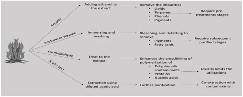
The nature of the chemical components, method of extraction, and interfering substances influence the extraction [24]. Most polysaccharides are extracted using water, acids, and organic solvents. Solvent extraction is difficult to implement owing to the complex nature of polymers [37]. The extraction procedure is a critical point in the purification of the target compound. The yield and structural alterations of sulphated polysaccharides are greatly influenced by the appropriate adjustment of parameters such as temperature, time, and pH [38]. This part of the review addresses the implemented extraction procedures that effectively enhance the purification of sulphated polysaccharides. The source of extraction of the sulphated polysaccharides is marine brown algae rather than marine invertebrates. The molecular weight, sulfation percentage, point of sulfation, and monosaccharide composition are vital to the bioactivity of fucoidan. Therefore, purification should be performed under mild conditions to avoid structural shifts [38]. The application of an ethanolic system helps remove co-extracting interferences, such as lipids, terpenes, and phenols Chlorophyll pigments are immediately removed using this method. Some researchers perform immersion and washing using acetone or hexane. Phenols are tightly bound to fucoidans or other polysaccharides during the extraction process. Formaldehyde treatment is a method of preventing cross-contamination by polymerizing phenols, proteins, and nucleic acids, and converting them into insoluble high-molecular-weight components [38,40,41]. Although it is useful in eradicating interfering modules, it may decrease fucoidan yield by interacting with polysaccharides and precipitating them into complexes [42].
Fig.3 “Effect of Different Solvents and Chemical Treatments on Aloe vera Extract Purification”
Herbal Bioactives In Nanocarrier Systems [43-47]
Herbal bioactives are widely used due to their pharmacological properties but face delivery challenges.
Anticoagulant and antithrombotic activity.
Antivirus activity
Antitumor and immunomodulatory activity
Antioxidant activity
Reducing blood lipids
Anticomplementary activity
Therapeutic potential in surgery
Anti-inflammatory
Gastric protection
Against hepatopathy
Against uropathy and renal Pathy
Common Bioactive: [48-53]
Curcumin → anticancer
Quercetin → antioxidant
Asiatic acid → wound healing
Resveratrol → anti-aging
Nanocarriers improve:
Stability
Solubility
Targeted delivery
Nano formulations significantly enhance the therapeutic efficiency of phytochemicals
Fucoidan-Based Hybrid Nanocarriers [54-61]
Table 1 - Types of Hybrid Systems
Type
Composition
Method
Application
Fucoidan–Chitosan NPs
Polyelectrolyte complex
Ionic gelation
Drug delivery
Fucoidan–Alginate Gel
Polymer blend
CaCl₂ crosslinking
Wound healing
Fucoidan–Metal NPs
Green synthesis
Reduction method
Antimicrobial
Fucoidan–Liposomes
Lipid + polysaccharide
Thin film hydration
Targeted therapy
Fucoidan’s sulphate groups enable strong interaction with polymers, forming stable nanostructures for drug delivery.
Mechanism Of Drug Delivery [62-68]
Hybrid Nanocarrier Mechanism
Curcumin, a bioactive phytoconstituent obtained from Curcuma longa, is widely known for its anti-inflammatory, antioxidant, and anticancer properties, but its clinical application is restricted due to poor aqueous solubility and low systemic bioavailability. To address these limitations, curcumin is encapsulated within a nanocarrier system, commonly prepared using natural polymers such as fucoidan and chitosan. These polymers form a stable polyelectrolyte complex matrix through electrostatic interaction between negatively charged sulphate groups of fucoidan and positively charged amino groups of chitosan, resulting in enhanced stability and protection of the encapsulated drug. Importantly, fucoidan imparts targeting capability by interacting with selectin receptors that are overexpressed on inflamed and tumor cells, enabling selective accumulation at the diseased site. Upon reaching the target, the nanocarrier binds to these receptors and undergoes receptor-mediated cellular uptake, primarily through endocytosis. Once internalized, the intracellular environment—characterized by acidic pH and enzymatic activity—triggers controlled and sustained release of curcumin from the nanocarrier system. This targeted and stimuli-responsive drug release enhances the therapeutic efficacy of curcumin while minimizing systemic toxicity, ultimately leading to improved anti-inflammatory and anticancer outcomes. Such fucoidan–chitosan based nanocarrier systems have been actively investigated in Indian pharmaceutical research for their potential in advanced herbal drug delivery applications.
Table – 2. Comparison: Fucoidan Vs Other Polymers [69-73]
Property
Fucoidan
Chitosan
Alginate
Charge
Negative
Positive
Negative
Source
Marine
Crustacean
Algae
Bioactivity
High
Moderate
Low
Drug Targeting
Excellent
Good
Moderate
Gel Formation
Moderate
Strong
Strong
Therapeutic Applications
1. Anticancer Activity [74,75,76]
Fucoidan, a sulphated polysaccharide isolated from brown seaweeds such as Sargassum weighted and Turbin aria conoids, has shown significant anticancer potential in various in vitro and in vivo studies conducted in India. It induces apoptosis in cancer cells through activation of caspase-dependent pathways and regulates key signalling mechanisms such as PI3K/Akt and MAPK. Indian studies have reported its effectiveness against breast, colon, and liver cancer cell lines, where it inhibits cell proliferation and metastasis while enhancing immune surveillance.
2. Antioxidant and Anti-inflammatory Effects [77,78,79]
Fucoidan exhibits strong antioxidant activity by scavenging free radicals and reducing oxidative stress. Research from institutions like CSIR-Central Food Technological Research Institute has demonstrated that fucoidan extracted from Indian seaweeds can significantly reduce lipid peroxidation and enhance endogenous antioxidant enzymes such as superoxide dismutase (SOD) and catalase. Additionally, its anti-inflammatory properties are attributed to the inhibition of pro-inflammatory mediators like TNF-α, IL-1β, and COX-2, making it useful in managing chronic inflammatory disorders.
3. Anticoagulant and Cardioprotective Activity [80,81,82]
Fucoidan possesses anticoagulant properties similar to heparin due to its sulphated structure. Studies conducted at Central Marine Fisheries Research Institute have reported that fucoidan from Indian brown algae can inhibit thrombin activity and reduce blood clot formation. This makes it a promising candidate for the prevention and management of cardiovascular diseases such as thrombosis and atherosclerosis, with potentially fewer side effects compared to synthetic anticoagulants.
4. Antidiabetic Potential [83,84]
Fucoidan has shown promising results in controlling blood glucose levels and improving insulin sensitivity. Indian experimental studies indicate that it can inhibit α-amylase and α-glucosidase enzymes, thereby reducing postprandial hyperglycaemia. Additionally, it helps in protecting pancreatic β-cells from oxidative damage, suggesting its therapeutic relevance in diabetes management.
5. Antiviral and Immunomodulatory Activity [85,86]
Fucoidan exhibits broad-spectrum antiviral activity by preventing viral attachment and entry into host cells. Research in India has explored its activity against viruses such as herpes simplex and influenza. It also enhances immune responses by stimulating macrophages, dendritic cells, and natural killer (NK) cells, thereby strengthening host defense mechanisms.
6. Wound Healing and Tissue Regeneration [87,88,89]
Fucoidan plays an important role in wound healing by promoting fibroblast proliferation, collagen synthesis, and angiogenesis. Studies from Central Salt and Marine Chemicals Research Institute have highlighted its potential in accelerating tissue repair and regeneration, particularly in burn and diabetic wounds. Its biocompatibility and ability to form hydrogels make it useful in advanced wound dressing systems. Fucoidan-based nanocarriers show improved therapeutic efficiency due to targeted delivery systems [90,91]
REFERENCES
McDowell, P.E. 15—Algal polysaccharides. In Methods in Plant Biochemistry, 1st ed.; Elsever: Amsterdam, The Netherlands, 1990; Volume 2, pp. 523–547.
Cui, K.; Tai, W.; Shan, X.; Hao, J.; Li, G.; Yu, G. Structural characterization and anti-thrombotic properties of fucoidan from Nemacystus decipiens. Int. J. Biol. Macromol. 2018, 120, 1817–1822.
Yokota,T.; Nomura,K.; Nagashima, M.;Kamimura,N. Fucoidan all eviates high-fatdiet-induced dyslipidemia and atherosclerosis in ApoEshl mice deficient in apolipoprotein E expression. J. Nutr. Biochem. 2016, 32, 46–54.
Mansour, M.B.; Balti, R.; Yacoubi, L.; Ollivier, V.; Chaubet, F.; Maaroufi, R.M. Primary structure and anticoagulant activity of fucoidan from the sea cucumber Holothuriapolii. Int. J.Biol. Macromol. 2019, 121, 1145–1153.
Koh, H.S.A.; Lu, J.; Zhou, W. Structure characterization and antioxidant activity of fucoidan isolated from Undaria pinnatifida grown in New Zealand. Carbohydr. Polym. 2019, 212, 178–185.
Jin, W.; Wu, W.; Tang, H.; Wei, B.; Wang, H.; Sun, J.; Zhang, W.; Zhong, W. Structure analysis and anti-tumor and anti-angiogenic activities of sulfated galactofucan extracted from Sargassum thunbergii. Mar. Drugs 2019, 17, 52.
Usoltseva,R.V.;Anastyuk,S.D.;Surits,V.V.;Shevchenko,.M.;Thinh,P.D.;Zadorozhny,P.A.;Ermakova,S.P. Comparison of structure and in vitro anticancer activity of native and modified fucoidans from Sargassum feldmannii and S. duplicatum. Int. J. Biol. Macromol. 2019, 124, 220–228.
Usoltseva, R.V.; Shevchenko, N.M.; Malyarenko, O.S.; Anastyuk, S.D.; Kasprik, A.E.; Zvyagintsev, N.V.; Ermakova, S.P. Fucoidans from brown algae Laminaria longipes and Saccharina cichorioides: Structural characteristics, anticancer and radio-sensitizing activity in vitro. Carbohydr. Polym. 2019, 221, 157–165.
Lahrsen, E.; Liewert, I.; Alban, S. Gradual degradation of fucoidan from Fucus vesiculosus and its effect on structure, antioxidant and antiproliferative activities. Carbohydr. Polym. 2018, 192,208–216.
Zhang,Z.;Till,S.;Jiang,C.;Knappe,S.;Reutterer,S.;Scheiflinger,F.;Szabo,C.M.;Dockal,M.Structure-activity relationship of the pro- and anticoagulant effects of Fucus vesiculosus fucoidan. Thromb. Haemost. 2014, 111, 429–437.
Bilan, M.I.; Grachev, A.A.; Ustuzhanina, N.E.; Shashkov, A.S.; Nifantiev, N.E.; Usov, A.I. Structure of a fucoidan from the brown seaweed Fucus evanescens C. Ag. Carbohydr. Res. 2002, 337, 719–730.
Bilan, M.I.; Grachev, A.A.; Ustuzhanina, N.E.; Shashkov, A.S.; Nifantiev, N.E.; Usov, A.I. A highly regular fraction of a fucoidan from the brown seaweed Fucus distichus L. Carbohydr. Res. 2004, 339, 511–517.
Bilan, M.I.; Grachev, A.A.; Shashkov, A.S.; Nifantiev, N.E.; Usov, A.I. Structure of a fucoidan from the brown seaweed Fucus serratus L. Carbohydr. Res. 2006, 341, 238–245.
Foley, S.A.; Szegezdi, E.; Mulloy, B.; Samali, A.; Tuohy, M.G. An unfractionated fucoidan from Ascophyllum nodosum: Extraction, characterization, and apoptotic effects in vitro. J. Nat. Prod. 2012, 75, 1674.
Descamps, V.; Colin, S.; Lahaye, M.; Jam, M.; Richard, C.; Potin, P.; Barbeyron, T.; Yvin, J.C.; Kloareg, B. Isolation and culture of a marine bacterium degrading the sulfated fucans from marine brown algae. Mar. Biotechnol. 2006, 8, 27–39.
Jabbar Mian, A.; Percival, E. Carbohydrates of the brown seaweeds Himanthalialorea and Bifurcariabifurcata: Part II. structural studies of the “fucans”. Carbohydr. Res. 1973, 26, 147–161.
Mak, W.; Hamid, N.; Liu, T.; Lu, J.; White, W.L. Fucoidan from New Zealand Undariapinnatifida: Monthly variations and determination of antioxidant activities. Carbohydr. Polym. 2013, 95, 606–614.
Fletcher, H.R.; Biller, P.; Ross, A.B.; Adams, J.M.M. The seasonal variation of fucoidan within three species of brown macroalgae. Algal Res. 2017, 22, 79–86.
Garcia-Vaquero, M.; Rajauria, G.; O’Doherty, J.V.; Sweeney, T. Polysaccharides from macroalgae: Recent advances, innovative technologies and challenges in extraction and purification. Food Res. Int. 2017, 99, 1011–1020.
Chevolot,L.;Foucault,A.;Chaubet,F.;Kervarec,N.;Sinquin,C.;Fisher,A.M.;BoissonVidal,C.Furtherdata on the structure of brown seaweed fucans: Relationships with anticoagulant activity. Carbohydr. Res. 1999, 319, 154–165.
Marais, M.F.; Joseleau, J.P. A fucoidan fraction from Ascophyllumnodosum. Carbohydr. Res. 2001, 336, 155–159.
Ponce, N.M.A.; Pujol, C.A.; Damonte, E.B.; Flores, M.L.; Stortz, C.A. Fucoidans from the brown seaweed Adenocystis utricularis: Extraction methods, antiviral activity and structural studies. Carbohydr. Res. 2003, 338, 153–165.
Irhimeh, M.R.; Fitton, J.H.; Lowenthal, R.M.; Kongtawelert, P. A quantitative method to detect fucoidan in human plasma using a novel antibody. Methods Find. Exp. Clin. Pharmacol. 2005, 27, 705–710.
Pozharitskaya, O.N.; Shikov, A.N.; Faustova, N.M.; Obluchinskaya, E.D.; Kosman, V.M.; Vuorela, H.; Makarov, V.G. Pharmacokinetic and tissue distribution of fucoidan from Fucus vesiculosus after oral administration to rats. Mar. Drugs 2018, 16, 132.
Torode, T.A.; Marcus, S.E.; Jam, M.; Tonon, T.; Blackburn, R.S.; Hervé, C.; Knox, J.P. Monoclonal antibodies directed to fucoidan preparations from brown algae. PLoS ONE 2015, 10, e0118366.
Li B et al. (2008). Fucoidan: Structure and bioactivity. Molecules, 13(8), 1671–1695. DOI: 10.3390/molecules13081671. Describes the L-fucose backbone, sulfate ester groups, molecular weight variability, and negative charge of fucoidan.
Ale MT et al. (2011). Important determinants for fucoidan bioactivity: A critical review of structure–function relations and extraction methods. Marine Drugs, 9(10), 2106–2130. DOI: 10.3390/md9102106. Explains structural complexity and source-dependent variations of fucoidan.
Fitton JH (2011). Therapies from Fucoidan; Multifunctional Marine Polymers. Marine Drugs, 9(10), 1731–1760. DOI: 10.3390/md9101731. Supports the role of sulfate groups in biological activity and electrostatic interactions.
Rinaudo M (2006). Chitin and chitosan: Properties and applications. Progress in Polymer Science, 31(7), 603–632. DOI: 10.1016/j.progpolymsci.2006.06.001. Describes positively charged amino groups of chitosan and its interaction with anionic polymers.
Huang YC et al. (2015). Development and characterization of mucoadhesive nanoparticles composed of fucoidan and chitosan for drug delivery. Carbohydrate Polymers, 117, 795–803. DOI: 10.1016/j.carbpol.2014.10.094. Reports formation of stable polyelectrolyte nanoparticles through electrostatic interaction between fucoidan and chitosan.
Ale MT, Mikkelsen JD, Meyer AS (2011). Designed optimization of a single-step extraction of fucose-containing sulfated polysaccharides from Sargassum sp. Journal of Applied Phycology, 24, 715–723. DOI: 10.1007/s10811-011-9690-3.
Wijesinghe WAJP & Jeon YJ (2012). Biological activities and potential industrial applications of fucose rich sulfated polysaccharides and fucoidans isolated from brown seaweeds. Carbohydrate Polymers, 88(1), 13–20. DOI: 10.1016/j.carbpol.2011.12.029.
Fernando IPS et al. (2019). A comprehensive review on fucoidan structure, extraction methods, bioactivities, and industrial applications. Marine Drugs, 17(10), 571. DOI: 10.3390/md17100571.
Kadam SU et al. (2015). Extraction, structure and biofunctional activities of laminarin and fucoidan from brown algae. International Journal of Food Science & Technology, 50(1), 24–31. DOI: 10.1111/ijfs.12629.
Wijesinghe, W.A.J.P.; Jeon, Y.-J. Biological activities and potential industrial applications of fucose rich sulfated polysaccharides and fucoidans isolated from brown seaweeds: A review. Carbohyd. Polym. 2012, 88, 13–20
Hahn, T.; Lang, S.; Ulber, R.; Muffler, K. Novel procedures for the extraction of fucoidan from brown algae. Process. Biochem 2012, 47, 1691–1698.
Chirinos, R.; Rogez, H.; Campos, D.; Pedreschi, R.; Larondelle, Y. Optimization of extraction conditions of antioxidant phenolic compounds from mashua (Tropaeolum tuberosum Ruíz & Pavón) tubers. Sep. Purif. Technol. 2007, 55, 217–225.
Black, W.A.P.; Dewar, E.T.; Woodward, F.N. Manufacture of algal chemicals. IV—Laboratory-scale isolation of fucoidin from brown marine algae. J. Sci. Food Agric. 1952, 3, 122–129.
Mian, A.J.; Percival, E. Carbohydrates of the brown seaweeds himanthalia lorea, bifurcaria bifurcata, and Padina pavonia. Carbohyd Res. 1973, 26, 133–146.
Rioux, L.E.; Turgeon, S.L.; Beaulieu, M. Characterization of polysaccharides extracted from brown seaweeds. Carbohyd Polym 2007, 69, 530–537.
Li B et al. (2008). Fucoidan: Structure and bioactivity. Molecules, 13(8), 1671–1695. DOI: 10.3390/molecules13081671. Describes broad biological activities including antioxidant, antiviral, anticoagulant, and lipid-lowering effects.
Wijesinghe WAJP & Jeon YJ (2012). Biological activities and potential industrial applications of fucoidans isolated from brown seaweeds. Carbohydrate Polymers, 88(1), 13–20. DOI: 10.1016/j.carbpol.2011.12.029. Reports antioxidant, anti-inflammatory, antitumor, and immunomodulatory properties.
Fernando IPS et al. (2019). A comprehensive review on fucoidan structure, extraction methods, bioactivities and industrial applications. Marine Drugs, 17(10), 571. DOI: 10.3390/md17100571. Reviews therapeutic applications including surgery-related healing, gastric protection, hepatoprotection, and nephroprotection.
Yallapu MM et al. (2012). Therapeutic applications of curcumin nanoformulations. The AAPS Journal, 14(1), 195–205. DOI: 10.1208/s12248-011-9336-7. Explains challenges in herbal bioactive delivery and the importance of nanocarrier systems for improved therapeutic efficacy.
Yallapu MM et al. (2012). Therapeutic applications of curcumin nanoformulations. The AAPS Journal, 14(1), 195–205. DOI: 10.1208/s12248-011-9336-7. Supports curcumin anticancer activity and enhancement of stability, solubility, and therapeutic efficacy through nanoformulations.
Boots AW et al. (2008). Health effects of quercetin: From antioxidant to nutraceutical. European Journal of Pharmacology, 585(2–3), 325–337. DOI: 10.1016/j.ejphar.2008.03.008. Supports antioxidant activity of quercetin.
James JT & Dubery IA (2009). Pentacyclic triterpenoids from the medicinal herb Centella asiatica (L.) Urban. Molecules, 14(10), 3922–3941. DOI: 10.3390/molecules14103922. Supports wound healing and therapeutic activity of asiatic acid.
Baur JA & Sinclair DA (2006). Therapeutic potential of resveratrol: The in vivo evidence. Nature Reviews Drug Discovery, 5(6), 493–506. DOI: 10.1038/nrd2060. Supports anti-aging and antioxidant properties of resveratrol.
Anitha A et al. (2014). Curcumin-loaded N,O-carboxymethyl chitosan nanoparticles for cancer drug delivery. Journal of Biomaterials Applications, 28(6), 843–854. DOI: 10.1177/0885328213496994. Explains how nanocarriers improve targeted delivery, stability, and bioavailability of phytochemicals.
Patra JK et al. (2018). Nano based drug delivery systems: recent developments and future prospects. Journal of Nanobiotechnology, 16, 71. DOI: 10.1186/s12951-018-0392-8. Supports the statement that nanoformulations significantly enhance therapeutic efficiency of phytochemicals.
Huang YC et al. (2015). Development of mucoadhesive nanoparticles composed of fucoidan and chitosan for drug delivery applications. Carbohydrate Polymers, 117, 795–803. DOI: 10.1016/j.carbpol.2014.10.094.
Sezer AD & Akbuga J (1999). Preparation and characterization of chitosan microspheres containing fucoidan. International Journal of Pharmaceutics, 182(2), 261–268. DOI: 10.1016/S0378-5173(99)00050-4.
Lee KY & Mooney DJ (2012). Alginate: properties and biomedical applications. Progress in Polymer Science, 37(1), 106–126. DOI: 10.1016/j.progpolymsci.2011.06.003.
Murakami K et al. (2003). Hydrogel blends of alginate and fucoidan for biomedical applications. Journal of Biomedical Materials Research Part A, 67A(4), 1272–1278. DOI: 10.1002/jbm.a.10106.
Rajeshkumar S et al. (2013). Synthesis of silver nanoparticles using marine brown seaweed and evaluation of antimicrobial activity. Journal of Nanostructure in Chemistry, 3, 44. DOI: 10.1186/2193-8865-3-44.
Venkatesan J et al. (2016). Green synthesis of metal nanoparticles using marine polysaccharides for biomedical applications. International Journal of Biological Macromolecules, 93, 1378–1383. DOI: 10.1016/j.ijbiomac.2016.03.038.
Deepa B et al. (2012). Surface modification of liposomes with polysaccharides for targeted drug delivery. Progress in Lipid Research, 51(4), 238–252. DOI: 10.1016/j.plipres.2012.06.001.
Thanou M et al. (2008). Polysaccharide-coated liposomes for enhanced drug targeting and stability. Journal of Controlled Release, 131(1), 1–10. DOI: 10.1016/j.jconrel.2008.06.015.
Mourya VK & Inamdar NN (2008). Chitosan-modifications and applications: Opportunities galore. Reactive and Functional Polymers, 68(6), 1013–1051. DOI: 10.1016/j.reactfunctpolym.2008.03.002.
Yallapu MM et al. (2012). Therapeutic applications of curcumin nanoformulations. The AAPS Journal, 14(1), 195–205. DOI: 10.1208/s12248-011-9336-7.
Huang YC et al. (2015). Development and characterization of mucoadhesive nanoparticles composed of fucoidan and chitosan for drug delivery. Carbohydrate Polymers, 117, 795–803. DOI: 10.1016/j.carbpol.2014.10.094.
Liu J et al. (2008). Fucoidan-based nanoparticles targeting P-selectin for enhanced drug delivery. Biomaterials, 29(30), 4122–4130. DOI: 10.1016/j.biomaterials.2008.07.009.
Anitha A et al. (2014). Curcumin-loaded N,O-carboxymethyl chitosan nanoparticles for cancer drug delivery. Journal of Biomaterials Applications, 28(6), 843–854. DOI: 10.1177/0885328213496994.
Sinha VR et al. (2004). Poly-ε-caprolactone microspheres and nanospheres: an overview. International Journal of Pharmaceutics, 278(1), 1–23. DOI: 10.1016/j.ijpharm.2004.01.044.
Fitton JH (2011). Therapies from Fucoidan; Multifunctional Marine Polymers. Marine Drugs, 9(10), 1731–1760. DOI: 10.3390/md9101731. Supports fucoidan properties such as negative charge, marine source, high bioactivity, and targeting ability.
Rinaudo M (2006). Chitin and chitosan: Properties and applications. Progress in Polymer Science, 31(7), 603–632. DOI: 10.1016/j.progpolymsci.2006.06.001. Supports chitosan characteristics including positive charge, crustacean origin, and strong gel-forming ability.
Lee KY & Mooney DJ (2012). Alginate: Properties and biomedical applications. Progress in Polymer Science, 37(1), 106–126. DOI: 10.1016/j.progpolymsci.2011.06.003. Supports alginate properties including algal source, negative charge, and strong gel formation.
Mourya VK & Inamdar NN (2008). Chitosan-modifications and applications: Opportunities galore. Reactive and Functional Polymers, 68(6), 1013–1051. DOI: 10.1016/j.reactfunctpolym.2008.03.002. Supports comparison of bioactivity and drug delivery performance among natural polymers.
Wijesinghe WAJP & Jeon YJ (2012). Biological activities and potential industrial applications of fucose-rich sulfated polysaccharides and fucoidans isolated from brown seaweeds. Carbohydrate Polymers, 88(1), 13–20. DOI: 10.1016/j.carbpol.2011.12.029. Supports the superior bioactivity and therapeutic potential of fucoidan.
Atashrazm F et al. (2015). Fucoidan and cancer: A multifunctional molecule with anti-tumor potential. Marine Drugs, 13(4), 2327–2346. DOI: 10.3390/md13042327.
CSIR-Central Food Technological Research Institute researchers reported bioactive potential of fucoidan isolated from Indian brown algae in cancer-related studies.
Ale MT et al. (2011). Important determinants for fucoidan bioactivity: A critical review of structure–function relations and extraction methods. Marine Drugs, 9(10), 2106–2130. DOI: 10.3390/md9102106.
Wijesinghe WAJP & Jeon YJ (2012). Biological activities and potential industrial applications of fucoidans isolated from brown seaweeds. Carbohydrate Polymers, 88(1), 13–20. DOI: 10.1016/j.carbpol.2011.12.029.
Fernando IPS et al. (2019). A comprehensive review on fucoidan structure, extraction methods, bioactivities and industrial applications. Marine Drugs, 17(10), 571. DOI: 10.3390/md17100571.
CSIR-Central Food Technological Research Institute studies demonstrated antioxidant enzyme enhancement and reduction of oxidative stress using fucoidan from Indian seaweeds.
Li B et al. (2008). Fucoidan: Structure and bioactivity. Molecules, 13(8), 1671–1695. DOI: 10.3390/molecules13081671.
Central Marine Fisheries Research Institute reported anticoagulant activity of fucoidan isolated from Indian brown algae species.
Kim KT et al. (2010). Protective effect of fucoidan against high glucose-induced oxidative stress. Journal of Food Science, 75(7), H173–H178. DOI: 10.1111/j.1750-3841.2010.01785.
Hwang PA et al. (2011). Inhibitory effect of fucoidan on α-glucosidase and α-amylase activities. Marine Drugs, 9(9), 1647–1659. DOI: 10.3390/md9091647.
Hayashi K et al. (2008). Antiviral activity of fucoidan from brown algae against herpes simplex virus. Planta Medica, 74(2), 174–178. DOI: 10.1055/s-2008-1034318.
Fitton JH et al. (2015). Fucoidan and immune modulation. Marine Drugs, 13(9), 5920–5946. DOI: 10.3390/md13095920.
Central Salt and Marine Chemicals Research Institute studies highlighted fucoidan-based hydrogels for wound healing and tissue regeneration.
Sezer AD & Cevher E (2012). Fucoidan-based biomaterials for wound healing applications. AAPS PharmSciTech, 13(3), 990–997. DOI: 10.1208/s12249-012-9816-3.
Murakami K et al. (2003). Accelerated wound healing by fucoidan-containing hydrogel. Journal of Biomedical Materials Research Part A, 67A(4), 1272–1278. DOI: 10.1002/jbm.a.10106.
Liu J et al. (2008). Fucoidan-based nanoparticles targeting P-selectin for enhanced drug delivery. Biomaterials, 29(30), 4122–4130. DOI: 10.1016/j.biomaterials.2008.07.009.
Yallapu MM et al. (2012). Therapeutic applications of curcumin nanoformulations. The AAPS Journal, 14(1), 195–205. DOI: 10.1208/s12248-011-9336-7.
Reference
McDowell, P.E. 15—Algal polysaccharides. In Methods in Plant Biochemistry, 1st ed.; Elsever: Amsterdam, The Netherlands, 1990; Volume 2, pp. 523–547.
Cui, K.; Tai, W.; Shan, X.; Hao, J.; Li, G.; Yu, G. Structural characterization and anti-thrombotic properties of fucoidan from Nemacystus decipiens. Int. J. Biol. Macromol. 2018, 120, 1817–1822.
Yokota,T.; Nomura,K.; Nagashima, M.;Kamimura,N. Fucoidan all eviates high-fatdiet-induced dyslipidemia and atherosclerosis in ApoEshl mice de?cient in apolipoprotein E expression. J. Nutr. Biochem. 2016, 32, 46–54.
Mansour, M.B.; Balti, R.; Yacoubi, L.; Ollivier, V.; Chaubet, F.; Maarou?, R.M. Primary structure and anticoagulant activity of fucoidan from the sea cucumber Holothuriapolii. Int. J.Biol. Macromol. 2019, 121, 1145–1153.
Koh, H.S.A.; Lu, J.; Zhou, W. Structure characterization and antioxidant activity of fucoidan isolated from Undaria pinnati?da grown in New Zealand. Carbohydr. Polym. 2019, 212, 178–185.
Jin, W.; Wu, W.; Tang, H.; Wei, B.; Wang, H.; Sun, J.; Zhang, W.; Zhong, W. Structure analysis and anti-tumor and anti-angiogenic activities of sulfated galactofucan extracted from Sargassum thunbergii. Mar. Drugs 2019, 17, 52.
Usoltseva,R.V.;Anastyuk,S.D.;Surits,V.V.;Shevchenko,.M.;Thinh,P.D.;Zadorozhny,P.A.;Ermakova,S.P. Comparison of structure and in vitro anticancer activity of native and modi?ed fucoidans from Sargassum feldmannii and S. duplicatum. Int. J. Biol. Macromol. 2019, 124, 220–228.
Usoltseva, R.V.; Shevchenko, N.M.; Malyarenko, O.S.; Anastyuk, S.D.; Kasprik, A.E.; Zvyagintsev, N.V.; Ermakova, S.P. Fucoidans from brown algae Laminaria longipes and Saccharina cichorioides: Structural characteristics, anticancer and radio-sensitizing activity in vitro. Carbohydr. Polym. 2019, 221, 157–165.
Lahrsen, E.; Liewert, I.; Alban, S. Gradual degradation of fucoidan from Fucus vesiculosus and its e?ect on structure, antioxidant and antiproliferative activities. Carbohydr. Polym. 2018, 192,208–216.
Zhang,Z.;Till,S.;Jiang,C.;Knappe,S.;Reutterer,S.;Schei?inger,F.;Szabo,C.M.;Dockal,M.Structure-activity relationship of the pro- and anticoagulant e?ects of Fucus vesiculosus fucoidan. Thromb. Haemost. 2014, 111, 429–437.
Bilan, M.I.; Grachev, A.A.; Ustuzhanina, N.E.; Shashkov, A.S.; Nifantiev, N.E.; Usov, A.I. Structure of a fucoidan from the brown seaweed Fucus evanescens C. Ag. Carbohydr. Res. 2002, 337, 719–730.
Bilan, M.I.; Grachev, A.A.; Ustuzhanina, N.E.; Shashkov, A.S.; Nifantiev, N.E.; Usov, A.I. A highly regular fraction of a fucoidan from the brown seaweed Fucus distichus L. Carbohydr. Res. 2004, 339, 511–517.
Bilan, M.I.; Grachev, A.A.; Shashkov, A.S.; Nifantiev, N.E.; Usov, A.I. Structure of a fucoidan from the brown seaweed Fucus serratus L. Carbohydr. Res. 2006, 341, 238–245.
Foley, S.A.; Szegezdi, E.; Mulloy, B.; Samali, A.; Tuohy, M.G. An unfractionated fucoidan from Ascophyllum nodosum: Extraction, characterization, and apoptotic e?ects in vitro. J. Nat. Prod. 2012, 75, 1674.
Descamps, V.; Colin, S.; Lahaye, M.; Jam, M.; Richard, C.; Potin, P.; Barbeyron, T.; Yvin, J.C.; Kloareg, B. Isolation and culture of a marine bacterium degrading the sulfated fucans from marine brown algae. Mar. Biotechnol. 2006, 8, 27–39.
Jabbar Mian, A.; Percival, E. Carbohydrates of the brown seaweeds Himanthalialorea and Bifurcariabifurcata: Part II. structural studies of the “fucans”. Carbohydr. Res. 1973, 26, 147–161.
Mak, W.; Hamid, N.; Liu, T.; Lu, J.; White, W.L. Fucoidan from New Zealand Undariapinnati?da: Monthly variations and determination of antioxidant activities. Carbohydr. Polym. 2013, 95, 606–614.
Fletcher, H.R.; Biller, P.; Ross, A.B.; Adams, J.M.M. The seasonal variation of fucoidan within three species of brown macroalgae. Algal Res. 2017, 22, 79–86.
Garcia-Vaquero, M.; Rajauria, G.; O’Doherty, J.V.; Sweeney, T. Polysaccharides from macroalgae: Recent advances, innovative technologies and challenges in extraction and puri?cation. Food Res. Int. 2017, 99, 1011–1020.
Chevolot,L.;Foucault,A.;Chaubet,F.;Kervarec,N.;Sinquin,C.;Fisher,A.M.;BoissonVidal,C.Furtherdata on the structure of brown seaweed fucans: Relationships with anticoagulant activity. Carbohydr. Res. 1999, 319, 154–165.
Marais, M.F.; Joseleau, J.P. A fucoidan fraction from Ascophyllumnodosum. Carbohydr. Res. 2001, 336, 155–159.
Ponce, N.M.A.; Pujol, C.A.; Damonte, E.B.; Flores, M.L.; Stortz, C.A. Fucoidans from the brown seaweed Adenocystis utricularis: Extraction methods, antiviral activity and structural studies. Carbohydr. Res. 2003, 338, 153–165.
Irhimeh, M.R.; Fitton, J.H.; Lowenthal, R.M.; Kongtawelert, P. A quantitative method to detect fucoidan in human plasma using a novel antibody. Methods Find. Exp. Clin. Pharmacol. 2005, 27, 705–710.
Pozharitskaya, O.N.; Shikov, A.N.; Faustova, N.M.; Obluchinskaya, E.D.; Kosman, V.M.; Vuorela, H.; Makarov, V.G. Pharmacokinetic and tissue distribution of fucoidan from Fucus vesiculosus after oral administration to rats. Mar. Drugs 2018, 16, 132.
Torode, T.A.; Marcus, S.E.; Jam, M.; Tonon, T.; Blackburn, R.S.; Hervé, C.; Knox, J.P. Monoclonal antibodies directed to fucoidan preparations from brown algae. PLoS ONE 2015, 10, e0118366.
Li B et al. (2008). Fucoidan: Structure and bioactivity. Molecules, 13(8), 1671–1695. DOI: 10.3390/molecules13081671. Describes the L-fucose backbone, sulfate ester groups, molecular weight variability, and negative charge of fucoidan.
Ale MT et al. (2011). Important determinants for fucoidan bioactivity: A critical review of structure–function relations and extraction methods. Marine Drugs, 9(10), 2106–2130. DOI: 10.3390/md9102106. Explains structural complexity and source-dependent variations of fucoidan.
Fitton JH (2011). Therapies from Fucoidan; Multifunctional Marine Polymers. Marine Drugs, 9(10), 1731–1760. DOI: 10.3390/md9101731. Supports the role of sulfate groups in biological activity and electrostatic interactions.
Rinaudo M (2006). Chitin and chitosan: Properties and applications. Progress in Polymer Science, 31(7), 603–632. DOI: 10.1016/j.progpolymsci.2006.06.001. Describes positively charged amino groups of chitosan and its interaction with anionic polymers.
Huang YC et al. (2015). Development and characterization of mucoadhesive nanoparticles composed of fucoidan and chitosan for drug delivery. Carbohydrate Polymers, 117, 795–803. DOI: 10.1016/j.carbpol.2014.10.094. Reports formation of stable polyelectrolyte nanoparticles through electrostatic interaction between fucoidan and chitosan.
Ale MT, Mikkelsen JD, Meyer AS (2011). Designed optimization of a single-step extraction of fucose-containing sulfated polysaccharides from Sargassum sp. Journal of Applied Phycology, 24, 715–723. DOI: 10.1007/s10811-011-9690-3.
Wijesinghe WAJP & Jeon YJ (2012). Biological activities and potential industrial applications of fucose rich sulfated polysaccharides and fucoidans isolated from brown seaweeds. Carbohydrate Polymers, 88(1), 13–20. DOI: 10.1016/j.carbpol.2011.12.029.
Fernando IPS et al. (2019). A comprehensive review on fucoidan structure, extraction methods, bioactivities, and industrial applications. Marine Drugs, 17(10), 571. DOI: 10.3390/md17100571.
Kadam SU et al. (2015). Extraction, structure and biofunctional activities of laminarin and fucoidan from brown algae. International Journal of Food Science & Technology, 50(1), 24–31. DOI: 10.1111/ijfs.12629.
Wijesinghe, W.A.J.P.; Jeon, Y.-J. Biological activities and potential industrial applications of fucose rich sulfated polysaccharides and fucoidans isolated from brown seaweeds: A review. Carbohyd. Polym. 2012, 88, 13–20
Hahn, T.; Lang, S.; Ulber, R.; Muffler, K. Novel procedures for the extraction of fucoidan from brown algae. Process. Biochem 2012, 47, 1691–1698.
Chirinos, R.; Rogez, H.; Campos, D.; Pedreschi, R.; Larondelle, Y. Optimization of extraction conditions of antioxidant phenolic compounds from mashua (Tropaeolum tuberosum Ruíz & Pavón) tubers. Sep. Purif. Technol. 2007, 55, 217–225.
Black, W.A.P.; Dewar, E.T.; Woodward, F.N. Manufacture of algal chemicals. IV—Laboratory-scale isolation of fucoidin from brown marine algae. J. Sci. Food Agric. 1952, 3, 122–129.
Mian, A.J.; Percival, E. Carbohydrates of the brown seaweeds himanthalia lorea, bifurcaria bifurcata, and Padina pavonia. Carbohyd Res. 1973, 26, 133–146.
Rioux, L.E.; Turgeon, S.L.; Beaulieu, M. Characterization of polysaccharides extracted from brown seaweeds. Carbohyd Polym 2007, 69, 530–537.
Li B et al. (2008). Fucoidan: Structure and bioactivity. Molecules, 13(8), 1671–1695. DOI: 10.3390/molecules13081671. Describes broad biological activities including antioxidant, antiviral, anticoagulant, and lipid-lowering effects.
Wijesinghe WAJP & Jeon YJ (2012). Biological activities and potential industrial applications of fucoidans isolated from brown seaweeds. Carbohydrate Polymers, 88(1), 13–20. DOI: 10.1016/j.carbpol.2011.12.029. Reports antioxidant, anti-inflammatory, antitumor, and immunomodulatory properties.
Fernando IPS et al. (2019). A comprehensive review on fucoidan structure, extraction methods, bioactivities and industrial applications. Marine Drugs, 17(10), 571. DOI: 10.3390/md17100571. Reviews therapeutic applications including surgery-related healing, gastric protection, hepatoprotection, and nephroprotection.
Yallapu MM et al. (2012). Therapeutic applications of curcumin nanoformulations. The AAPS Journal, 14(1), 195–205. DOI: 10.1208/s12248-011-9336-7. Explains challenges in herbal bioactive delivery and the importance of nanocarrier systems for improved therapeutic efficacy.
Yallapu MM et al. (2012). Therapeutic applications of curcumin nanoformulations. The AAPS Journal, 14(1), 195–205. DOI: 10.1208/s12248-011-9336-7. Supports curcumin anticancer activity and enhancement of stability, solubility, and therapeutic efficacy through nanoformulations.
Boots AW et al. (2008). Health effects of quercetin: From antioxidant to nutraceutical. European Journal of Pharmacology, 585(2–3), 325–337. DOI: 10.1016/j.ejphar.2008.03.008. Supports antioxidant activity of quercetin.
James JT & Dubery IA (2009). Pentacyclic triterpenoids from the medicinal herb Centella asiatica (L.) Urban. Molecules, 14(10), 3922–3941. DOI: 10.3390/molecules14103922. Supports wound healing and therapeutic activity of asiatic acid.
Baur JA & Sinclair DA (2006). Therapeutic potential of resveratrol: The in vivo evidence. Nature Reviews Drug Discovery, 5(6), 493–506. DOI: 10.1038/nrd2060. Supports anti-aging and antioxidant properties of resveratrol.
Anitha A et al. (2014). Curcumin-loaded N,O-carboxymethyl chitosan nanoparticles for cancer drug delivery. Journal of Biomaterials Applications, 28(6), 843–854. DOI: 10.1177/0885328213496994. Explains how nanocarriers improve targeted delivery, stability, and bioavailability of phytochemicals.
Patra JK et al. (2018). Nano based drug delivery systems: recent developments and future prospects. Journal of Nanobiotechnology, 16, 71. DOI: 10.1186/s12951-018-0392-8. Supports the statement that nanoformulations significantly enhance therapeutic efficiency of phytochemicals.
Huang YC et al. (2015). Development of mucoadhesive nanoparticles composed of fucoidan and chitosan for drug delivery applications. Carbohydrate Polymers, 117, 795–803. DOI: 10.1016/j.carbpol.2014.10.094.
Sezer AD & Akbuga J (1999). Preparation and characterization of chitosan microspheres containing fucoidan. International Journal of Pharmaceutics, 182(2), 261–268. DOI: 10.1016/S0378-5173(99)00050-4.
Lee KY & Mooney DJ (2012). Alginate: properties and biomedical applications. Progress in Polymer Science, 37(1), 106–126. DOI: 10.1016/j.progpolymsci.2011.06.003.
Murakami K et al. (2003). Hydrogel blends of alginate and fucoidan for biomedical applications. Journal of Biomedical Materials Research Part A, 67A(4), 1272–1278. DOI: 10.1002/jbm.a.10106.
Rajeshkumar S et al. (2013). Synthesis of silver nanoparticles using marine brown seaweed and evaluation of antimicrobial activity. Journal of Nanostructure in Chemistry, 3, 44. DOI: 10.1186/2193-8865-3-44.
Venkatesan J et al. (2016). Green synthesis of metal nanoparticles using marine polysaccharides for biomedical applications. International Journal of Biological Macromolecules, 93, 1378–1383. DOI: 10.1016/j.ijbiomac.2016.03.038.
Deepa B et al. (2012). Surface modification of liposomes with polysaccharides for targeted drug delivery. Progress in Lipid Research, 51(4), 238–252. DOI: 10.1016/j.plipres.2012.06.001.
Thanou M et al. (2008). Polysaccharide-coated liposomes for enhanced drug targeting and stability. Journal of Controlled Release, 131(1), 1–10. DOI: 10.1016/j.jconrel.2008.06.015.
Mourya VK & Inamdar NN (2008). Chitosan-modifications and applications: Opportunities galore. Reactive and Functional Polymers, 68(6), 1013–1051. DOI: 10.1016/j.reactfunctpolym.2008.03.002.
Yallapu MM et al. (2012). Therapeutic applications of curcumin nanoformulations. The AAPS Journal, 14(1), 195–205. DOI: 10.1208/s12248-011-9336-7.
Huang YC et al. (2015). Development and characterization of mucoadhesive nanoparticles composed of fucoidan and chitosan for drug delivery. Carbohydrate Polymers, 117, 795–803. DOI: 10.1016/j.carbpol.2014.10.094.
Liu J et al. (2008). Fucoidan-based nanoparticles targeting P-selectin for enhanced drug delivery. Biomaterials, 29(30), 4122–4130. DOI: 10.1016/j.biomaterials.2008.07.009.
Anitha A et al. (2014). Curcumin-loaded N,O-carboxymethyl chitosan nanoparticles for cancer drug delivery. Journal of Biomaterials Applications, 28(6), 843–854. DOI: 10.1177/0885328213496994.
Sinha VR et al. (2004). Poly-ε-caprolactone microspheres and nanospheres: an overview. International Journal of Pharmaceutics, 278(1), 1–23. DOI: 10.1016/j.ijpharm.2004.01.044.
Fitton JH (2011). Therapies from Fucoidan; Multifunctional Marine Polymers. Marine Drugs, 9(10), 1731–1760. DOI: 10.3390/md9101731. Supports fucoidan properties such as negative charge, marine source, high bioactivity, and targeting ability.
Rinaudo M (2006). Chitin and chitosan: Properties and applications. Progress in Polymer Science, 31(7), 603–632. DOI: 10.1016/j.progpolymsci.2006.06.001. Supports chitosan characteristics including positive charge, crustacean origin, and strong gel-forming ability.
Lee KY & Mooney DJ (2012). Alginate: Properties and biomedical applications. Progress in Polymer Science, 37(1), 106–126. DOI: 10.1016/j.progpolymsci.2011.06.003. Supports alginate properties including algal source, negative charge, and strong gel formation.
Mourya VK & Inamdar NN (2008). Chitosan-modifications and applications: Opportunities galore. Reactive and Functional Polymers, 68(6), 1013–1051. DOI: 10.1016/j.reactfunctpolym.2008.03.002. Supports comparison of bioactivity and drug delivery performance among natural polymers.
Wijesinghe WAJP & Jeon YJ (2012). Biological activities and potential industrial applications of fucose-rich sulfated polysaccharides and fucoidans isolated from brown seaweeds. Carbohydrate Polymers, 88(1), 13–20. DOI: 10.1016/j.carbpol.2011.12.029. Supports the superior bioactivity and therapeutic potential of fucoidan.
Atashrazm F et al. (2015). Fucoidan and cancer: A multifunctional molecule with anti-tumor potential. Marine Drugs, 13(4), 2327–2346. DOI: 10.3390/md13042327.
CSIR-Central Food Technological Research Institute researchers reported bioactive potential of fucoidan isolated from Indian brown algae in cancer-related studies.
Ale MT et al. (2011). Important determinants for fucoidan bioactivity: A critical review of structure–function relations and extraction methods. Marine Drugs, 9(10), 2106–2130. DOI: 10.3390/md9102106.
Wijesinghe WAJP & Jeon YJ (2012). Biological activities and potential industrial applications of fucoidans isolated from brown seaweeds. Carbohydrate Polymers, 88(1), 13–20. DOI: 10.1016/j.carbpol.2011.12.029.
Fernando IPS et al. (2019). A comprehensive review on fucoidan structure, extraction methods, bioactivities and industrial applications. Marine Drugs, 17(10), 571. DOI: 10.3390/md17100571.
CSIR-Central Food Technological Research Institute studies demonstrated antioxidant enzyme enhancement and reduction of oxidative stress using fucoidan from Indian seaweeds.
Li B et al. (2008). Fucoidan: Structure and bioactivity. Molecules, 13(8), 1671–1695. DOI: 10.3390/molecules13081671.
Central Marine Fisheries Research Institute reported anticoagulant activity of fucoidan isolated from Indian brown algae species.
Kim KT et al. (2010). Protective effect of fucoidan against high glucose-induced oxidative stress. Journal of Food Science, 75(7), H173–H178. DOI: 10.1111/j.1750-3841.2010.01785.
Hwang PA et al. (2011). Inhibitory effect of fucoidan on α-glucosidase and α-amylase activities. Marine Drugs, 9(9), 1647–1659. DOI: 10.3390/md9091647.
Hayashi K et al. (2008). Antiviral activity of fucoidan from brown algae against herpes simplex virus. Planta Medica, 74(2), 174–178. DOI: 10.1055/s-2008-1034318.
Fitton JH et al. (2015). Fucoidan and immune modulation. Marine Drugs, 13(9), 5920–5946. DOI: 10.3390/md13095920.
Central Salt and Marine Chemicals Research Institute studies highlighted fucoidan-based hydrogels for wound healing and tissue regeneration.
Sezer AD & Cevher E (2012). Fucoidan-based biomaterials for wound healing applications. AAPS PharmSciTech, 13(3), 990–997. DOI: 10.1208/s12249-012-9816-3.
Murakami K et al. (2003). Accelerated wound healing by fucoidan-containing hydrogel. Journal of Biomedical Materials Research Part A, 67A(4), 1272–1278. DOI: 10.1002/jbm.a.10106.
Liu J et al. (2008). Fucoidan-based nanoparticles targeting P-selectin for enhanced drug delivery. Biomaterials, 29(30), 4122–4130. DOI: 10.1016/j.biomaterials.2008.07.009.
Yallapu MM et al. (2012). Therapeutic applications of curcumin nanoformulations. The AAPS Journal, 14(1), 195–205. DOI: 10.1208/s12248-011-9336-7.
Harshita Srivastava
Corresponding author
Shree Krishna College of pharmacy, Sitapur AKTU, Lucknow, UP, India
Arun Prakash
Co-author
Shree Krishna College of pharmacy, Sitapur AKTU, Lucknow, UP, India
Anurag Kumar
Co-author
Shree Krishna College of pharmacy, Sitapur AKTU, Lucknow, UP, India
Harshita Srivastava*, Arun Prakash, Anurag Kumar, Design and Applications of Fucoidan-based Nanocarriers for herbal bio-actives:, Int. J. of Pharm. Sci., 2026, Vol 4, Issue 5, 6635-6648. https://doi.org/10.5281/zenodo.20390301



 10.5281/zenodo.20390301