

We use cookies to ensure our website works properly and to personalise your experience. Cookies policy
1*, 2 Sudhakarrao Naik Institute of Pharmacy, Pusad, Yavatmal, Maharashtra-445204 India
Cystic fibrosis is a progressive genetic disorder associated with defective CFTR-mediated ion transport, viscous airway secretions, and chronic pulmonary complications. The present study aimed to develop Ivacaftor-loaded polymeric nanoparticles for sustained pulmonary drug delivery. Ivacaftor was characterized by preformulation, FT-IR, and DSC studies, followed by formulation using the solvent evaporation method with PLGA, PLA, chitosan, and PEG. The prepared nanoparticles were evaluated for particle size, polydispersity index (PDI), nanoparticle yield, drug content, entrapment efficiency, zeta potential, morphology, in-vitro drug release, and physical stability. Among the six formulations, F5 was identified as the optimized formulation, exhibiting a particle size of 198.3 ± 3.2 nm, PDI of 0.214, yield of 87.71%, drug content of 88.9 ± 1.3%, entrapment efficiency of 85.6 ± 0.9%, and zeta potential of ?31.6 ± 1.0 mV. Morphological evaluation confirmed the formation of spherical and non-aggregated nanoparticles. F5 demonstrated a biphasic and sustained drug-release profile, achieving approximately 98% drug release over 24 h. The optimized formulation remained physically stable for six months under both storage conditions, with comparatively better stability at 4 ± 2 °C. The findings demonstrate that Ivacaftor-loaded polymeric nanoparticles can provide a nanosized, stable, and sustained-release platform with potential for pulmonary drug delivery in cystic fibrosis. Further aerosolization and in-vivo studies are required to establish pulmonary deposition and therapeutic performance.
Cystic Fibrosis is a life-threatening autosomal recessive disorder caused by mutations in the CFTR gene, leading to impaired chloride ion transport and the production of thick, viscous mucus in various organs, particularly the lungs.1 This abnormal mucus accumulation results in chronic respiratory infections, inflammation, and progressive lung damage, significantly affecting patient morbidity and mortality. Despite advances in diagnosis and management, cystic fibrosis continues to pose a substantial global health burden, with increasing prevalence reported across different populations, including India. The disease is associated with multiple mutations, among which the ΔF508 mutation is the most common, affecting the structure and function of the CFTR protein.2
The pathophysiology of cystic fibrosis involves disrupted ion transport across epithelial cells, leading to dehydration of airway surfaces, reduced mucociliary clearance, and persistent bacterial infections, particularly by Pseudomonas aeruginosa. Clinically, patients present with respiratory complications, pancreatic insufficiency, malabsorption, and reduced quality of life. Current treatment strategies focus on symptom management, including airway clearance, antimicrobial therapy, anti-inflammatory agents, and CFTR modulators such as Ivacaftor, which enhances chloride channel function in specific mutations. However, limitations such as poor drug solubility, systemic side effects, and inadequate pulmonary targeting restrict the therapeutic effectiveness of conventional dosage forms.3-5
Pulmonary drug delivery has emerged as a promising approach for the treatment of respiratory diseases due to its ability to deliver drugs directly to the site of action, providing rapid onset, improved bioavailability, and reduced systemic exposure. The unique anatomical and physiological characteristics of the lungs, including large surface area and high permeability, make them an attractive route for drug administration. In this context, nanoparticulate drug delivery systems have gained significant attention due to their ability to enhance drug solubility, provide controlled release, and improve targeted delivery.6-7
Polymeric nanoparticles, particularly those prepared using biodegradable polymers such as PLGA, PLA, and chitosan, offer several advantages including enhanced stability, protection of drug molecules, and prolonged residence time in the pulmonary system. These nanoscale carriers can be engineered to optimize particle size, surface properties, and drug release profiles, thereby improving therapeutic outcomes. The incorporation of poorly soluble drugs like Ivacaftor into polymeric nanoparticles can significantly enhance their dissolution behavior and bioavailability.8
Therefore, the present study focuses on the design and development of Ivacaftor-loaded polymeric nanoparticles for pulmonary delivery as an innovative strategy to improve drug delivery efficiency and therapeutic efficacy in cystic fibrosis. This approach aims to overcome the limitations of conventional therapies and provide a targeted, sustained, and effective treatment option for better disease management.9-10
MATERIALS AND METHODS:
MATERIALS:
Ivacaftor, a selective CFTR potentiator used for the treatment of Cystic Fibrosis, was selected as the active pharmaceutical ingredient (API) for the present study. Biodegradable and biocompatible polymers, including chitosan, poly (lactic-co-glycolic acid) (PLGA), and polylactic acid (PLA), were employed for the preparation of polymeric nanoparticles due to their proven suitability in controlled drug delivery systems. Polyethylene glycol (PEG) was utilized as a surface-modifying and coating agent to enhance nanoparticle stability, reduce aggregation, and improve pulmonary compatibility.
Various analytical-grade solvents were used during formulation and characterization processes. Organic solvents such as methanol, ethanol, dichloromethane, and dimethyl sulfoxide (DMSO) were employed for dissolving the drug and polymers, while distilled water was used as the aqueous phase during nanoparticle preparation. All chemicals and reagents used in the study were of analytical grade and were used without further purification.
METHODOLOGY:
Preformulation Studies
Preformulation studies were carried out to evaluate the physicochemical properties of Ivacaftor and its compatibility with selected excipients to ensure the development of a stable and effective nanoparticulate dosage form for Cystic Fibrosis. The drug was characterized for physical appearance, color, odor, and nature using standard descriptive methods. The melting point was determined using a capillary method to confirm purity and identity. Solubility studies were performed in various solvents to assess the drug’s dissolution behavior and select suitable formulation media.11-13
Fourier Transform Infrared (FT-IR) spectroscopy
Fourier Transform Infrared (FT-IR) spectroscopy was conducted to identify characteristic functional groups and evaluate drug–excipient compatibility.14
Analytical Method
Quantitative analysis of Ivacaftor was performed using UV-visible spectrophotometry. A stock solution was prepared in ethanol and further diluted to obtain working concentrations. The solution was scanned in the wavelength range of 200–400 nm, and the maximum absorbance (λmax) was found to be 215 nm. A calibration curve was constructed using concentrations of 1–5 µg/ml, and absorbance was measured at 215 nm to establish linearity for drug quantification.15
Method of Preparation of Nanoparticles (Solvent Evaporation Method)
Ivacaftor polymeric nanoparticles were prepared by the solvent evaporation method using different polymers and solvent systems. Accurately weighed quantities of Ivacaftor, polymers, and PEG were dissolved in 10 mL of respective organic solvent systems to obtain a clear organic phase. Among all formulations, formulation F5 containing 10 mg of Ivacaftor, 25 mg of PLGA, 25 mg of PLA, and 10 mg of PEG dissolved in ethanol was selected as the optimized batch.
Separately, 50 mL of purified water containing Tween 80 was prepared as the aqueous phase. The organic phase was added dropwise into the aqueous phase under continuous magnetic stirring at 1200 rpm to form an oil-in-water emulsion. The emulsion was further homogenized at 11,000 rpm for 13 minutes to obtain uniform nanoparticles. The formulation was then stirred continuously at room temperature for complete evaporation of the organic solvent, resulting in the formation of polymeric nanoparticles entrapping Ivacaftor.
The nanoparticle dispersion was centrifuged at 15,000 rpm for 25 minutes and the obtained pellet was washed twice with distilled water to remove unentrapped drug and excess surfactant. The purified nanoparticles were redispersed in distilled water and spray dried using mannitol as cryoprotectant to obtain dry free-flowing nanoparticulate powder suitable for inhalable delivery.16-20
Evaluation Parameters of Nanoparticles
The prepared Ivacaftor-loaded polymeric nanoparticles were evaluated for various physicochemical and performance characteristics to assess their suitability for pulmonary delivery in Cystic Fibrosis.21-30
Particle size and polydispersity index (PDI)
Particle size and polydispersity index (PDI) were determined using a zeta sizer to evaluate the average size and uniformity of the nanoparticle dispersion. The nanoparticle yield was assessed by comparing the amount of nanoparticles recovered after preparation with the total amount of materials used, indicating the efficiency of the formulation process.
Drug content and entrapment efficiency
Drug content and entrapment efficiency were determined using an indirect method. The nanoparticle suspension was centrifuged at 5000 rpm for 30 minutes at 4 °C to separate the unentrapped drug. The supernatant was collected, suitably diluted, and analyzed using a UV spectrophotometer at 215 nm to estimate the amount of free drug, from which drug loading and entrapment efficiency were determined.
Zeta potential
Zeta potential was measured using a zeta sizer to evaluate the surface charge and stability of the nanoparticles. Morphological characteristics were examined using transmission electron microscopy (TEM), which provided detailed information on particle shape and surface features. Optical microscopy was also employed to observe the size, distribution, and aggregation behavior of nanoparticles under 100 × magnifications.
In-vitro drug release studies
In-vitro drug release studies were performed using a Franz diffusion cell with a dialysis membrane separating the donor and receptor compartments. The receptor compartment contained phosphate buffer (pH 6.8) maintained under constant stirring and temperature conditions. Samples were withdrawn at predetermined intervals over 24 hours and analyzed using UV spectrophotometry at 215 nm.
Stability studies
Stability studies were conducted according to ICH guidelines by storing the formulations at room temperature (approximately 20 °C) and refrigerated conditions (4 °C) for a period of six months. Samples were evaluated at regular intervals for changes in particle size, zeta potential, and physical appearance, including sedimentation, to assess formulation stability.
RESULTS AND DISCUSSION:
Pre-formulation Studies:
Pre-formulation studies of Ivacaftor were performed to evaluate its physicochemical properties for the development of a nanoparticulate system intended for Cystic Fibrosis. The drug was identified as a white to off-white, odorless crystalline powder, confirming its purity and suitability for formulation. The melting point (281–284 °C) closely matched reported values, indicating high purity, crystalline nature, and good thermal stability.
Solubility studies revealed that Ivacaftor is practically insoluble in aqueous media but highly soluble in organic solvents such as methanol, ethanol, and DMSO. This behavior confirms its hydrophobic nature and supports the need for nanoparticle-based delivery to enhance solubility and pulmonary bioavailability. Overall, the results demonstrate that Ivacaftor is suitable for incorporation into polymeric nanoparticles.
FT-IR Spectroscopy
FT-IR spectral analysis was performed to confirm the identity of Ivacaftor and to evaluate its compatibility with selected excipients used in nanoparticle formulation for Cystic Fibrosis. The FT-IR spectrum of Ivacaftor exhibited characteristic peaks corresponding to key functional groups, including C–H stretching, C=O stretching, C=N stretching, and aromatic C=C vibrations, confirming the structural integrity of the drug.
Similarly, the spectra of polymers such as PLGA, chitosan, and PEG showed their respective characteristic peaks, indicating their chemical identity. Importantly, comparison of the drug and excipient spectra revealed no significant shifts, disappearance, or formation of new peaks. This indicates the absence of chemical interaction between Ivacaftor and the selected excipients.
Overall, the FT-IR results confirm that the drug is compatible with the formulation components, supporting their suitability for the development of stable polymeric nanoparticles.

Figure 1: FTIR Spectra of Ivacaftor
Drug Excipient Compatibility Study:
This shows the compatibility of drugs and excipients utilizing FTIR spectra, so there is no proof of any incompatibility between the drugs and excipients.

Figure 2: FTIR Spectra of Physical Mixture
Differential Scanning Calorimetry (DSC)
Differential scanning calorimetry was performed to evaluate the thermal behavior and compatibility of Ivacaftor for its application in nanoparticulate systems targeting Cystic Fibrosis. The DSC thermogram of pure Ivacaftor exhibited a sharp endothermic peak at 164.75 °C, confirming its crystalline nature and high purity. The absence of additional thermal events indicated that the drug is thermally stable and does not undergo degradation within the studied temperature range.
The DSC thermogram of the physical mixture showed a similar melting endotherm of Ivacaftor at 164.27 °C with a slight reduction in peak intensity, along with a minor broad peak at lower temperature attributed to excipient-related transitions. The preservation of the characteristic drug peak without significant shifting or disappearance confirms the absence of drug–excipient interaction.
Overall, DSC analysis demonstrated that Ivacaftor is a thermally stable and crystalline drug, and the selected excipients are compatible, supporting their suitability for polymeric nanoparticle formulation.
Analytical Method
Determination of λ max
The stock solution of Ivacaftor (10μg/ml) was analyzed using UV-visible spectrophotometer and absorption maximum (λ max) was recorded to be 215nm.

Figure 3: Maximum wavelength detection of Ivacaftor
Development of standard calibration curve for the Ivacaftor in ethanol
The calibration curve of Ivacaftor was performed and graph plotted concentration vs. absorbance. The absorbance values of different concentration were noted. The regression equation was found to be y = 0.0144x+0.0969, with R2 value of 0.9997. The graph was found to be linear.

Figure 4: Standard Curve for Ivacaftor
Nanoparticle Preparation
Ivacaftor-loaded polymeric nanoparticles were successfully prepared using the solvent evaporation method for targeted pulmonary delivery in Cystic Fibrosis. The drug Ivacaftor was effectively dissolved in organic solvents, ensuring uniform distribution within the polymeric phase. Biodegradable polymers such as chitosan, PLGA, and PLA formed stable matrices, enabling efficient drug entrapment during nanoparticle formation.
The emulsification followed by solvent evaporation resulted in uniform, stable, and non-aggregated nanoparticle dispersions without phase separation. The incorporation of polyethylene glycol (PEG) improved dispersion stability and prevented aggregation, contributing to enhanced colloidal stability. The use of suitable organic solvents facilitated efficient drug–polymer interaction and rapid solvent removal, leading to successful nanoparticle formation.
Overall, the method proved to be simple, reproducible, and effective for encapsulating poorly water-soluble drugs. The prepared nanoparticles were stable and suitable for further characterization and development of pulmonary drug delivery systems.
Table 1: Composition of Ivacaftor Polymeric Nanoparticle Formulations
|
Components |
F1 |
F2 |
F3 |
F4 |
F5 |
F6 |
|
Ivacaftor (mg) |
10 |
10 |
10 |
10 |
10 |
10 |
|
Chitosan (mg) |
50 |
– |
– |
25 |
– |
– |
|
PLGA (mg) |
– |
50 |
– |
25 |
25 |
50 |
|
PLA (mg) |
– |
– |
50 |
– |
25 |
– |
|
PEG (mg) |
10 |
10 |
10 |
10 |
10 |
20 |
|
Organic Solvent System* |
Methanol : DCM (1:1) |
DCM |
Ethanol : DCM (1:1) |
Methanol |
Ethanol |
DCM : DMSO (9:1) |
|
Aqueous Phase |
Purified Water |
Purified Water |
Purified Water |
Purified Water |
Purified Water |
Purified Water |
*Organic solvent system used for dissolving Ivacaftor and polymer(s) prior to emulsification
.Evaluation of Ivacaftor Polymeric Nanoparticles
1. Particle Size and Polydispersity Index (PDI)
Particle size is a crucial parameter influencing lung deposition, cellular uptake, and drug release in pulmonary delivery systems for Cystic Fibrosis. All formulations exhibited particle sizes below 350 nm, indicating their suitability for nanoparticulate delivery. Among them, F5 showed the smallest particle size and lowest PDI, indicating a highly uniform and monodisperse system. PDI values below 0.3 confirm good homogeneity, which is essential for reproducible aerosolization and consistent therapeutic performance.

Figure 5: Particle Size of Ivacaftor Nanoparticles
2. Nanoparticle Yield
Nanoparticle yield reflects the efficiency of the preparation method and the extent of material recovery. The yield ranged from 73.71% to 87.71%, demonstrating the effectiveness of the solvent evaporation technique. Formulation F5 exhibited the highest yield, suggesting optimal processing conditions and minimal loss of materials. High yield is important for scalability and cost-effective production of nanoparticle formulations.
3. Drug Content and Entrapment Efficiency
Drug content indicates the amount of drug present in the nanoparticles, while entrapment efficiency reflects the ability of the polymeric system to encapsulate the drug. A gradual increase in both parameters was observed across formulations, with F5 showing the highest values. This suggests improved interaction between Ivacaftor and the polymer matrix. High entrapment efficiency is particularly beneficial for pulmonary delivery, as it ensures sufficient drug loading and sustained release at lower doses.
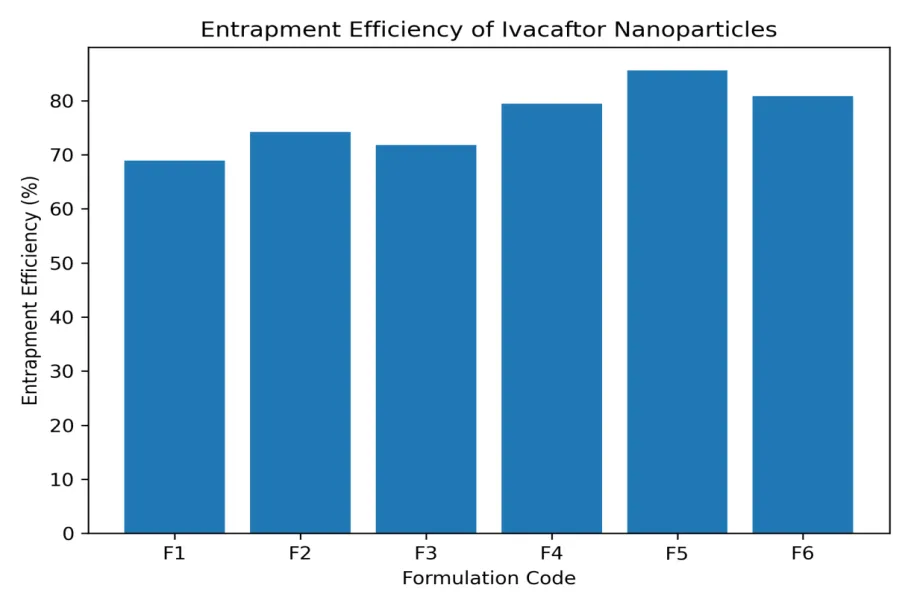
Figure 6: Entrapment Efficiency of Ivacaftor Nanoparticles
4. Zeta Potential
Zeta potential is an indicator of surface charge and colloidal stability of nanoparticle dispersions. All formulations exhibited negative zeta potential values, indicating good stability. Formulation F5 showed the highest negative value, suggesting strong electrostatic repulsion between particles and reduced aggregation. This enhances formulation stability during storage and improves aerosol performance during pulmonary administration.
Among all formulations, F5 demonstrated superior performance across all evaluation parameters, including smallest particle size, lowest PDI, highest yield, maximum drug entrapment, and optimal zeta potential. These characteristics make F5 the most promising formulation for effective nanoparticulate pulmonary delivery.
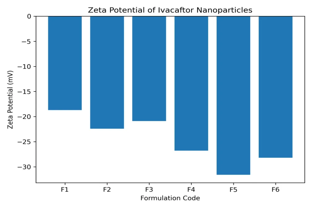
Figure 7: Zeta Potential of Ivacaftor Nanoparticles
Table 2: Evaluation parameters of prepare nanoparticles
|
Formulation |
Particle Size (nm) |
PDI |
Yield (%) |
Drug Content (%) |
Entrapment Efficiency (%) |
Zeta Potential (mV) |
|
F1 |
312.4 ± 6.8 |
0.321 |
73.71 |
72.4 ± 1.8 |
68.9 ± 1.5 |
−18.7 ± 1.2 |
|
F2 |
246.7 ± 4.3 |
0.278 |
79.71 |
78.6 ± 1.6 |
74.2 ± 1.3 |
−22.4 ± 1.4 |
|
F3 |
268.9 ± 5.1 |
0.295 |
77.42 |
75.9 ± 1.4 |
71.8 ± 1.2 |
−20.9 ± 1.3 |
|
F4 |
221.6 ± 3.9 |
0.241 |
84.14 |
82.7 ± 1.5 |
79.4 ± 1.1 |
−26.8 ± 1.1 |
|
F5 |
198.3 ± 3.2 |
0.214 |
87.71 |
88.9 ± 1.3 |
85.6 ± 0.9 |
−31.6 ± 1.0 |
|
F6 |
235.8 ± 4.6 |
0.262 |
84.00 |
84.3 ± 1.2 |
80.8 ± 1.0 |
−28.2 ± 1.2 |
In-vitro Drug Release Study
In-vitro drug release studies were carried out using a Franz diffusion cell with phosphate buffer (pH 6.8) to simulate pulmonary conditions. All formulations demonstrated a biphasic release pattern, consisting of an initial release followed by sustained drug release over 24 hours. The initial phase is attributed to drug present on or near the nanoparticle surface, while the sustained phase corresponds to controlled diffusion from the polymeric matrix.
Among all formulations, F5 exhibited the highest cumulative drug release, reaching approximately 98% at 24 hours. This enhanced release behavior can be attributed to its optimized particle size, uniform distribution, and higher drug entrapment efficiency. In contrast, formulations with larger particle sizes showed comparatively slower release profiles. The sustained and near-complete release from F5 indicates its potential to maintain prolonged therapeutic levels in pulmonary tissues, which is beneficial for effective cystic fibrosis management.
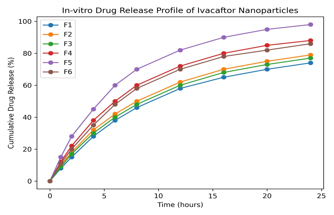
Figure 8: In-vitro Drug Release Profile of Ivacaftor Nanoparticles (pH 6.8)
Overall Evaluation of Nanoparticles
Based on comprehensive evaluation, formulation F5 was identified as the optimized formulation. It exhibited the smallest particle size, lowest polydispersity index, highest nanoparticle yield, maximum drug entrapment efficiency, and the most favorable zeta potential. Additionally, it showed uniform morphology and excellent dispersion stability. These combined properties indicate that the developed nanoparticulate system is highly suitable for pulmonary drug delivery applications.
Stability Study of Ivacaftor-Loaded Nanoparticles
The physical stability of optimized ivacaftor-loaded nanoparticles was evaluated for 6 months under room temperature (20 ± 2 °C) and refrigerated conditions (4 ± 2 °C), with samples analyzed monthly for sedimentation, particle size, and zeta potential. Results are expressed as mean ± SD (n = 3).
At 20 ± 2 °C, no sedimentation was observed up to 2 months, followed by slight, readily redispersible sedimentation from the third month. Particle size increased gradually from 198.3 ± 3.2 to 224.6 ± 5.3 nm, while zeta potential changed from −31.6 ± 1.0 to −27.2 ± 1.6 mV, indicating acceptable colloidal stability.
At 4 ± 2 °C, no sedimentation was observed throughout the study. Particle size showed only a minor increase from 198.3 ± 3.2 to 206.9 ± 4.1 nm, while zeta potential remained relatively stable (−31.6 ± 1.0 to −29.7 ± 1.3 mV), demonstrating superior physical stability.
Table 3: Stability study of optimized ivacaftor-loaded nanoparticles under different storage conditions
|
Storage condition |
Time (month) |
Sedimentation |
Particle size (nm) |
Zeta potential (mV) |
|
20 ± 2 °C |
0 |
Absent |
198.3 ± 3.2 |
−31.6 ± 1.0 |
|
1 |
Absent |
201.1 ± 3.6 |
−30.9 ± 1.1 |
|
|
2 |
Absent |
204.7 ± 3.9 |
−30.1 ± 1.2 |
|
|
3 |
Slight |
208.9 ± 4.2 |
−29.3 ± 1.3 |
|
|
4 |
Slight |
213.5 ± 4.6 |
−28.6 ± 1.4 |
|
|
5 |
Slight |
218.8 ± 4.9 |
−27.9 ± 1.5 |
|
|
6 |
Slight |
224.6 ± 5.3 |
−27.2 ± 1.6 |
|
|
4 ± 2 °C |
0 |
Absent |
198.3 ± 3.2 |
−31.6 ± 1.0 |
|
1 |
Absent |
199.4 ± 3.4 |
−31.3 ± 1.1 |
|
|
2 |
Absent |
200.7 ± 3.5 |
−31.0 ± 1.1 |
|
|
3 |
Absent |
202.1 ± 3.7 |
−30.7 ± 1.2 |
|
|
4 |
Absent |
203.6 ± 3.8 |
−30.3 ± 1.2 |
|
|
5 |
Absent |
205.2 ± 4.0 |
−30.0 ± 1.3 |
|
|
6 |
Absent |
206.9 ± 4.1 |
−29.7 ± 1.3 |
Overall, the optimized ivacaftor nanoparticles remained physically stable for 6 months under both conditions, with refrigerated storage providing better stability, as evidenced by minimal changes in particle size, zeta potential, and absence of sedimentation.
CONCLUSION:
The present study successfully developed Ivacaftor-loaded polymeric nanoparticles using the solvent evaporation method for potential pulmonary drug delivery in cystic fibrosis. Preformulation, FT-IR, and DSC studies confirmed the identity, thermal stability, and compatibility of Ivacaftor with the selected formulation components. Among the developed formulations, F5 was identified as the optimized batch, exhibiting a particle size of 198.3 ± 3.2 nm, low PDI, high nanoparticle yield, 85.6 ± 0.9% entrapment efficiency, and a favorable zeta potential of −31.6 ± 1.0 mV. The optimized formulation demonstrated spherical morphology and a biphasic sustained-release profile, with approximately 98% drug release over 24 h. Six-month stability studies further confirmed acceptable physical stability, with superior stability observed under refrigerated conditions. Overall, the developed Ivacaftor-loaded nanoparticles represent a promising platform for sustained pulmonary drug delivery. However, further studies involving aerodynamic characterization, aerosolization performance, in-vivo pulmonary deposition, pharmacokinetics, and pharmacodynamic evaluation are necessary to establish their suitability for clinical application in cystic fibrosis.
CONFLICTS OF INTERESTS:
All authors declared that the there is no conflict of interest.
REFERENCES

Pooja Durke*, Dr. R. S. Wanare, Design And Development Of Nanoparticulate Dosage Form For Treatment Of Cystic Fibrosis., Int. J. of Pharm. Sci., 2026, Vol 4, Issue 8, 2784-2797. https://doi.org/10.5281/zenodo.21988928
 10.5281/zenodo.21988928