

We use cookies to ensure our website works properly and to personalise your experience. Cookies policy
IPS Academy College of Pharmacy, Rajendra Nagar, A.B. Road, Indore–452012, (M.P) India.
Theranostics — the integration of diagnostic imaging with targeted therapy using a single agent or platform — has become one of the more clinically meaningful developments in modern oncology. The core appeal is straightforward: by using the same molecule to both locate and treat disease, clinicians can directly confirm target expression before committing to therapy, and monitor response during it. This review covers the major theranostic imaging techniques currently in clinical and investigational use, including PET/SPECT-paired radionuclide therapies, nanoparticle-based multimodal platforms, photoacoustic and photothermal approaches, and targeted alpha therapy. The two FDA-approved agents — ¹??Lu-DOTATATE for neuroendocrine tumours and ¹??Lu-PSMA-617 for metastatic castration-resistant prostate cancer — are reviewed in detail, drawing on data from the VISION and TheraP trials.We also examine Actinium-225 as the leading alpha-emitting candidate, the growing interest in biomimetic nanotheranostic platforms, and the early integration of AI into dosimetry workflows. On the practical side, the field still faces real constraints: radiopharmaceutical supply is limited, patient access is uneven, dosimetry remains technically demanding, and long-term safety data are still accumulating. Taken together, the evidence suggests theranostics is moving from niche nuclear medicine into mainstream precision oncology — but getting there will require coordinated work across multiple disciplines.
In 2002, John Funkhouser introduced the term "theranostic" to describe a unified approach combining diagnosis and therapy into a single clinical tool.(3) The concept builds on three components that must work together: a diagnostic function to identify and characterise disease, a therapeutic payload to treat it, and a monitoring mechanism to track the response. None of these is new on its own — what theranostics offers is their integration within a single agent or platform.
Historically, cancer care has been reactive. A patient develops symptoms, gets imaged, receives a diagnosis, and is then treated — usually weeks later with a different agent than the one used
imaging. Theranostics shortens and simplifies this by using one compound to do both. The concept is not new to nuclear medicine; radioiodine therapy for thyroid cancer has been in use since 1946 and remains the clearest example of the theranostic principle in clinical practice.(4)
What has changed recently is scale. The FDA approvals of (Lutathera) in 2018 for metastatic neuroendocrine tumours and ¹⁷⁷Lu-PSMA-617 (Pluvicto) in 2022 for metastatic castration-resistant prostate cancer(1) brought theranostics into the mainstream and triggered a wave of new clinical development. This review covers the current state of the field — the imaging modalities used, the clinical evidence behind the major radiopharmaceutical pairs, the nanoparticle and alpha therapy frontiers, and the barriers that still need to be addressed.
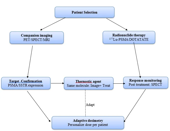
Fig 1: Theranostic Closed loop concept
2. TYPES OF RADIATION IN THERANOSTICS
Radionuclides used in theranostics fall into two broad categories based on their emission type. Diagnostic isotopes emit gamma rays (used in SPECT) or positrons (used in PET). Therapeutic isotopes emit either beta-minus particles or alpha particles, which deposit their energy within tissue to kill tumour cells.(5)
¹⁷⁷Lutetium occupies a unique position because it emits both: therapeutic beta-minus particles and gamma rays of sufficient energy for post-treatment scintigraphic imaging.(5) This means a single
¹⁷⁷Lu-labelled compound can treat and image simultaneously — which is why it has become the dominant radionuclide in current theranostic practice.
Alpha particles work differently. Their linear energy transfer (LET) is approximately 100 keV/μm, and their range in tissue is only 50–100 μm.(6) Within that tiny path length they deposit a concentrated burst of ionising energy that produces clustered DNA double-strand breaks — the most lethal form of radiation damage. Because the range is so short, adjacent healthy tissue is largely spared. This makes alpha emitters particularly well-suited to micrometastatic disease, where small clusters of tumour cells sit within normal tissue.
3. IMAGING MODALITIES IN THERANOSTICS
3.1 PET Imaging
PET detects coincident 511 keV photon pairs produced when a positron emitted from a radiotracer annihilates with a local electron.(7) The high sensitivity and spatial accuracy of this technique has made it the standard method for assessing whether a patient is a suitable candidate for a given radiopharmaceutical therapy. Common PET tracers in theranostics include ⁶⁸Ga, ¹⁸F, ⁸⁹Zr, and ⁶⁴Cu. When combined with CT (PET/CT) or MRI (PET/MRI), it provides complementary anatomical context — PET/MRI being especially useful for soft tissue tumours and vascularised pelvic masses where MRI contrast is superior.(1)
At the preclinical level, ⁶⁴Cu-DOTA-labelled iron oxide nanoparticles have been used to demonstrate that combined PET and near-infrared fluorescence imaging can achieve higher signal- to-noise ratios than MRI alone for distribution mapping — though MRI/micro-CT still provides better spatial resolution when that is the priority.(8)
3.2 SPECT Imaging
Unlike PET, SPECT detects gamma rays emitted directly by the radioisotope without requiring positron annihilation. Commonly used SPECT isotopes include ⁹⁹ᵐTc, ¹¹¹In, and ¹²³I. Compared to PET, SPECT has broader isotope availability and lower cost, and it can image longer-lived radionuclides that PET cannot handle well.(7)
Combining SPECT with PET in the same session — or with MRI or CT — allows more information to be extracted from a single imaging encounter.(7) In the context of ¹⁷⁷Lu-based therapies specifically, post-treatment SPECT using the gamma emissions of lutetium is now routinely used to verify tumour uptake, calculate absorbed dose, and assess early treatment response.(4)
3.3 MRI-Based Theranostics
MRI offers excellent soft tissue contrast without ionising radiation, and has been integrated into theranostics primarily through superparamagnetic iron oxide nanoparticles (SPIONs). These act as T2/T2* contrast agents by influencing the relaxation of surrounding water protons.(9) SPIONs are versatile — they have been used in theranostic platforms for drug delivery, magnetic hyperthermia (where an external alternating magnetic field heats the particles to kill nearby tumour cells), and ferroptosis induction.(10)
The Nanotherm system, an intratumoral SPION formulation activated by an external magnetic field, has received both FDA and EMA approval for glioblastoma treatment via magnetic hyperthermia — one of the clearer examples of a nanotheranostic system reaching clinical use.(10) More complex multimodal systems have also been developed, such as nanocarriers combining MRI contrast (Gd-DOTA), NIR fluorescence (IRDye), SPECT (⁹⁹ᵐTc), and PET (⁶⁴Cu) in a single platform.(8)
3.4 Optical and Photoacoustic Imaging
Photoacoustic imaging (PAI) uses pulsed laser light to generate ultrasound waves from tissue chromophores and nanoparticles — producing images with optical-level contrast at ultrasound- level depth.(11) Gold nanoparticles (AuNPs) are among the more studied agents here. Folic acid- conjugated AuNPs have shown stable, pH-independent behaviour in cervical cancer cell lines, with significantly enhanced reactive oxygen species production — relevant for photodynamic killing of tumour cells.(7)
Phthalocyanine (PC) and naphthalocyanine (NC) dyes have attracted attention as combined PAI/photothermal agents due to their NIR absorption and photostability.(11) Studies testing roughly 40 different NC/PC dye formulations in polymeric micelles have identified several candidates capable of achieving effective photothermal tumour ablation alongside PAI contrast — though most of this work remains preclinical.
3.5 RADIOPHARMACEUTICAL THERANOSTIC PAIRS: CLINICAL APPLICATIONS
3.6 Thyroid Cancer: ¹²³I / Na¹³¹I
The radioiodine pair for thyroid cancer has been in clinical use since 1946 and is, in many ways, the original theranostic system.(4) Both diagnostic (¹²³I or ⁹⁹ᵐTc) and therapeutic (Na¹³¹I) agents exploit the sodium-iodide symporter expressed on thyroid follicular cells. Diagnostic imaging confirms symporter expression and maps the extent of disease; Na¹³¹I then delivers beta-minus radiation via the same mechanism to ablate residual thyroid tissue and any metastases.
What makes this pair historically important is not just its efficacy — it's the conceptual clarity it demonstrates. Confirm the target with a diagnostic agent, then destroy it with a therapeutic one using the same molecular pathway. That principle has guided every theranostic system developed since. Seventy-plus years of clinical data from radioiodine therapy have also provided a safety and efficacy foundation that newer platforms are still building toward.
3.7 Neuroendocrine Tumours: ⁶⁸Ga-DOTATATE / ¹⁷⁷Lu-DOTATATE
Gastroenteropancreatic neuroendocrine tumours (GEP-NETs) express somatostatin receptors at high surface density, which makes them well suited to receptor-targeted theranostics. ⁶⁸Ga- DOTATATE PET/CT provides whole-body SSTR mapping with high sensitivity and is used to confirm receptor expression before treatment.(12)
Lutathera (¹⁷⁷Lu-DOTATATE) shares the same DOTATATE peptide backbone but is labelled with ¹⁷⁷Lutetium rather than ⁶⁸Gallium, delivering beta-minus radiation to SSTR-expressing tumour cells. The FDA approved it in 2018 for metastatic pancreatic and midgut NETs based on the phase III NETTER-1 trial.(1) The low-energy gamma co-emissions of ¹⁷⁷Lu allow post- treatment SPECT for biodistribution verification and dosimetry calculation,(13) completing the imaging-therapy loop.
3.8 Prostate Cancer: PSMA-Based Theranostics
3.8.1 Molecular Target
Prostate-specific membrane antigen (PSMA) is a transmembrane glycoprotein overexpressed in prostate cancer cells at levels roughly 100–1000 times higher than normal tissue, with expression increasing further in castration-resistant and poorly differentiated disease.(14) It is rapidly internalised upon ligand binding, which concentrates a therapeutic payload intracellularly. These properties — high expression, tumour specificity, and efficient internalisation — make PSMA the most clinically exploited molecular target in prostate cancer theranostics.
3.8.2 ⁶⁸Ga-PSMA-11 PET
⁶⁸Ga-PSMA-11 PET/CT is now the standard imaging approach for staging and restaging PSMA- expressing prostate cancer, and it serves as the gating criterion for radioligand therapy — patients who are not PSMA-avid on PET are not eligible for ¹⁷⁷Lu-PSMA treatment. Quantitative PET
metrics also appear to carry prognostic value; higher SUVmean on whole-body ⁶⁸Ga-PSMA-11 PET/CT has been associated with better therapeutic outcomes from PSMA-targeted therapy.(15)
3.8.3 The VISION Trial
The phase III VISION trial (NCT03511664) enrolled patients with mCRPC who were PSMA- positive on ⁶⁸Ga-PSMA-11 PET/CT and had received at least one androgen receptor inhibitor and one or two prior taxane regimens. Patients were randomised to ¹⁷⁷Lu-PSMA-617 (7.4 GBq every six weeks for four to six cycles) plus standard care, or standard care alone.(16)
¹⁷⁷Lu-PSMA-617 produced significant improvements in both imaging-based progression-free survival and overall survival.(16) Quality-of-life analyses showed delayed worsening of HRQOL scores, pain, and time to first symptomatic skeletal event.(17) These results were significant not just for the efficacy data but because they confirmed that a companion diagnostic — PSMA PET — could be used to select patients for a targeted radionuclide therapy and that this selection strategy worked.
3.8.4 The TheraP Trial
The TheraP phase II randomised trial (NCT03392428), run across 11 Australian sites, took a different approach: it directly compared ¹⁷⁷Lu-PSMA-617 against cabazitaxel in mCRPC patients who had progressed on docetaxel. Unusually, it required dual PET imaging — both ⁶⁸Ga-PSMA- 11 and ¹⁸F-FDG PET/CT — to confirm PSMA-avid disease and exclude patients with discordant FDG-avid lesions that might indicate disease unlikely to respond to PSMA-targeted treatment.(2)
¹⁷⁷Lu-PSMA-617 showed superior PSA response rates, better quality of life scores, and a considerably more favourable toxicity profile compared to cabazitaxel.(2) The dual-imaging selection strategy used in TheraP has since become an important reference point for how PSMA theranostic candidates should be identified — particularly because it explicitly defined a subgroup of scordant FDG-avid patients who were unlikely to benefit.
Table 1 — FDA-approved theranostic agents: key characteristics
|
Parameter |
¹⁷⁷Lu-DOTATATE (Lutathera) |
¹⁷⁷Lu-PSMA-617 (Pluvicto) |
|
Indication |
Metastatic GEP-NETs (midgut, pancreatic) |
Metastatic castration-resistant prostate cancer (mCRPC) |
|
FDA approval |
2018 |
2022 |
|
Molecular target |
Somatostatin receptor (SSTR2/5) |
Prostate-secific membrane antigen (PSMA) |
|
Companion diagnostic |
⁶⁸Ga-DOTATATE PET/CT |
⁶⁸Ga-PSMA-11 PET/CT |
|
Key trial |
NETTER-1 (Phase III) |
VISION (Phase III); TheraP (Phase II) |
|
Dosing regimen |
7.4 GBq every 8 weeks × 4 cycles |
7.4 GBq every 6 weeks × 4–6 cycles |
|
Post-treatment imaging |
¹⁷⁷Lu gamma emission → SPECT dosimetry |
¹⁷⁷Lu gamma emission → SPECT biodistribution |
4. NANOPARTICLE-BASED THERANOSTIC PLATFORMS
4.1 Overview
Nanotheranostics brings diagnostic imaging and therapeutic capabilities together within a single nanoscale construct.(18) The primary advantage over conventional small molecules is the capacity to carry multiple functional payloads simultaneously — an imaging agent, a therapeutic drug, and a targeting ligand — within the same particle. Tumour accumulation occurs partly through the enhanced permeability and retention (EPR) effect, whereby nanoparticles leak preferentially through the abnormally large pores in tumour vasculature.(19) Surface chemistry can also be tuned to attach targeting ligands, further improving selectivity.(19)
The main categories of nanotheranostic platform are: metal nanoparticles (gold, iron oxide, platinum, silver); lipid-based systems (liposomes); polymeric nanoparticles (PLGA, PEG-PCL micelles); and biomimetic systems. Each has different properties suited to specific imaging modalities and therapeutic combinations.
4.2 Iron Oxide Nanoparticles (SPIONs)
SPIONs function as T2/T2* MRI contrast agents and can simultaneously produce magnetic particle imaging (MPI) signals and generate heat for hyperthermia therapy.(20) In preclinical work, SPIONs can be loaded with anticancer drugs or siRNA to serve as both imaging agents and drug delivery vehicles. Cross-linked iron oxide (CLIO) nanoparticles conjugated to an angiogenesis inhibitor (ICT2552) have been tested in glioblastoma mouse models, allowing real-time MRI tracking of nanoparticle delivery alongside radiation therapy.(20) The Nanotherm product — already approved by the FDA and EMA for glioblastoma — represents the most direct clinical translation of this technology.
4.3 Gold Nanoparticles
AuNPs are stable, biocompatible, and can be tuned in size and shape to exploit surface plasmon resonance for near-infrared light absorption — making them useful for photoacoustic contrast and
photothermal ablation.(7) Gold nanorods (AuNRs) co-loaded with the NIR dye IR780 and doxorubicin, and functionalised with an RGD peptide targeting αvβ3 integrin, have demonstrated synergistic chemo-photothermal therapy combined with PAI. The temperature increase in these composites was more than 2.5-fold greater than individual components, and photoacoustic signal was roughly three times stronger.(7)
4.4 Polymeric Nanoparticles and Liposomes
Polymeric nanoparticles made from materials like PLGA or PEGylated polymers offer controlled drug release, extended circulation time, and low immunogenicity.(21) Liposomes have been used as theranostic carriers by co-encapsulating radionuclides alongside therapeutic agents. Polymeric micelles — self-assembling nanostructures with a hydrophobic drug-carrying core and a PEG corona — range from 10 to 100 nm and accumulate in tumours via the EPR effect. When radiolabelled, they can provide simultaneous PET/SPECT imaging and drug delivery within a single system.(21)
4.5 Biomimetic Nanotheranostics
Biomimetic platforms try to solve several persistent problems with conventional nanoparticles — multidrug resistance, poor tumour penetration, and immune-mediated clearance — by drawing on biological design principles.(22) Approaches include nanoparticles coated with cancer cell membranes or erythrocyte membranes to evade immune detection, exosomes used as inherently biocompatible drug carriers, and bioinspired pretargeting ligands to improve radiotherapeutic uptake at the tumour site.(22)
Biomimetic systems have been applied in breast, prostate, and skin cancers for applications ranging from immune checkpoint blockade to photothermal ablation.(23) The translational challenge is scalable, consistent manufacturing — biological sourcing of membrane components and exosomes introduces variability that will need to be addressed before these systems move into routine clinical use.
Table 2 - Approved cancer drug therapies based on nanotechnology(24)
|
Product |
Nanoparticle Material |
Company |
Drug Mechanism |
Approval Year |
References |
|
Hensify (NBTXR3) |
Hafnium oxide nanoparticle |
Nanobiotix (Paris, France) |
Radiotherapy |
EMA (2019) |
(25) |
|
Pazenir |
Nanoparticlebound albumin |
Ratiopharm GmbH (Ulm, Germany) |
Paclitaxel |
EMA (2019) |
(26) |
|
Vyxeos |
Liposome |
Celator/Jazz Pharma (NJ, USA) |
Cytarabine/ Daunorubicin |
FDA (2017) EMA (2018) |
(26) |
|
Onivyde |
Liposome |
Merrimack Pharma (MA, USA) |
Irinotecan |
FDA (2015) |
(27) |
|
NanoTherm |
Iron oxide nanoparticles |
MagForce Nanotechnologies AG (Berlin, Germany) |
Thermal ablation with magnetic field |
EMA (2010, 2013) |
(27) |
|
Marqibo |
Liposome |
Talon Therapeutics/Spectrum Pharmaceuticals (MA, USA) |
Vincristine |
FDA (2012) |
(26) |
|
Oncaspar |
Polymer protein conjugate |
Les Laboratoires Servier (Suresnes, France) |
Pegaspargase/Lasparaginase |
FDA (1994, 2006) |
(28) |
|
Genexol-PM |
PEG-PLA polymeric micelle |
Samyang Biopharmaceuticals (Gyeonggi-do, South Korea) |
Paclitaxel |
South Korea (2007) |
(28) |
|
Abraxane |
Nanoparticlebound albumin |
Abraxis/Celgene (NJ, USA) |
Paclitaxel |
FDA (2005) |
(27) |
5. TARGETED ALPHA THERAPY
5.1 Why Alpha Emitters?
The case for alpha particles in cancer therapy comes down to physics. A LET of ~100 keV/μm deposited over a 50–100 μm path in tissue means concentrated, highly lethal DNA damage within a microscopic footprint.(33) The resulting double-strand breaks are dense enough to overwhelm cellular repair mechanisms. And because the range of an alpha particle barely extends beyond a few cell diameters, surrounding healthy tissue is largely spared.(4) This is particularly relevant for micrometastatic disease and haematological malignancies, where tumour cells are dispersed among normal tissue — exactly the setting where the crossfire effect of beta emitters becomes a liability.
5.2 Actinium-225
Among the alpha emitters in clinical development, ²²⁵Ac has attracted the most attention. Its 9.92- day half-life is logistically workable, and each decay generates four alpha particles, three beta-like emissions, and two gamma emissions before the chain ends at stable ²⁰⁹Bismuth.(30) This makes
²²⁵Ac a kind of in vivo nanogenerator: one administered atom produces a cascade of cytotoxic radiation events at the tumour site.(34) The gamma emissions, while limited, also allow SPECT imaging of biodistribution — preserving the theranostic principle.
Clinical trials of ²²⁵Ac in prostate cancer are primarily using PSMA-targeted agents: [²²⁵Ac]Ac- PSMA-617, [²²⁵Ac]Ac-PSMA-I&T, and [²²⁵Ac]Ac-J591.(27) The idea of targeting cancer with ²²⁵Ac has been around since 1993, and three decades of work have moved it from concept to active clinical evaluation.(29)
5.3 Other Alpha Emitters
²²³Radium (t½ = 11.4 days) is already FDA-approved for bone-metastatic prostate cancer. Its calcium-mimetic chemistry drives natural bone uptake, and its short path length limits bone marrow exposure — a meaningful safety advantage in a site that is inherently radiation- sensitive.(32) A range of other alpha emitters are also under active investigation: ²¹¹At, ²¹³Bi, ²¹²Pb,
²²⁷Th, and ¹⁴⁹Tb each have distinct half-lives, energy profiles, and daughter isotope characteristics
suited to different clinical contexts.(29) Phase I trials are ongoing across multiple tumour types for most of these, with Phase III recruitment for neuroendocrine tumours beginning for some.
5.4 Challenges
The main bottleneck for ²²⁵Ac is supply — global production is estimated at around 1,000 Ci/year, which is far below what would be needed for widespread clinical use. Production via thorium-229 decay or particle accelerators is geographically concentrated and logistically complex. Daughter recoil — the displacement of progeny isotopes from the target site after alpha emission — carries a risk of off-target irradiation. The limited gamma signal from small administered quantities also makes SPECT-based dosimetry difficult. Cyclotron-based regional production of ²²⁵Ac and ²¹¹At is in development as a way to reduce dependence on centralised supply, and progress in chelation chemistry and dosimetry modelling will be necessary before the full clinical potential of alpha therapy can be realised.
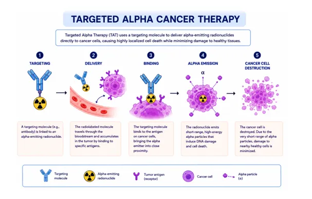
Fig 2: Targeted alpha cancer theraphy mechanism
6. DOSIMETRY AND TREATMENT PERSONALISATION
Dosimetry in radiopharmaceutical therapy is more complicated than in external beam radiotherapy. With external beam, the geometry is fixed and dose can be precisely modelled; with a radiopharmaceutical, the source of radiation moves through the body over time according to patient-specific pharmacokinetics and biodistribution.(4) For longer-lived agents, multiple imaging sessions over several days may be needed to characterise the kinetic curve adequately. The accuracy of any dosimetric estimate also depends on careful calibration of imaging equipment — dose calibrators, SPECT and PET scanners — and appropriate corrections for scatter and attenuation.(4)
The field is moving toward real-time adaptive dosimetry: serial imaging during a treatment course is used to track changing biodistribution, adjust plans, and optimise the dose delivered to tumour while protecting kidneys and bone marrow.(35) This is a meaningful shift from fixed population- based dosing toward something genuinely personalised. Experts in the field have consistently identified improved dosimetry as one of the most important next steps for theranostics broadly,(1) and the clinical case for it is clear — variability in pharmacokinetics between patients is large enough that one-size-fits-all dosing almost certainly undertreats some patients while overtreating others.
7. ARTIFICIAL INTELLIGENCE IN THERANOSTICS
AI is being integrated into theranostics at several points in the workflow — a convergence sometimes referred to as 'TherAInostics'.(34) Current applications include automated tumour segmentation and PSMA quantification from PET images, predictive modelling of treatment response from baseline imaging features, and longitudinal dosimetry monitoring across treatment cycles.(37)
Making this work at scale requires structured data. Large, well-annotated multicentre theranostic registries — integrating dosimetric, genomic, imaging, and outcomes data — will be needed to train models that are useful in individual patient decisions.(35) The 2025 ICPO Forum highlighted multi-tracer imaging combined with AI analytics as a priority area for improving patient selection and response monitoring.(30) It is worth noting that most current AI tools in this space are still early- stage; the clinical validation work required to embed them into standard practice is largely ahead of us.
8. CHALLENGES AND FUTURE DIRECTIONS
8.1 Patient Access
¹⁷⁷Lu-PSMA treatments are currently available mainly at large tertiary cancer centres.(36) Ongoing efforts to scale up production have helped at the supply end, but patients in rural areas still face barriers that have nothing to do with supply: travel costs, multiple required visits, and limited local infrastructure for handling radiopharmaceuticals. The relationship between travel distance and likelihood of pursuing specialty care is well established in the literature,(36) and it directly applies here. Addressing this will require deliberate investment in establishing regional theranostic hubs, not just increasing production of existing agents.
8.2 Expanding Indications
PSMA is not exclusively a prostate cancer target — it is expressed in the neovasculature of several solid tumour types, which opens the door to PSMA-targeted theranostics in other malignancies.(32) A range of newer molecular targets are in clinical trials: FAP, CAIX, GPC3, Trop2, and DLL3 are each expressed in distinct cancer types and being evaluated with matched diagnostic/therapeutic agent pairs.(30) CXCR4 — overexpressed in pancreatic, breast, lung, prostate, and colon adenocarcinoma — has been targeted with [⁶⁸Ga]Ga-Pentixafor and [¹⁷⁷Lu]Lu-Pentixather, showing early promise in multiple myeloma and as pre-transplant conditioning in diffuse large B- cell lymphoma.(5)
8.3 Combination Strategies
Single-agent theranostics will not be enough for many patients. Tumour heterogeneity and resistance mechanisms mean that most durable responses will likely require combinations. Alpha and beta emitters are being paired; ¹⁷⁷Lu-PSMA is being combined with androgen receptor inhibitors, PARP inhibitors, and chemotherapy.(29) Data from TheraP and related analyses suggest that ¹⁷⁷Lu-PSMA-617 outcomes are driven primarily by tumour biology rather than prior treatment history,(33) which is an argument for using it as a backbone in combination regimens rather than reserving it for heavily pre-treated patients.
8.4 Harmonisation and Trial Design
Getting theranostics adopted globally requires standardised practice — consistent dosimetry protocols, patient selection criteria, and quality assurance frameworks across centres.(37) There is growing consensus that multidisciplinary theranostics tumour boards and investment in clinical infrastructure are necessary, not optional.(37)
Trial design also needs to evolve. Standard oncology trial frameworks were built around systemic chemotherapy and do not map well onto radiopharmaceutical therapy, where companion imaging is central to patient selection and response assessment.(38) Adaptive trial designs that incorporate imaging biomarker endpoints alongside traditional survival outcomes are increasingly seen as the right approach for the next generation of theranostic studies.
CONCLUSION
Theranostics has moved a long way from its origins in radioiodine therapy. The FDA approvals of¹⁷⁷Lu-DOTATATE and ¹⁷⁷Lu-PSMA-617(1) were not just regulatory milestones — they validated a way of thinking about cancer treatment that had been theoretical for a long time. By using the same molecular target for both imaging and therapy, theranostics makes patient selection explicit and response monitoring inherent to the treatment itself.
The next phase of development will likely involve alpha emitters offering greater cytotoxic precision for micrometastatic disease, nanotheranostic platforms capable of carrying multiple therapeutic and imaging payloads simultaneously, biomimetic delivery systems that can evade the immune defences that have limited earlier nanoparticle approaches, and AI-assisted dosimetry that allows treatment protocols to adapt to individual patient pharmacokinetics rather than relying on population averages.
The obstacles are real and should not be minimised: supply chains for radiopharmaceuticals remain constrained, access is geographically uneven, dosimetry is technically demanding, and long-term safety data for newer agents are still limited. These are problems that require investment and coordination across academia, industry, and health systems — not just continued research at the bench.
What the evidence reviewed here makes clear is that theranostics is no longer a specialty application within nuclear medicine. It is developing into a core component of precision oncology
— one that offers something genuinely distinct: a closed loop between diagnosis and therapy, where imaging informs treatment and treatment is monitored by imaging in real time. That is a meaningful clinical advance, and it is worth the work required to make it widely available.
REFERENCES

Uzma Khan, Dr. Dinesh Kumar Jain, Theranostics In Oncology: A Review of Imaging Modalities, Radiopharmaceuticals, And Emerging Approaches, Int. J. of Pharm. Sci., 2026, Vol 4, Issue 8, 2840-2852, https://doi.org/10.5281/zenodo.22014983
 10.5281/zenodo.22014983