

We use cookies to ensure our website works properly and to personalise your experience. Cookies policy
Department of Pharmaceutics, Bharath institute of pharmacy college, Mangalpally, Hyderabad, Ranga reddy, Telangana - 501510, India.
The present study was aimed at the design, preparation, and in vitro evaluation of Deoxycortisone-loaded solid lipid nanoparticles (SLNs) using the hot homogenization technique to improve drug delivery and achieve sustained drug release. Preformulation studies, including organoleptic evaluation, melting point determination, solubility studies, calibration curve preparation, and Fourier Transform Infrared (FTIR) spectroscopy, were carried out to assess the physicochemical properties of the drug and its compatibility with the selected excipients. The FTIR results confirmed the absence of significant drug–excipient interactions. Deoxycortisone-loaded SLNs were prepared using glyceryl monostearate as the lipid, Poloxamer 407 and Tween 80 as stabilizers, and methanol–chloroform as the organic solvent system by the hot homogenization technique. The prepared formulations were evaluated for particle size, zeta potential, entrapment efficiency, surface morphology by scanning electron microscopy (SEM), in vitro drug release, and stability. Among the four formulations, F4 was identified as the optimized formulation, exhibiting the smallest particle size (132 nm), satisfactory zeta potential (?23 mV), highest entrapment efficiency (85.16%), and maximum cumulative drug release (98.55%) over 12 hours. SEM analysis revealed spherical nanoparticles with a smooth surface and no significant aggregation, confirming successful nanoparticle formation. Stability studies carried out under ICH-recommended storage conditions for three months demonstrated that the optimized formulation remained physically and chemically stable, with no significant changes in drug release characteristics.
Solid Lipid Nanoparticles are colloidal drug delivery systems composed of a solid lipid matrix in which the active pharmaceutical ingredient is incorporated or entrapped. SLNs generally possess particle sizes in the nanometer range, commonly between approximately 50 and 1000 nm. The lipid matrix remains solid at both room temperature and body temperature, providing a stable environment for incorporation of the drug. The lipid component may include physiological or biocompatible lipids such as glyceryl monostearate, stearic acid, compritol, cetyl palmitate and triglycerides, while surfactants or stabilizers are used to prevent aggregation and improve the physical stability of the formulation.
The development of SLNs was proposed as an alternative to conventional colloidal carriers such as liposomes, emulsions and polymeric nanoparticles. Although conventional systems offer several advantages, they may suffer from problems such as poor physical stability, drug leakage, oxidation and limitations associated with the use of organic solvents. SLNs combine some of the advantages of emulsions and liposomes while providing a solid lipid matrix that can improve drug protection and controlled release. The use of physiologically acceptable lipids and surfactants further increases their suitability for pharmaceutical and biomedical applications.
One of the major advantages of SLNs is their ability to improve the solubility and bioavailability of poorly water-soluble drugs. Drugs incorporated into the lipid matrix may be protected from chemical or enzymatic degradation and can exhibit improved dissolution and absorption characteristics. Depending on the composition and manufacturing method, SLNs can provide controlled and sustained release of the incorporated drug, thereby maintaining therapeutic drug concentrations for a prolonged period and potentially reducing dosing frequency. Their nanoscale particle size also provides a large surface area, which can influence drug dissolution and interaction with biological membranes.
SLNs can also be designed for site-specific or targeted drug delivery. Their small particle size and modifiable surface properties can facilitate interaction with biological barriers and enhance drug deposition in selected tissues. In addition, lipid-based nanoparticles may improve the cellular uptake of certain therapeutic agents. SLNs have therefore been investigated for oral, parenteral, topical, ocular, pulmonary and transdermal drug delivery. They have also attracted interest in the delivery of anticancer agents, antimicrobial drugs, peptides, proteins, vitamins and various natural bioactive compounds.
Several methods have been developed for the preparation of SLNs, including high-pressure homogenization, hot homogenization, cold homogenization, solvent evaporation, solvent emulsification-diffusion, microemulsion technique and ultrasonication. The selection of preparation method depends on the characteristics of the drug and lipid, desired particle size, thermal stability of the drug and intended route of administration. High-pressure homogenization is one of the commonly employed techniques because it can produce nanoparticles with relatively uniform particle size and is suitable for large-scale production.
MATERIALS
Deoxycortisone was procured from Synpharma Research Labs, Hyd, Glyceryl monostearate, Tween 80, Span 20, Poloxamer 407, Dichloromethane and Methanol were obtained from Synpharma Research Labs, Hyderabad. Other chemicals and the reagents used were of analytical grade.
METHODOLOGY
Compatibility Study (IR spectroscopy)
The compatibility between Deoxycortisone and the selected excipients was evaluated using Fourier Transform Infrared (FTIR) spectroscopy. The spectra of the pure drug, excipients, and their physical mixture were recorded over the range of 4000–400 cm⁻¹ and compared to detect any possible chemical interaction.
Method of Preparation of Deoxycortisone Loaded Solid Lipid Nanoparticles:
Deoxycortisone-loaded solid lipid nanoparticles (SLNs) were prepared by the hot homogenization technique. Accurately weighed quantities of Deoxycortisone (10 mg) and glyceryl monostearate (GMS) were dissolved in a mixture of methanol and chloroform (1:2 v/v). The organic solvents were removed using a rotary flash evaporator to obtain a thin lipid film. The lipid film was then heated to approximately 5°C above the melting point of glyceryl monostearate to obtain a homogeneous molten lipid phase. The aqueous phase was prepared by dissolving the required quantity of Poloxamer 407 and Tween 80 (as per the formulation) in 30 mL of distilled water. The aqueous phase was heated to the same temperature as the lipid phase to prevent premature solidification during emulsification. The hot aqueous phase was added slowly to the molten lipid phase under continuous homogenization using a hot homogenizer at 3000 rpm for 30 minutes while maintaining the temperature at 50°C, resulting in the formation of a coarse oil-in-water emulsion. The emulsion was then subjected to probe sonication for 25 minutes to reduce the particle size and obtain a uniform Nano suspension. The resulting hot nanoemulsion was allowed to cool gradually to room temperature, during which the lipid solidified to form solid lipid nanoparticles. The prepared Deoxycortisone-loaded SLN dispersion was transferred into airtight glass containers and stored under refrigerated conditions (4 ± 2°C) until further evaluation.
Table 3: Composition of Deoxycortisone for Preparation of Solid Lipid Nanoparticles
| Ingredients | F1 | F2 | F3 | F4 |
|---|---|---|---|---|
| Deoxycortisone | 10 | 10 | 10 | 10 |
| Glyceryl monostearate | 50 | 50 | 50 | 50 |
| Poloxamer 407 | 20 | 20 | 20 | 20 |
| Tween 80 | 1 | 1.5 | 2 | 2.5 |
| Methanol | 10 | 10 | 10 | 10 |
| Chloroform | 20 | 20 | 20 | 20 |
Evaluation of Deoxycortisone Loaded Nanoparticles:
Particle Size and Zeta Potential
The particle size of the formulation was determined by Dynamic Light Scattering (DLS) with a zeta master (Malvern Instruments, UK) equipped with the Malvern PCS software. Every sample was diluted with distilled water. The surface charge (Zeta potential) was determined by measuring the electrophoretic mobility of the nanoparticles using a Malvern zeta sizer (Malvern Instruments, UK). Samples were prepared by diluting with distilled water.31
SEM Analysis:
The morphology of NPs was studied by a scanning electron microscope. For this purpose, the sample was lyophilized and placed on aluminium stubs and the surface was coated with a layer of gold particles using a sputter coater. The shape of the NPs was determined by scanning electron microscopy (SEM) (XL30, Philips, the Netherlands) at 15 kV and 750 mA.
Drug Encapsulation Efficiency:
Lyophilized nanoparticles 50mg were dissolved in 100ml of phosphate buffer and the drug amount was determined by UV analysis. The encapsulation efficiency was determined as the mass ratio of entrapped Deoxycortisone in nanoparticles to the theoretical amount of the drug used in the preparation.
Amount entrapped
Entrapment Efficiency (%) = --------------------------------- × 100
Total drug loaded
In-Vitro Drug Release Studies:
The in vitro drug release study was carried out using a Franz diffusion cell. The receptor compartment was filled with 10 mL of phosphate-buffered saline (PBS, pH 7.4) and maintained at 37 ± 0.5°C with continuous stirring using a magnetic stirrer. Dialysis membrane was taken and one end of the membrane was sealed. After separation of non-entrapped Deoxycortisone dispersion was filled in the dialysis membrane and other end was closed. The dialysis membrane containing the sample was suspended in the medium. 1ml of aliquots were withdrawn at specific intervals, filtered after withdrawal and the apparatus was immediately replenished with same quantity of fresh buffer medium.
Percentage of drug release was determined using the following formula.
Where, Dt = Total amount of the drug
Da = The amount of drug released
Stability Studies:
Selected Formulation was subjected to stability studies as per ICH guidelines.
Following conditions were used for Stability Testing.
1. 250C/60% RH analyzed every month for period of three months.
2. 300C/75% RH analyzed every month for period of three months.
3. 400C/75% RH analyzed every month for period of three months.
RESULTS AND DISCUSSION
FT-IR Spectrum of Deoxycortisone
FT-IR Spectra of Deoxycortisone and excipients were recorded. All these peaks have appeared in formulation and physical mixture, indicating no chemical interaction between Drug and lipids. It also confirmed that the stability of drug during encapsulation process.
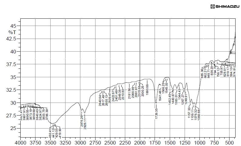
Figure 1: FTIR Studies of Deoxycortisone
Table 2: Characteristic Peaks for Deoxycortisone
| S. No. | Characteristic Peaks | Frequency range (cm-1) |
Frequency (cm-1) |
|---|---|---|---|
| 1 | OH Bending | 4000-3500 | 3907.91 |
| 2 | C- C stretching | 2500-2000 | 2422.67 |
| 3 | C=O stretching | 1500-1000 | 1440.87 |
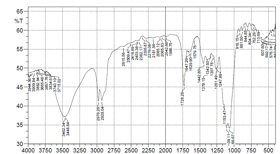
Figure 2: FTIR Studies of Optimized Formulation
Table 3: Characteristic Peaks for Optimized Formulation
| S.No. | Characteristic Peaks | Frequency range (cm-1) |
Frequency (cm-1) |
|---|---|---|---|
| 1 | OH Bending | 4000-3500 | 3857.76 |
| 2 | C- C stretching | 2500-2000 | 2335.87 |
| 3 | C=O stretching | 1500-1000 | 1342.50 |
EVALUATION PARAMETERS
Particle Size:
Particle size analysis revealed that all formulations were within the nanometer range. Among them, formulation F4 exhibited the smallest particle size (132 nm), indicating better particle size distribution and stability.
Zeta Potential
The zeta potential values ranged from −22 to −28 mV, indicating moderate colloidal stability. Formulation F3 exhibited the highest negative zeta potential (−28 mV), while F4 showed satisfactory stability with a zeta potential of −23 mV.
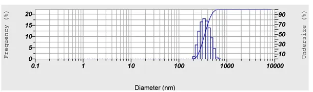
Figure 3: Particle Size Analysis of Optimized Formulation
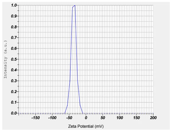
Figure 4: Zeta Potential of Optimized Formulation
Drug Entrapment Efficiency:
Entrapment efficiency of all formulations ranged from 76.93% to 85.16%. Among all formulations, F4 exhibited the highest entrapment efficiency (85.16%), which may be attributed to the optimized concentration of surfactant, resulting in efficient drug incorporation within the lipid matrix.
Table 4: Evaluation Studies of Prepared Solid Lipid Nanoparticles: Entrapment Efficiency and Particle Size
| Batch No | Particle size (nm) | Zeta potential (mV) | Entrapment Efficiency (%) |
|---|---|---|---|
| F1 | 149 | -26 | 76.93 |
| F2 | 151 | -22 | 81.20 |
| F3 | 148 | -28 | 79.86 |
| F4 | 132 | -23 | 85.16 |
Surface Morphology:
According to scanning electron microscopy (SEM), the solid lipid nanoparticles were round, smooth, and free of any aggregation.
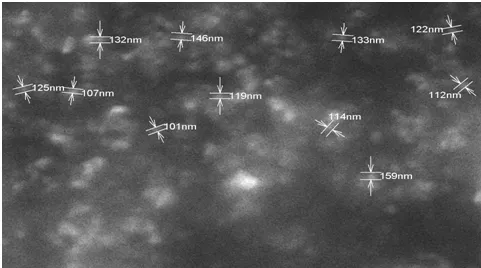
Figure 5: SEM Analysis of Optimized Solid Lipid Nanoparticle
In Vitro Drug Release Studies
In vitro drug release studies demonstrated a sustained release pattern for all formulations over 12 hours. Among the formulations, F4 showed the highest cumulative drug release (98.55%), indicating the optimized lipid–surfactant composition and enhanced drug diffusion from the solid lipid nanoparticles.
Table 5: In Vitro Drug Release Profiles of SLN (F1-F4)
| Time | F1 | F2 | F3 | F4 |
|---|---|---|---|---|
| 0 | 0 | 0 | 0 | 0 |
| 1 | 18.63 | 19.67 | 16.39 | 19.68 |
| 2 | 26.35 | 22.35 | 21.14 | 26.98 |
| 3 | 37.10 | 36.79 | 35.95 | 39.67 |
| 4 | 55.69 | 55.51 | 54.72 | 48.55 |
| 6 | 69.86 | 66.39 | 68.18 | 63.59 |
| 8 | 78.91 | 75.52 | 74.96 | 79.82 |
| 10 | 84.13 | 82.15 | 83.29 | 85.22 |
| 12 | 93.69 | 94.63 | 95.56 | 98.55 |
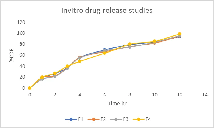
Figure 6: Drug Release for (F1-F4) Formulations
Stability Studies:
The optimized formulation (F4) remained stable under all storage conditions for three months. No significant changes were observed in physical appearance or drug release. The cumulative drug release remained above 95%, indicating good physical and chemical stability of the formulation.
Table 6: Results of Stability Studies of Optimized Formulation F-4
| Formulation Code | Parameters | Initial | 1st Month | 2nd Month | 3rd Month | Limits as per Specifications |
|---|---|---|---|---|---|---|
| F-4 | 250C/60%RH | 98.55 | 97.56 | 96.35 | 95.85 | Not less than |
| F-4 | 300C/75% RH | 98.55 | 97.21 | 96.20 | 95.48 | Not less than |
| F-4 | 400C/75% RH | 98.55 | 97.26 | 96.13 | 95.36 | Not less than |
SUMMARY AND CONCLUSION
The present study successfully formulated and evaluated Deoxycortisone-loaded solid lipid nanoparticles (SLNs) using the hot homogenization technique. Preformulation studies confirmed the purity and suitability of Deoxycortisone for formulation development. FTIR analysis demonstrated the compatibility of the drug with the selected excipients, indicating the absence of any significant chemical interaction.
The prepared SLNs were successfully characterized for particle size, zeta potential, entrapment efficiency, surface morphology, and in vitro drug release. SEM analysis revealed that the nanoparticles were spherical with a smooth surface and showed no significant aggregation. Among all the formulations, F4 was identified as the optimized formulation, exhibiting the smallest particle size (132 nm), satisfactory zeta potential (−23 mV), the highest entrapment efficiency (85.16%), and the maximum cumulative drug release (98.55% at 12 hours).
Stability studies performed under different storage conditions demonstrated that the optimized formulation remained stable for three months without any significant changes in drug release characteristics. These findings indicate that the developed solid lipid nanoparticle formulation is a promising carrier for Deoxycortisone, offering sustained drug release, improved formulation stability, and the potential to enhance therapeutic efficacy. Further in vivo studies are recommended to establish its pharmacokinetic performance and clinical applicability.
REFERENCES

P. V. Pavan Kumar, Sazeeda, S. Alekhya, S. Swetha, S. Sudarshan, S. Charani, Design Prepare and in Vitro Evaluation of Solid Lipid Nanoparticles Loaded Deoxycortisone by Using Hot Homogenisation Technique, Int. J. of Pharm. Sci., 2026, Vol 4, Issue 11, 1377-1384. https://doi.org/10.5281/zenodo.24003706
 10.5281/zenodo.24003706