

We use cookies to ensure our website works properly and to personalise your experience. Cookies policy
M Pharm Pharmaceutics, Karnataka college of pharmacy.
Glaucoma, a chronic, progressive optic neuropathy characterized by retinal ganglion cell degeneration and elevated intraocular pressure (IOP), can cause blindness and permanent vision impairment if treatment is not received. Conventional ocular drug delivery methods, like eye drops, have low bioavailability and often lead to poorer patient compliance because of frequent dosage requirements, poor corneal permeability, and rapid tear turnover. To overcome these limitations, new drug delivery strategies that integrate liposomal carriers with in-situ gel systems have emerged as practical approaches for long-term, targeted ocular treatment.By encasing both hydrophilic and lipophilic drugs, liposomes—biocompatible vesicular structures—improve drug stability, controlled release, and corneal penetration. By going through a sol-to-gel transition in response to physiological cues like temperature, pH, or ionic strength, in-situ gels lengthen precorneal residency and decrease drug loss. Combining liposomes with in-situ gels has synergistic benefits that include longer drug release, improved ocular bioavailability, fewer doses, and increased therapeutic efficacy.This study focuses on the formulation strategies, optimization techniques, polymer selection, production procedures, and physicochemical characterization of liposomal in-situ gel systems for the treatment of glaucoma. Among the important aspects discussed are particle size, stability, gelation behavior, encapsulation efficacy, and zeta potential. The therapeutic potential of the delivery platform and recent developments in lowering IOP and improving patient adherence are also highlighted. All things considered, liposomal in-situ gel formulations are a viable and effective replacement for conventional therapy because they provide longer drug delivery, improved safety, and superior long-term glaucoma control.
The progressive loss of retinal ganglion cells and the ensuing abnormalities in the vision field characterize glaucoma, a complex group of optic neuropathies. Globally, glaucoma is the leading cause of permanent blindness [1] The reason glaucoma is referred to as "the silent thief of sight" is that in its early stages, patients rarely exhibit any symptoms [2].This is the reason the illness has its name. Peripheral and mid-peripheral vision gradually deteriorates due to glaucoma, but the patient is not aware of this loss because the visual cortex fills in the gaps .Both the center and the periphery of the field of vision gradually lose their vision due to glaucoma [3].Despite being the leading cause of irreversible vision impairment worldwide, glaucoma is frequently misdiagnosed. The loss of retinal ganglion cells and weakening of the retinal nerve fiber layer are the hallmarks of glaucoma [4].which is now recognized as a typical progressive degeneration of the optic nerve that may eventually result in specific visual field deficits. The prevalence of glaucoma rises with age, despite the fact that its pathophysiology is not fully understood [5]. Patients often use multiple medications and have multiple comorbidities. Moreover, the most significant risk factor for glaucoma is rising intraocular pressure (IOP)[6] It has been shown that the only proven method of effectively treating the condition is to reduce intraocular pressure. Usually, laser therapy or antiglaucoma medications can accomplish this [7].
Liposomes are synthetic copies of natural membranes. These particles, made up of phospholipids, cholesterol, and transporters, resemble cell membranes and are highly compatible with biological systems. These systems are typically spherical, with polar lipid layers and an aqueous region between them. Liposomes have diameters that range from nanometers to micrometers.Liposomes' physicochemical features enable them to transfer chemicals relevant to medications and food production to different locations. Liposomes can retain hydrophilic therapeutic ingredients in their interior areas, as well as high-fat solubility and amphiphilic compounds in their membranes [8].
The ocular drug delivery system is seen as both crucial and challenging because the human eye is an isolated organ. Additionally, typical opthalmic formulations exhibit a short pre-corneal residence period and low bioavailability due to the rapid and complete removal of medications from pre-corneal lachrymal fluid by solution drainage, lachrymation [9]. and ineffective conjunctival absorption. To address the drawbacks of conventional ophthalmic formulations, numerous attempts have been made to develop stable sustained release in-situ gels [10]. More recent studies in ophthalmic drug delivery systems concentrate on the integration of multiple drug delivery techniques. This entails creating systems that not only increase the length of time the vehicle is in contact with the ocular surface [11].
OBJECTIVE OF THE STUDY
The formulation techniques, polymer types, and therapeutic potential of liposomal in situ gel systems for glaucoma management are assessed in this study. Improving patient compliance, reducing intraocular pressure, and administering ocular medications more effectively are the main goals.
ANATOMY AND PHYSIOLOGY OF EYE
The sclera, choroid, ciliary body, iris, and retina—which is composed of nerve tissue—are the three layers that make up the spherical structure of the eye. The white internal tissues of the eye are shielded by a fibrous layer called the sclera. Light can enter through the cornea, a transparent part at the front. The pigmented iris, the colourful area of the eye (blue, green, brown, hazel, or gray), is formed by numerous blood vessels located in the choroid layer of the sclera [12].
Pictures are sent to the nerve system in the back of the eye by the cornea, a transparent protrusion in the front of the eye. The mature cornea has a radius of 7-8 mm and is a vascular tissue. At the cornea-sclera junction, lachrymal fluid, aqueous humor, and blood vessels supply it with nutrients and oxygen. The epithelium, Bowman's layer, stroma, Descemet's membrane, and endothelium are the five layers that make up the cornea. The main pathways through which drugs are absorbed into the eye are these layers.The corneal epithelium is the main barrier to drug absorption into the eye, in contrast to many other relatively impermeable epithelial tissues (intestinal, nasal, bronchial, and tracheal). The epithelium is squamous stratified, with five to six layers of cells, and has a thickness of 50–100 μm. The tight junctions of the basal cells not only act as an effective barrier against dust particles and most bacteria, but they also facilitate drug absorption. The transcellular or paracellular pathway is the main way that medications pass through the corneal epithelium. Hydrophilic drugs choose the paracellular pathway (passive or altered diffusion via the cells' intercellular gaps), whereas lipophilic drugs choose the transcellular channel for penetration [13].
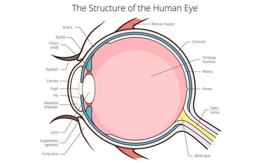
FIGURE. 1 ANATOMY OF EYE
PATHOPHYSIOLOGY OF EYE
Retinal ganglion cell loss is closely linked to intraocular pressure, despite the fact that the exact cause of glaucoma is unknown. Mendelian inheritance is the main mechanism of heredity, but genetics may be involved even though it presents in the typical adult-onset form.[5, 11] Glaucoma can be categorized as "closed-angle" or "open-angle" depending on where the iris and ocular lens are in relation to the trabecular meshwork[14]. The open-angle type is a problem with the trabecular meshwork because drainage is either insufficient or blocked, while the closed-angle type is a problem with the iris's anatomical adhesion to the cornea, which obstructs both the trabecular meshwork and uveoscleral drainage [15].
GLAUCOMA
HISTORY OF GLAUCOMA
Glaucoma changes in the neuroretinal rim tissue in the optic nerve head (ONH) and a subsequent narrowing of the visual field (VF) are the hallmarks of glaucoma [16] .One Glaucoma, a group of eye disorders, is the most common cause of permanent blindness worldwide .Two in addition to the risk of developing glaucoma, there is also a chance that it will go undiagnosed and result in irreversible vision loss. In a sample of 5000 urban Greek adults over 59, it was discovered that 57.1% of cases of glaucoma went undiagnosed [17] .According to a study of 3654 mostly white Australians (90.2% over 60 and 24% over 80), the prevalence of POAG was 3.0%, and 51% had never received a diagnosis [18]. Studies carried out in hospitals or specialty clinics, for example, may be biased to particular classes of referred patients because so many cases of glaucoma may go undiagnosed [19]. As a result, they may not be representative of the glaucoma cohort, which should ideally include all of the undiagnosed cases.
To determine the risk factors for the development of glaucoma [20] .population-based research is required.
GENDER
In the Hypertension Treatment (OHT) study, univariate analysis showed that male gender was a good predictor of the onset of primary open angle glaucoma (POAG) [21]. A Bayesian meta-analysis found that men were more likely to have OAG; however, the gender effect varies depending on the definition of glaucoma [22]. .For example, a review of the literature revealed that women are more likely to develop angle-closure glaucoma (ACG), but there is no clear gender predisposition for OAG .These findings might only be revelant to the groups being studied .Because women usually live longer than men, they are more likely to develop glaucoma and glaucoma blindness[23] .
GLAUCOMA: PATHOPHYSIOLOGY AND CURRENT THERAPEUTIC APPROACH
PATHOPHYSIOLOGY AND TYPES OF GLAUCOMA
Retinal ganglion cell loss is linked to glaucoma, although the precise cause is unknown. A more thorough understanding of the pathophysiological mechanisms underlying the initial.
The advancement of glaucomatous optic neuropathy is crucial to the development of better therapeutic options .The natural physiological balance between the secretion and outflow of aqueous humor is impacted by this disorder. Aqueous humor secreted by the ciliary body drains via the trabecular meshwork and the uveoscleral outflow channel [24].Filtration is impacted by pressure gradients, high IOP, and blood pressure. The humor can move from the ciliary stromal ultrafiltrate pool into the posterior chamber, where it forms watery humor, thanks to osmotic gradients produced by the active release of solutes like sodium and bicarbonate ions [25]. Numerous receptors and transmitters are found in the smooth muscle tissues and ciliary epithelium of the eye. The normal function of the eye involves prostaglandins, muscarinic receptors, sodium and potassium-activated triphosphates, α- and β-adrenergic receptors, and carbonic anhydrase [26].
TYPES OF GLAUCOMA
Glaucoma can be classified as 2 types
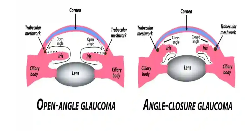
ANGLE-CLOSURE GLAUCOMA
A German ophthalmologist by the name of von Graefe developed a treatment for angle-closure glaucoma more than a century ago, in 1857. Interestingly, it took over 50 years to determine the reason behind the operation's success. A key factor in angle closure glaucoma was discovered in the 1920s: pupillary block, which is the buildup of aqueous humor behind the iris that causes the iris to expand forward like a sail in the wind [27].
Aqueous typically travels from the posterior to the anterior chamber of the pupil.
Water must squeeze between the lens and the iris in order to enter the pupil and, by extension, the anterior chamber. Because of the unusually tight contact between the iris and lens in pupillary block, aqueous cannot enter the pupil. In severe circumstances, the iris enlarges into the trabecular meshwork and the "angle" formed by the iris, trabecular meshwork, and cornea closes because the production of aqueous humor is autonomous. When intraocular pressure increases to the point where blood flow into the eye is compromised, peripheral iridectomy allows aqueous to avoid the pupil and enter the anterior chamber through a short circuit, a surgically made hole in the iris [28].
An eye that has had an attack of angle-closure glaucoma is extremely vulnerable to more attacks if an iridectomy is not performed. Pilocarpine and other miotics offer temporary protection against angle-closure attacks, but they are not a long-term solution. When one eye has angle-closure glaucoma, the contralateral eye is also more likely to develop angle closure. In one study, a 50% incidence of acute angle-closure glaucoma was observed in other eyes within five years after an incident in the first eye.5 The other eye should frequently undergo a prophylactic iridectomy or iridotomy[29].
OPEN ANGLE GLAUCOMA
Due to its extreme discomfort, redness, and blindness, acute angle-closure glaucoma was the most prevalent and dangerous type of glaucoma. However, because open-angle glaucoma is subtle and causes a slow, painless loss of vision in an otherwise normal-looking eye, it was much more difficult to diagnose. Its progression is usually measured in years rather than days, in contrast to angle-closure glaucoma. In actuality, open-angle glaucoma was not recognized as a separate illness until 1862 [30].Open-angle glaucoma results in the irreversible loss of axons and damage to the optic nerve due to the intraocular pressure. Blind spots develop above and below the area of central vision because of this damage. These blind areas gradually increase and merge and develop into arcs, which correspond to the distribution area of the retinal axons because of the progression of the disease. Even though there are highly sensitive psychophysical methods that would detect the vision loss earlier, the central vision usually remains intact until the very end stage of the disease.7 ^{11} Most sufferers do not even realize that their eyesight is lost until late in the course of the illness, when many axons have suffered irreversible damage. A very small center and peripheral island of vision is left in extreme cases ("tunnel vision") [31].So, even if a person has "normal" vision of 20/20, they may still be legally blind because they can't see things on the sides of their eyes. Treatment may generally halt or postpone the progression of the disease; however, it cannot recover lost vision, thus early detection is imperative [32].
TREATEMENTS TO CURE GLAUCOMA
Traditional Treatment Approaches
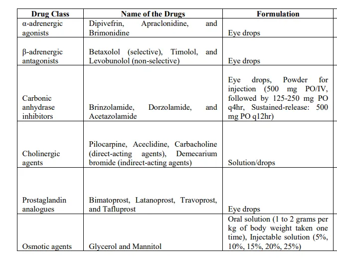
Traditional glaucoma therapy frequently involves the use of medications designed to lower intraocular pressure, a major risk factor for glaucoma. These medications either increase fluid outflow or reduce fluid production in the eye to maintain a healthy pressure level [33].
Anti- Glaucoma Medications
Common glaucoma medications include carbonic anhydrase inhibitors, beta-blockers, alpha adrenergic agonists, and prostaglandin analogues.
SURGICAL OPTIONS
Trabeculectomy
If prescribed eye drops don't significantly reduce intraocular pressure (IOP), your doctor might suggest traditional glaucoma surgery. The most popular surgical procedure is trabeculectomy, also referred to as filtration surgery.
uring this procedure, a small hole is created in the sclera and covered with a thin trapdoor. This makes it possible for the excess aqueous humour—the clear fluid that sits between the cornea and lens of the eye—to drain down the trapdoor into a small reservoir that is situated just below the surface of the eye and concealed by the eyelid. This reduces intraocular pressure and delays the onset of glaucoma [34] .
Approximately 50% of patients do not need glaucoma medication for a significant period of time following trabeculectomy. Even though the procedure effectively reduces intraocular pressure, it's crucial to keep in mind that there is no cure and that patients may still experience visual loss after the procedure [35].
Minimally invasive glaucoma surgery (MIGS) offers a safer and less invasive method of lowering intraocular pressure (IOP) than traditional surgery, potentially eliminating the need for topical medications. It is often used in conjunction with phacoemulsification cataract surgery, according to multiple clinical studies. These procedures currently target patients with mild-to-moderate glaucoma. It lowers IOP in four main ways: it improves trabecular outflow by avoiding the juxtacanalicular trabecular meshwork (TM); it facilitates uveoscleral suprachoroidal outflow through pathways; it reduces aqueous production from the ciliary body; and it creates a sub conjunctival drainage pathway [36]
It has renewed interest in lowering intraocular pressure (IOP) in eyes affected by glaucoma through laser trabeculoplasty. The safety profile of selective laser trabeculoplasty (SLT) includes eye discomfort, mild and temporary inflammation, and a small risk of a significant increase in IOP after the procedure. Although the exact mechanism of SLT is not fully understood, it causes less damage to angle tissues by delivering less energy to the trabecular meshwork. This treatment can be used as primary or additional therapy for glaucoma-affected eyes, and it is a safe and effective method to lower intraocular pressure (IOP) [37].
Both patients with angle-closure glaucoma and those at risk of developing it can benefit from this operation. Laser energy is used to create a tiny hole in the iris, which is the colorful part at the front of the eye. This helps open the drainage angle and manage or prevent angle-closure glaucoma. The unaided eye cannot see this hole. The goal of this treatment is to lower the risk of vision loss from glaucoma and to avoid high eye pressure. There is a 66 to 75% chance of "curing" the disease if the operation is done in its early stages. If the operation is performed later, it can help slow down or stop the disease's progression [38].
Anti-glaucoma medications that are currently available need help getting past the blood-retinal barrier or achieving good systemic bioavailability. Because of this, using medications with lower therapeutic indices requires concentrated solutions to build up in the eye. This can lead to harmful effects and damage cells. To improve the effectiveness of anti-glaucoma treatments, new drug delivery methods such as in-situ gels, liposomes, niosomes, hydrogels, dendrimers, nanoparticles, solid lipid nanoparticles, microneedles, or ocular inserts are necessary[35] .
OVERVIEW OF LIPOSOMAL DELIVERY
Both hydrophilic and hydrophobic medications can be encapsulated in liposomal drug delivery systems, which are flexible nanoscale carriers composed of phospholipid bilayers. Liposomes have attracted a lot of attention since they were developed in the 1960s because of their special ability to improve bioavailability, prevent drug degradation, and enable controlled drug release. To enable customized drug delivery, the size, charge, and lipid content of these vesicles can be altered.
Liposomal formulations provide an ideal platform for brain-targeting therapies because they are especially good at delivering drugs through biological barriers like the blood-brain barrier and the nasal mucosa. The phospholipid bilayer structure of liposomes, which resembles the normal cell membrane, enhances their ability to adhere to cellular membranes and transport therapeutic substances straight into cells. Furthermore, liposomes have longer circulation times and can avoid immune system detection, which boosts the efficacy of medications that would typically be quickly absorbed or eliminated from the body [39]
ADVANTAGES OF LIPOSOMAL DELIVERY
Liposomal drug delivery systems have gained significant attention due to their ability to encapsulate both hydrophilic and hydrophobic medicines, improve drug stability, and prolong drug circulation in the body. Phospholipid bilayers form small, round vesicles called liposomes. These liposomes can enhance a drug's ability to cross biological barriers like the nasal mucosa while also protecting it from enzyme breakdown. When combined with in situ gel technology, liposomal formulations offer additional benefits. These include delayed release, a longer time at the administration site, and improved patient compliance [40].
STRUCTURE OF LIPOSOMES
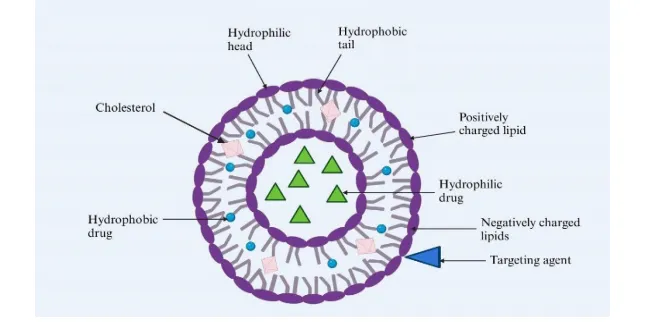
Liposomes are used in many areas today. These include gene therapy, targeted delivery as an antitumor and anticancer medicine, prolonged release, cosmetics like face serum and liposomal lotions, improved penetration and agriculture.
Phosphatidylcholine, whether natural or synthetic, and cholesterol combine to form liposomes. Cholesterol, being a sterol, helps maintain the stability of liposomes.
Liposomes are spherical vesicles that measure between 50 and 500 nm in diameter. Their size can vary from 0.025 to 2.5 µm [41].
TYPES OF LIPOSOMES
Types of Liposomes
Liposomes can be broadly classified based on size and number of bilayers, composition, and method of preparation.
1. Based on Size and Number of Bilayers
2. Based on Composition
3. Based on Method of Preparation
PREPARATION OF LIPOSOMES
1 .Thin-Film Hydration Method
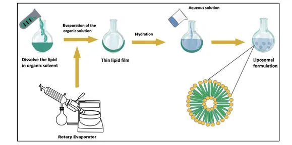
Thin-film hydration, which dissolves the phospholipids in organic solvents like dichloromethane, chloroform, ethanol, and chloroform-methanol mixtures (2:1 v/v, 9:1 v/v, and 3:1 v/v), is the most widely used and simple method for producing MLV. A thin, homogeneous lipid layer is produced when the solvent evaporates under vacuum at a temperature of 45 to 600 C. Nitrogen gas is used to completely remove any remaining solvent. Distilled water, phosphate buffer, phosphate saline buffer at pH 7.4, and regular saline buffer 2-4 are used in the hydration stage [42] .
The hydration process took one to two hours at 60 to 700 C. To achieve full lipid hydration, the liposomal solution is maintained at 4 0 C throughout the entire night. Any kind of lipid combination can be treated with the thin-film hydration method. The method's poor encapsulation, uneven size distribution, and challenges with scaling up are its main drawbacks[43]
2 .Ether Injection Method
The ether injection method involves gradually injecting a solution of lipids dissolved in ether or diethyl ether/methanol into an aqueous solution of the item to be capsulated. When the organic solvent is subsequently extracted at a lower pressure, liposomes are produced. The main disadvantages of the method are the heterogeneous population and the chemicals to be encapsulated being exposed to organic solvents or high temperatures [44].
3. Ethanol Injection Method
The ethanol injection method involves quickly injecting a large excess of heated distilled water or TRIS-HCl buffer with an ethanolic lipid solution. The drug's hydrophilic or hydrophobic nature determines whether it can be incorporated into liposomal vesicles. Compared to 5-fluorouracil, which migrates to the external aqueous phase, nimesulide, a lipid-soluble component, integrates better in liposomes. The primary benefits of the ethanol injection method are its easy scaling up and use of a safe solvent like ethanol. Its applicability is diminished by the potential for azeotrope formation with water [45].
4. Sonication method
Based on size transformation, the sonication technique involves using sonic energy to sonicate MLVs created by the thin-film hydration approach, usually in an inert environment containing nitrogen or argon. The sonication technique enables the uniform dispersion of small vesicles with the potential for increased tissue penetration using a bath-type or probe-type sonicator. The probe tip sonicator provides a substantial amount of energy to the lipid solution. The possibility of the lipid suspension overheating causes degradation. Sonication tips frequently release titanium particles into the lipid mixture; these particles need to be separated by centrifugation before being utilized. The bath sonicator is the most widely used tool for SUV preparation. Large volumes of diluted lipids are treated with them [46].
5.High-Pressure Extrusion Method
In the high-pressure extrusion technique, the MLVs, which are produced through the thin-film hydration technique, are passed through filters consisting of polycarbonate membranes to decrease the sizes of the liposomes. In the thin-film hydration technique, liposomes are produced, and they undergo extrusion techniques for a period of 10 cycles to ensure uniform sizes of liposomes [47].
6. Dehydration-Rehydration Method
This process of dehydration-rehydration is also utilized for the preparation of liposomes. The sonication creates the small unilamellar vesicles, and these are composed of phosphatidylcholine, 1,2-dioleoyl-3-trimethylammonium-propane, cholesterol, and plasmid DNA. The mixture is frozen and freeze-dried overnight. The controlled rehydration of the resultant dry powders resulted in the formation of multilamellar dehydration-rehydration vesicles containing DNA within their structure due to the binding of cationic charges of the inner bilayers [48].
7. Microfluidization
A microfluidization or microemulsification-based technique is applied for large-scale production of liposomes. Boltič et al. described the preparation of antimicrobial liposomes by thin-layer hydration approach, sonication using bath-type sonicator, and microfluidization for partial homogenization.
Microfluidization represents one of the few repeatable methods for producing liposomes with high aqueous phase encapsulation [49].
CHARACTERIZATION AND EVALUATION OF LIPOSOME
Size and Size distribution
In liposomes, the average size depends highly on the manufacturing method and the lipid concentrate used. For the determination of the average liposome sizes within an aqueous solution, the Dynamic Light Scattering technique is employed. These methods are well-known for their ability to provide information on the sizes dispersing. Indirect methods involving the imaging of liposomes can be made possible through Transmission and Scanning Electron Microscopy, which give information qualitatively on sizes, shape, and presence or absence of fusion/aggregations. Key information on thickness and distance between the layers can also be generated by this imaging. After the manufacturing process, liposomes will on average be 0.1–0.2 µm. Size metrics become crucial as they can influence the penetration as well as the absorption by cells, as well as blood circulation time, which influence the efficacy of therapy [50].
Surface charge
The presence of charge on the surface of liposomes is of prime importance for them to be stable, well-encapsulated, and responsive to biological tissues.
Liposomes can also become positively or negatively charged, depending on the type of lipids used, or they can remain neutral.
Increased levels of zeta potential, which describe electrostatic repulsion between like-charged particles, are an indication that there are lower levels of particle aggregation. For increased levels of liposomes in drug delivery formulations and to enhance their efficacy and reduce side effects, charge modification on the surface of liposomes has to be performed [51].
Zeta potential
The suspension medium, the adsorbed layers, and the surface charge of the lipid vesicles all influence the zeta potential. Though it cannot be determined directly, it can be determined using theoretical equations based on the electrophoretic mobility of the particles. By inhibiting the processes of fusion, coalescence, and the precipitate, a high zeta potential can, therefore, ensure the integrity of the liposome. Steady circulation in the bloodstream can also depend on the surface charge of the liposome. Levels of the zeta potential can, therefore, be dependent on the lipid concentration. Filion and Phillips employed a zetasizer, as well as Doppler electrophoretic light scatting, with the use of laser Doppler electrophoresis, to determine the zeta potential of cationic liposomes [52].
Morphology: Transmission electron microscopy (TEM) and cryogenic transmission electron
The morphology and internal structure of liposomes can also be visualized at the nanoscale dimension through powerful imaging methods such as microscopy, also referred to as cryo-TEM. TEM provides detailed images of liposome morphology. These techniques help explain the liposome size, shape, lamellarity, and integrity of the vesicles [53].
Determination of Lamellarity
The number of lipid bilayers present in liposomes can be quantified by electron microscopy or spectroscopic analysis for lamellarity. Verifications of layering can also be supported by investigations of encapsulation efficacy performed by using hydrophilic labels. An understanding of lamellarity is critical for understanding the stability and drug carrier capabilities of liposomes [54].
APPLICATION OF LIPOSOME
Liposomes in medicines
Liposomes, as customized drug delivery carriers, have become a crucial part of the medicinal field. They have fewer side effects as well as the capability of delivering medicated drugs to the targeted organs. It is notable that the use of liposomal formulations in the treatment of cancer is effective, with fewer toxicities compared to traditional medicines, although the problem of bioavailability prevails. Liposomes can cross the blood-brain barrier for the treatment of neurological disorders [55] .They also help in the treatment of lung-related disorders. Liposomes can be administered using multiple techniques, which enhances their use in the treatment of a disease [56].
Liposomes in Infectious and Parasitic diseases
With the ability to be taken up by phagocytic cells, liposomes turn out to be powerful carriers in the management of parasitic and infectious diseases. This "Trojan horse" mechanism allows them to deliver drugs directly to infected cells, thus enhancing therapeutic efficacy against parasites and pathogens. They also enhance the stability and bioavailability of therapeutic agents with minimal toxicity; all these features make the liposomal formulations useful in the treatment of diseases such as leishmaniasis and tuberculosis [57].
Liposomes in Anticancer therapy
Because they can deliver drug medication, thereby enhancing the effectiveness of chemotherapeutic medications, liposomes are critical components of anti-cancer medication.
Liposomes help to reduce toxicity and improve stability by encapsulating medications, thus allowing high doses of medications to reach cancerous areas while preserving other body parts from any damage. Such a targeted treatment might lead to fewer side effects and improved patient compliance. There are currently several liposomal medications approved that have improved pharmacological properties compared to current chemotherapy, including Doxil. However, problems associated with absorption and efficacy remain, requiring more research in this area [58].
Liposomes for Respiratory Drug Delivery System
There is also increased recognition of the effectiveness of liposomes as a drug-delivery system for the treatment of respiratory ailments. They can also be aerosolized for inhalation, which can allow for precise targeting of the medication to the lungs. This is opposed to current inhalation drug-delivery systems that have more adverse effects and are often uncomfortable. Additionally, liposomes can enclose hydrophilic, hydrophobic, and other types of drugs [59].
Liposomes are Brain Targeted Drug Delivery
Since the ability of liposomes to cross the blood-brain barrier (BBB) has been established, they have been considered very promising as the targeted approach in the delivery of medications into the brain. The approach consists of encapsulating the desired medication molecules into liposomes. As a result, medications can be administered effectively to the brain tissues with reduced adverse effects in the body. The approach is very effective when considering the treatment of neurological disorders as well as brain malignancies because it enhances the targeting of medication towards brain cells by changing the surfaces of the liposomes with specific ligands [60].
INSITU GEL TECHNOLOGY
The modes of drug delivery have had a tremendous change, especially in the area of ophthalmology, due to the development of in situ gel technologies for the controlled or extended release of drugs. In a reaction, the polymer employed here can change from a sol to a gel. This occurs due to changes in the physiological factors of temperature, pH, or ionic strength.
MECHANISM OF INSITU GEL FORMULATION
Temperature-triggered gelation: At certain temperatures, such as above body temperature, polymers change from liquid to gel.
pH- sensitive gelation: When pH levels shift, polymers undergo a phase transition (e.g., from acidic to alkaline).
Ion-activated gelation: Ionic interactions with certain ions (like calcium ions) in the environment cause gelation.
Swelling controlled: Gels expand as they absorb water, creating a gel matrix that holds medications.
Diffusion controlled: Drug release is regulated by the diffusion of solvent into the polymer matrix.
Chemically controlled: involves chemical processes like precipitation from supersaturated solutions or enzymatic degradation.
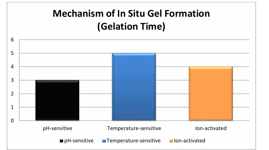
Fig. The gelation times for a number of methods of gel formation in situ are presented in this bar graph. Ion-activated and temperature-sensitive gels come next after pH-sensitive gels in terms of gelation rates.
ADVANTAGES OF INSITU GEL
POLYMERS USED IN THE INSITU GEL DELIVERY
Enhancing the effectiveness of pharmaceutical products requires careful selection of the appropriate polymer for the formulation. Materials that show a sol to gel transition in aqueous solution are used in in-situ gelation. Polymers that may gel in-situ include poloxamer, pluronics, and other co-polymers such as PEO-PLLA and PEG-PLGA-PEG. Gellan gum, cellulose acetophalate latex, pectin, gelrite, alginate, matrigel, carbopol, and chitin. Temperature variations result in gel formation for poloxamer, cellulose, and acetophalate latex; pH variations result in gelation for carbopol [62].
CONCLUSION
Glaucoma remains a major cause of irreversible blindness worldwide, primarily due to poor patient adherence and limited bioavailability associated with conventional ocular dosage forms such as eye drops. Rapid tear turnover, short precorneal residence time, and low corneal permeability significantly reduce therapeutic efficacy. The integration of liposomal drug delivery with in-situ gel technology represents a promising strategy to overcome these limitations and enhance glaucoma management.
Liposomal carriers provide excellent biocompatibility, the ability to encapsulate both hydrophilic and lipophilic drugs, protection of drugs from degradation, and controlled release characteristics. Meanwhile, in-situ gels undergo a sol-to-gel transition in response to physiological stimuli such as temperature, pH, or ionic strength, thereby increasing ocular residence time and minimizing drug loss through nasolacrimal drainage. The synergistic combination of these systems results in prolonged drug release, improved corneal penetration, enhanced bioavailability, and reduced dosing frequency, ultimately improving patient compliance.
Optimization parameters including particle size, zeta potential, encapsulation efficiency, viscosity, gelation time, and stability play a critical role in achieving effective therapeutic performance. Recent studies demonstrate that liposomal in-situ gel formulations can significantly lower intraocular pressure while minimizing systemic side effects.
Overall, liposomal in-situ gel systems offer a safe, efficient, and patient-friendly platform for sustained ocular drug delivery in glaucoma therapy. Continued research, scale-up studies, and clinical validation are essential to translate these advanced formulations into commercially viable products and improve long-term clinical outcomes for glaucoma patients.
REFERENCES

Punith Gowda L, S Swetha Malika Devi, Dr Beny Baby, Harish Gowda S M, Sumanth N, Development And Characterization of Liposomal Insitu Gel for Enhanced Glaucoma Therapy, Int. J. of Pharm. Sci., 2026, Vol 4, Issue 9, 2297-2314, https://doi.org/10.5281/zenodo.22867570
 10.5281/zenodo.22867570