

We use cookies to ensure our website works properly and to personalise your experience. Cookies policy
Maharaja Agrasen School of Pharmacy, Maharaja Agrasen University, Baddi, Himachal Pradesh, India
Background: Allergic disorders are among the most prevalent chronic inflammatory diseases worldwide and are primarily mediated by immunoglobulin E (IgE), mast cell activation, and the release of inflammatory mediators such as histamine. Although conventional antiallergic drugs provide symptomatic relief, their prolonged use is frequently associated with adverse effects and poor patient compliance. Nepeta cataria L. (family: Lamiaceae) is a medicinal herb rich in bioactive phytoconstituents including nepetalactones, rosmarinic acid, flavonoids, and phenolic compounds, which possess significant anti-inflammatory, antioxidant, and antiallergic activities. However, the therapeutic application of herbal extracts is often limited by poor stability, low bioavailability, and inadequate skin permeation. Proniosomal drug delivery systems have emerged as promising vesicular carriers capable of enhancing the stability, encapsulation efficiency, and controlled release of herbal bioactives. Objective: The present study aimed to develop and characterize proniosomal formulations containing Nepeta cataria leaf extract and to evaluate their physicochemical properties and potential antiallergic activity for improved therapeutic performance. Methods: Nepeta cataria leaves were successively extracted using suitable solvents and subjected to preliminary phytochemical screening and preformulation studies, including UV-visible spectroscopy and FTIR analysis. Proniosomes were prepared by the slurry method using Tween 80, cholesterol, and suitable carrier materials. The prepared formulations were evaluated for vesicle size, polydispersity index, zeta potential, entrapment efficiency, morphology, drug content, in vitro drug release, and stability. The optimized formulation was further assessed for its antiallergic potential using suitable in vitro and/or in vivo experimental models and compared with the crude extract. Results: The prepared proniosomal formulations exhibited satisfactory physicochemical characteristics with uniform vesicle formation, high entrapment efficiency, acceptable particle size distribution, and good stability. The optimized formulation demonstrated sustained and significantly enhanced drug release compared with the crude extract, indicating successful encapsulation of the phytoconstituents. Morphological studies confirmed the formation of spherical vesicles, while FTIR analysis revealed the absence of significant drug–excipient interactions. Evaluation of antiallergic activity demonstrated that the optimized proniosomal formulation produced superior inhibition of allergic responses compared with the plain extract, suggesting enhanced therapeutic efficacy due to improved bioavailability and controlled release. Conclusion: The developed proniosomal formulation of Nepeta cataria extract successfully improved the physicochemical properties, stability, and release characteristics of the herbal bioactives while enhancing their antiallergic activity. The findings suggest that proniosomal vesicular delivery represents a promising approach for the effective delivery of herbal antiallergic agents and may serve as a potential alternative to conventional dosage forms.
Allergic disorders are among the most prevalent chronic inflammatory diseases worldwide and represent a significant public health concern because of their increasing incidence and socioeconomic burden. According to the World Allergy Organization (WAO), nearly 20–30% of the global population is affected by one or more allergic diseases, including allergic rhinitis, bronchial asthma, atopic dermatitis, urticaria, conjunctivitis, and food allergies [1]. The prevalence of these disorders has increased considerably during the past few decades due to rapid urbanization, industrialization, environmental pollution, climate change, dietary modifications, and increased exposure to allergens [2]. Allergic diseases are characterized by exaggerated immune responses against otherwise harmless environmental antigens. These reactions are primarily mediated by immunoglobulin E (IgE), which binds to high-affinity FcεRI receptors on mast cells and basophils. Upon subsequent exposure to allergens, cross-linking of IgE molecules triggers mast cell degranulation and the release of numerous inflammatory mediators, including histamine, leukotrienes, prostaglandins, platelet-activating factor, proteases, and pro-inflammatory cytokines. These mediators are responsible for vasodilation, increased vascular permeability, mucus hypersecretion, bronchoconstriction, itching, erythema, and tissue inflammation that characterize allergic reactions [2–4].
Current pharmacological management of allergic disorders primarily involves antihistamines, corticosteroids, leukotriene receptor antagonists, mast-cell stabilizers, and monoclonal antibodies. Although these therapeutic agents effectively relieve allergic symptoms, prolonged administration is often associated with several adverse effects, including sedation, dry mouth, immunosuppression, osteoporosis, metabolic abnormalities, gastrointestinal disturbances, and reduced patient compliance [5]. Furthermore, these therapies generally provide symptomatic relief rather than addressing the underlying inflammatory processes. Consequently, there has been growing interest in identifying naturally derived therapeutic agents possessing anti-inflammatory, antioxidant, immunomodulatory, and mast cell-stabilizing properties with improved safety profiles. Medicinal plants have therefore emerged as promising alternatives because they contain structurally diverse phytochemicals capable of modulating multiple signaling pathways involved in allergic inflammation [6].
Among medicinal plants, Nepeta cataria L., commonly known as catnip or catmint, belongs to the family Lamiaceae and has gained considerable scientific attention owing to its extensive traditional medicinal applications and diverse pharmacological activities. The genus Nepeta comprises nearly 250 species distributed throughout Europe, Asia, North Africa, and North America. Traditionally, N. cataria has been employed in various systems of medicine for the treatment of fever, cough, bronchitis, asthma, common cold, gastrointestinal disorders, anxiety, insomnia, headache, muscle spasms, inflammatory conditions, and infectious diseases [7,8]. Ethnopharmacological reports indicate that infusions and decoctions prepared from the aerial parts of the plant have been widely used as sedative, antispasmodic, carminative, diaphoretic, expectorant, and digestive remedies. The increasing scientific validation of these traditional uses has stimulated extensive investigations into the phytochemical composition and pharmacological potential of N. cataria [7–9].
Phytochemical investigations have demonstrated that N. cataria contains a wide spectrum of biologically active constituents, including iridoid monoterpenes (particularly nepetalactones), flavonoids, phenolic acids, tannins, triterpenoids, sterols, alkaloids, and volatile oils [8,10]. Nepetalactone is recognized as the major constituent of the essential oil and contributes significantly to the plant's characteristic aroma and biological activities. Other important phytochemicals include rosmarinic acid, chlorogenic acid, caffeic acid, luteolin, apigenin, quercetin derivatives, and various polyphenolic compounds possessing remarkable antioxidant and anti-inflammatory properties [10,11].
These phytoconstituents have been reported to inhibit reactive oxygen species generation, suppress lipid peroxidation, downregulate nuclear factor-kappa B (NF-κB), and reduce the production of inflammatory mediators such as tumor necrosis factor-alpha (TNF-α), interleukin-4 (IL-4), interleukin-5 (IL-5), and interleukin-13 (IL-13). In addition, several flavonoids isolated from medicinal plants exhibit mast cell-stabilizing activity by preventing calcium influx and inhibiting histamine release, thereby reducing immediate hypersensitivity reactions [11,12].
Several pharmacological studies have confirmed that extracts and essential oils of N. cataria possess antioxidant, antimicrobial, anti-inflammatory, analgesic, antispasmodic, anxiolytic, bronchodilatory, and immunomodulatory activities [8–10]. The bronchodilatory and smooth muscle relaxant effects of the plant support its traditional use in respiratory disorders associated with allergic conditions. Moreover, the strong antioxidant potential of N. cataria protects tissues from oxidative stress, which plays a pivotal role in the pathogenesis of allergic inflammation. These pharmacological properties collectively indicate that N. cataria represents a promising herbal candidate for the development of novel antiallergic formulations. However, despite its therapeutic potential, the clinical application of the herbal extract remains limited because many of its bioactive constituents exhibit poor aqueous solubility, limited membrane permeability, chemical instability, and low oral bioavailability. These limitations necessitate the development of advanced drug delivery systems capable of enhancing the stability, bioavailability, and therapeutic efficacy of the herbal bioactive compounds [13–15].
2. MATERIALS AND METHODS
2.1 Materials
The aerial parts of Nepeta cataria L. were collected from Palampur, Kangra district, Himachal Pradesh, India, during September 2025 and authenticated at the Council of Scientific and Industrial Research–Institute of Himalayan Bioresource Technology (CSIR-IHBT), Palampur. Phospholipid, cholesterol, Tween 80, Span 60, Carbopol 934, methanol, chloroform, petroleum ether, ethyl acetate, and all analytical-grade chemicals were procured from standard commercial suppliers and used without further purification.
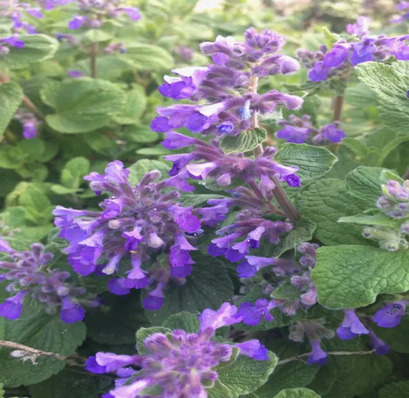
Figure 2.1: Aerial parts of Nepeta cataria
2.2 Preparation of Plant Extract
The dried aerial parts were powdered and subjected to successive Soxhlet extraction using petroleum ether, ethyl acetate, and distilled water. Each extraction was continued until complete exhaustion of the solvent. The extracts were concentrated under reduced pressure using a rotary vacuum evaporator and dried under vacuum. The dried extracts were stored in airtight containers at 4°C until further use.
2.3 Preliminary Phytochemical Screening
The petroleum ether, ethyl acetate, and aqueous extracts were qualitatively screened for alkaloids, flavonoids, phenolics, tannins, saponins, glycosides, steroids, terpenoids, proteins, carbohydrates, and coumarins using standard phytochemical methods.
2.4 UV–Visible Spectrophotometric Analysis
The extract was dissolved in methanol to prepare an appropriate stock solution. The absorption spectrum was recorded between 200 and 400 nm using a UV–Visible spectrophotometer with methanol as the blank. The wavelength of maximum absorbance (λmax) was selected for quantitative estimation according to Beer–Lambert's law.
2.5 Fourier Transform Infrared (FTIR) Analysis
The dried extract was mixed with potassium bromide (KBr), compressed into pellets, and scanned using an FTIR spectrophotometer over the range of 4000–400 cm⁻¹ to identify characteristic functional groups.
2.6 Preparation of Proniosomes
Proniosomes containing Nepeta cataria extract were prepared by the slurry method. Briefly, Tween 80 or Span 60, cholesterol, and the extract were dissolved in chloroform: methanol (1:1). The lipid solution was added to sucrose or sorbitol carrier particles and evaporated under reduced pressure using a rotary evaporator at 45°C. The resulting dry proniosomal powder was stored in airtight containers at 4°C until further evaluation.
2.7 Preparation of Niosomes
Proniosomal powder was hydrated with phosphate buffer (pH 6.8) at 80°C and vortexed for 2–3 min. The dispersion was sonicated to obtain uniformly distributed niosomal vesicles for characterization.
Table 2.1: Composition of different type of surfactant & carrier in proniosomal formulations containing Nepeta cataria
|
Sr. No. |
Formulation code |
Nepeta cataria Extract (mg) |
Surfactant: Cholesterol ratio |
Surfactant (mg) |
Cholesterol (mg) |
Carrier (mg) |
||
|
Span 60 |
Tween 80 |
Sorbitol |
Sucrose |
|||||
|
1 |
F1 |
10 |
1:1 |
250 |
- |
250 |
1250 |
- |
|
2 |
F2 |
10 |
1:1 |
250 |
- |
250 |
- |
1250 |
|
3 |
F3 |
10 |
1:1 |
- |
250 |
250 |
- |
1250 |
|
4 |
F4 |
10 |
1:1 |
- |
250 |
250 |
1250 |
- |
Table 2.2: Composition of different proniosomes formulations containing Nepeta cataria
|
Sr. No. |
Formulation code |
Nepeta cataria Extract (mg) |
Surfactant: Cholesterol ratio |
Tween 80 (mg) |
Cholesterol (mg) |
Sucrose (mg) |
|
1 |
F5 |
10 |
2.5:1 |
625 |
250 |
1250 |
|
2 |
F6 |
10 |
1.5:1 |
375 |
250 |
1250 |
|
3 |
F7 |
10 |
1:1.5 |
250 |
375 |
1250 |
|
4 |
F8 |
10 |
1:2.5 |
250 |
625 |
1250 |
2.8 Evaluation of Proniosomes
The prepared proniosomes were evaluated for physical appearance, particle morphology by optical microscopy, flow properties (angle of repose, bulk density, tapped density, Carr's index, and Hausner ratio), vesicle size, polydispersity index (PDI), zeta potential, entrapment efficiency, and transmission electron microscopy (TEM). Particle size and zeta potential were determined using dynamic light scattering, while vesicle morphology was examined using TEM.
2.9 In Vitro Drug Release
In vitro release studies were performed using USP Dissolution Apparatus II containing 900 mL of phosphate buffer (pH 6.8) maintained at 37 ± 0.5°C and stirred at 50 rpm. Samples were withdrawn at predetermined intervals and replaced with fresh medium. Drug concentration was determined spectrophotometrically.
2.10 Drug Release Kinetics
The release data were fitted to Zero-order, First-order, Higuchi, and Korsmeyer–Peppas kinetic models to determine the release mechanism and kinetics of the optimized proniosomal formulation.
2.11 Statistical Analysis
All experiments were performed in triplicate (n = 3), and results were expressed as mean ± standard deviation (SD). Statistical analysis was carried out using one-way ANOVA followed by Tukey's post hoc test. A value of p < 0.05 was considered statistically significant.
2.12 Percentage Yield of Various Extracts of Nepeta cataria Leaves
The extraction of phytoconstituents from medicinal plants is a crucial step in herbal drug research, as the efficiency of extraction directly influences the recovery of bioactive compounds responsible for pharmacological activity. In the present investigation, dried leaves of Nepeta cataria were subjected to successive Soxhlet extraction using solvents of increasing polarity, namely petroleum ether (60–80°C), ethyl acetate, and distilled water. The successive extraction method was adopted to ensure the selective extraction of compounds based on their polarity and to maximize the recovery of phytochemicals.
The percentage yield obtained from different solvent extracts is presented in Table 2.3. Among the three extracts, the aqueous extract exhibited the highest percentage yield (15.8% w/w), followed by the ethyl acetate extract (10.6% w/w), whereas the petroleum ether extract showed the lowest yield (4.2% w/w).
Table 2.3 Percentage yield of various extracts of Nepeta cataria leaves
|
Sr. No. |
Extract |
Percentage Yield (% w/w) |
|
1 |
Petroleum ether extract |
4.2 |
|
2 |
Ethyl acetate extract |
10.6 |
|
3 |
Water extract |
15.8 |
The relatively low yield observed with petroleum ether can be attributed to its non-polar nature, which mainly extracts lipophilic constituents such as fixed oils, waxes, chlorophyll pigments, and certain terpenoids. Since Nepeta cataria contains comparatively lower amounts of non-polar constituents, the recovery obtained with petroleum ether was limited. Similar observations have been reported in previous phytochemical investigations of members of the Lamiaceae family, where petroleum ether extracts generally exhibit lower extraction efficiency than semi-polar and polar solvents.
The ethyl acetate extract produced a moderate extraction yield (10.6% w/w), indicating the presence of a considerable amount of semi-polar phytoconstituents. Ethyl acetate is particularly effective for extracting flavonoids, phenolic acids, tannins, coumarins, and triterpenoids. These classes of compounds are recognized for their antioxidant, anti-inflammatory, antihistaminic, and immunomodulatory activities. Since the present research aimed to develop a proniosomal formulation for antiallergic activity, the recovery of these biologically active constituents was considered more important than obtaining the highest extraction yield.
The aqueous extract demonstrated the highest extraction yield (15.8% w/w), reflecting the abundance of hydrophilic constituents such as carbohydrates, proteins, amino acids, glycosides, polysaccharides, and certain phenolic compounds. Although the aqueous extract yielded the greatest quantity of extractable material, phytochemical screening revealed that many of these constituents are not directly responsible for the desired antiallergic activity.
The difference in extraction yield clearly demonstrates the influence of solvent polarity on the extraction process. Polar solvents generally possess higher penetration capacity into plant tissues and effectively dissolve polar metabolites through hydrogen bonding interactions. In contrast, non-polar solvents are selective for lipophilic constituents and therefore produce lower extraction yields.
The present findings are consistent with previous studies, which reported that methanol, ethanol, and water extracts of Nepeta cataria exhibit significantly higher extraction yields than petroleum ether extracts because of the predominance of phenolic compounds and flavonoids in the plant. Similar extraction behaviour has also been reported for other medicinal plants belonging to the family Lamiaceae.
Overall, the extraction study indicates that although the aqueous extract produced the highest percentage yield, the ethyl acetate extract contained a greater concentration of pharmacologically active constituents. Therefore, the ethyl acetate extract was selected for further phytochemical investigation, isolation of bioactive compounds, and proniosomal formulation development.
2.13 Preliminary Phytochemical Screening of Various Extracts
Preliminary phytochemical screening is one of the most important steps in herbal drug research because it provides qualitative information regarding the chemical constituents present in plant extracts. Identification of phytochemical classes helps establish a relationship between the observed pharmacological activity and the bioactive constituents responsible for the therapeutic effect.
In the present study, qualitative phytochemical analysis was carried out using standard chemical tests to detect the presence of alkaloids, carbohydrates, glycosides, steroids, triterpenoids, flavonoids, tannins, proteins, saponins, coumarins, and fixed oils in different solvent extracts of Nepeta cataria. The results are summarized in Table 2.4.
The petroleum ether extract tested positive only for fixed oils, while all other phytochemical tests were negative. This observation indicates that petroleum ether primarily extracted non-polar constituents, including lipids and fatty substances. These compounds contribute little to the intended antiallergic activity and therefore the petroleum ether extract was not considered suitable for further investigation.
The ethyl acetate extract showed positive reactions for flavonoids, triterpenoids, tannins, and saponins. The presence of these phytoconstituents is of considerable pharmacological significance because each class is associated with important biological activities.
Flavonoids are among the most extensively studied natural antioxidants. They inhibit mast-cell degranulation, suppress histamine release, reduce eosinophil infiltration, and inhibit the synthesis of inflammatory mediators such as prostaglandins and leukotrienes. Their ability to stabilize mast cells makes them valuable candidates for the treatment of allergic disorders.
Triterpenoids possess potent anti-inflammatory properties through inhibition of cyclooxygenase (COX) and lipoxygenase pathways. They also suppress the production of pro-inflammatory cytokines including TNF-α, IL-1β, and IL-6. These mechanisms contribute significantly to the reduction of allergic inflammation.
Tannins exhibit excellent antioxidant activity by scavenging reactive oxygen species generated during inflammatory responses. Oxidative stress plays a major role in allergic diseases, and therefore tannins indirectly contribute to the reduction of tissue damage associated with chronic inflammation.
Saponins possess immunomodulatory activity and are reported to regulate both humoral and cell-mediated immune responses. Several studies have demonstrated that saponins reduce inflammatory edema and inhibit the release of allergic mediators from activated mast cells.
The aqueous extract showed positive tests for carbohydrates, proteins, and saponins. These constituents contribute to the nutritional and medicinal value of the plant but are considered less significant for the targeted antiallergic activity when compared with phenolic compounds and flavonoids.
Among all three extracts, the ethyl acetate fraction demonstrated the richest composition of pharmacologically important phytoconstituents. Therefore, it was selected for further chromatographic isolation of active compounds and subsequent formulation studies.
The phytochemical profile obtained in the present investigation agrees with previously published reports on Nepeta cataria. Several researchers have identified flavonoids, rosmarinic acid, caffeic acid derivatives, nepetalactones, iridoids, and phenolic compounds as the major bioactive constituents responsible for the plant's antioxidant, anti-inflammatory, antimicrobial, and antiallergic activities.
These findings validate the selection of the ethyl acetate extract as the most suitable candidate for the development of a proniosomal drug delivery system. The abundance of flavonoids and phenolic compounds in this extract is expected to contribute significantly to improved therapeutic efficacy against allergic disorders.
Table 2.4: Preliminary phytochemical screening of various extracts of Nepeta cataria leaves
|
Sr. No. |
Class of phytoconstituents |
Petroleum ether extract |
Ethyl acetate extract |
Water extract |
|
1. |
Alkaloids |
- |
- |
- |
|
2. |
Carbohydrates |
- |
- |
+ |
|
3. |
Anthraquinone glycosides |
- |
- |
- |
|
4. |
Cyanogenetic glycosides |
- |
- |
- |
|
5. |
Cardiac glycosides |
- |
- |
- |
|
6. |
Steroids / Triterpenoids |
-/- |
-/+ |
-/- |
|
7. |
Saponins |
- |
+ |
+ |
|
8. |
Coumarins |
- |
- |
- |
|
9. |
Flavonoids |
- |
+ |
- |
|
10. |
Tannins |
- |
+ |
- |
|
11. |
Proteins |
- |
- |
+ |
|
12. |
Fixed oils |
+ |
- |
- |
+: present, -: absent
2.14 Characterization of the Isolated Compound (NC-1)
Following phytochemical screening, the ethyl acetate extract was subjected to column chromatography for the isolation of bioactive constituents. Fractions showing similar thin-layer chromatographic (TLC) profiles were pooled, concentrated, and purified to obtain the isolated compound designated as NC-1.
The isolated compound was characterized using ultraviolet (UV) spectroscopy and Fourier Transform Infrared (FTIR) spectroscopy. These analytical techniques provide valuable information regarding chromophoric groups and functional groups present in the isolated molecule.
The UV spectrum of NC-1 exhibited a characteristic absorption maximum corresponding to conjugated aromatic systems, indicating the presence of a phenolic compound. The observed UV absorption pattern was consistent with hydroxycinnamic acid derivatives reported in the literature.
FTIR spectral analysis further confirmed the chemical identity of the isolated compound. A broad absorption band observed around 3398 cm⁻¹ indicated hydroxyl (–OH) stretching vibration, confirming the presence of phenolic hydroxyl groups. The absorption peak at 1670 cm⁻¹ corresponded to carbonyl (C=O) stretching of the carboxylic acid group. Characteristic aromatic C=C stretching bands were observed at 1601 cm⁻¹ and 1510 cm⁻¹, while peaks between 1314–1212 cm⁻¹ represented C–O stretching vibrations. Additional absorption bands in the fingerprint region further supported the presence of substituted aromatic structures.
Comparison of the obtained spectral data with published literature revealed excellent agreement with the reported spectra of p-coumaric acid. The melting point of the isolated compound also corresponded to the standard melting range of p-coumaric acid, confirming its identity.
The isolation of p-coumaric acid is of particular significance because this naturally occurring phenolic acid exhibits strong antioxidant, anti-inflammatory, and antihistaminic properties. It has been reported to inhibit mast-cell activation, suppress histamine release, reduce oxidative stress, and regulate inflammatory signaling pathways such as NF-κB. These pharmacological actions strongly support its role as one of the active constituents responsible for the traditional medicinal use of Nepeta cataria in allergic and inflammatory disorders.
The successful isolation and characterization of p-coumaric acid also validated the effectiveness of the extraction and chromatographic procedures employed in the present study. Moreover, the identification of this bioactive marker compound provides scientific evidence for the therapeutic potential of Nepeta cataria and justifies its incorporation into a proniosomal drug delivery system aimed at enhancing bioavailability and sustained drug release
3. Preformulation Studies
3.1 UV–Visible Spectral Analysis
The UV–visible spectrum of the isolated compound (NC-1) exhibited a characteristic absorption maximum corresponding to the conjugated phenolic structure of the molecule (Figure 3.1). The observed λmax was consistent with the reported spectral characteristics of p-coumaric acid, indicating the presence of an aromatic cinnamic acid derivative. The UV spectral data, together with the melting point and FTIR analysis, supported the preliminary identification of NC-1 as p-coumaric acid.
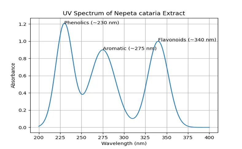
Figure 3.1: UV spectra of Nepeta Cataria Extract
3.2 FTIR Analysis
The FTIR spectrum of NC-1 (Figure 3.2) confirmed the presence of the characteristic functional groups of p-coumaric acid. A broad absorption band at 3398.16 cm⁻¹ corresponded to hydroxyl (–OH) stretching, while peaks at 2819.69 and 2576.05 cm⁻¹ were assigned to the O–H stretching vibration of the carboxylic acid group. The strong peak at 1670.88 cm⁻¹ represented carbonyl (C=O) stretching of the carboxylic acid. Aromatic C=C stretching vibrations appeared at 1601.83 and 1510.15 cm⁻¹, whereas peaks between 1314.46–1212.98 cm⁻¹ corresponded to C–O stretching vibrations. The fingerprint region showed characteristic aromatic C–H bending peaks at 977.73, 940.08, 831.70, and 517.70 cm⁻¹ (Table 3.1). These spectral features were in agreement with published data for p-coumaric acid, confirming the identity of NC-1.
3.3 FTIR analysis
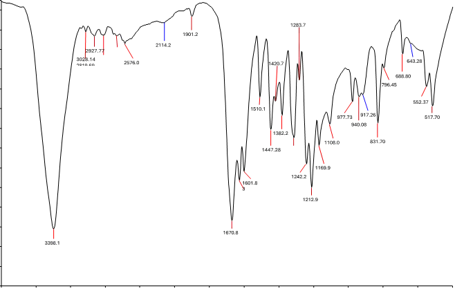
Figure 3.2: IR Spectra of NC-1
Table 3.1: IR spectral data results of NC-1
|
Functional group |
Intensity of peak (cm-1) |
|
OH stretching |
3398.16, 2819.69, 2576.05 |
|
=C-H stretching |
2927.77 |
|
C=O stretching of COOH |
1670.88 |
|
C=C aromatic ring stretching |
1601.83, 1510.15 |
|
O-H bending |
1447.28 |
|
C-O stretching of COOH |
1314.46 |
|
C-O-C stretching |
1242.27 |
|
C-O stretching of alcohol |
1212.98 |
|
aromatic bending |
977.73, 940.08, 831.70, 517.70 |
The chemical structure of NC-1 was characterized as p-coumaric acid based on melting point, UV, IR spectral data.
4. Evaluation of Nepeta cataria Proniosomes
4.1 Physical Appearance and Morphology
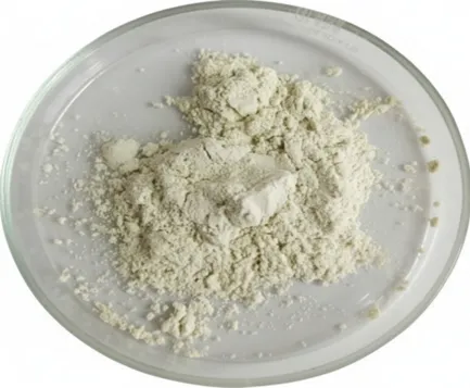
Figure 4.1: Nepeta cataria proniosome powder
Discussion: The above figure 4.1 shows, that Nepeta cataria proniosome is off white, free flowing powder.
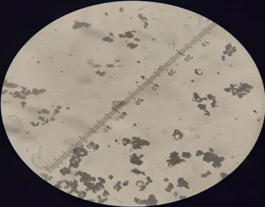
Figure 4.2: Optical Microscopy of Nepeta cataria proniosome powder
The prepared proniosomal formulation was obtained as an off-white, dry, and free-flowing powder without evidence of aggregation (Figure 4.2). Optical microscopic examination revealed predominantly spherical particles with a relatively uniform size distribution and slightly rough surface morphology. The absence of particle agglomeration suggested successful preparation of stable proniosomal powder.
4.3 Entrapment Efficiency
Table 4.1: Drug Entrapment of formulations (F1-F8)
|
Sr. No. |
Formulation Code |
% Entrapment Efficiency |
|
1 |
F1 |
65.280 ± 0.152 |
|
2 |
F2 |
72.846 ± 0.118 |
|
3 |
F3 |
88.965 ± 0.165 |
|
4 |
F4 |
79.432 ± 0.136 |
|
5 |
F5 |
83.217 ± 0.124 |
|
6 |
F6 |
86.904 ± 0.042 |
|
7 |
F7 |
76.518 ± 0.109 |
|
8 |
F8 |
70.334 ± 0.115 |
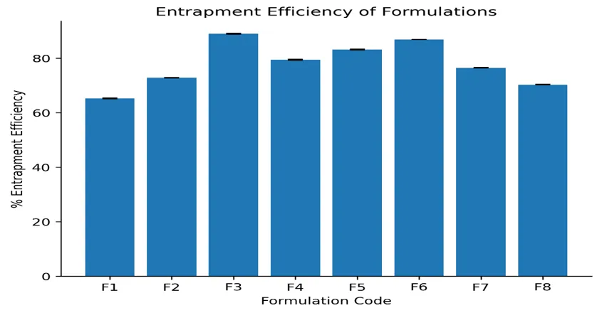
Figure 4.3: Drug Entrapment of formulations (F1-F8)
The percentage entrapment efficiency of formulations F1–F8 ranged from 65.28 ± 0.15% to 88.97 ± 0.17%. Formulation F3 exhibited the highest entrapment efficiency (88.97 ± 0.17%), whereas F1 showed the lowest (65.28 ± 0.15%). The enhanced entrapment observed in F3 may be attributed to the optimized surfactant-to-cholesterol ratio, which improves bilayer organization and minimizes drug leakage. The results indicate that formulation composition significantly influenced drug encapsulation efficiency, with F3 demonstrating superior vesicle-forming ability.
4.4 Particle Size and Zeta Potentia
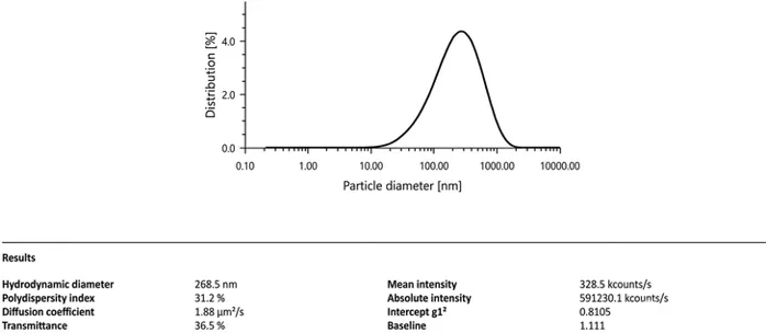
Figure 4.4: Particle size of proniosomes
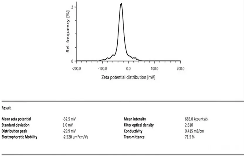
Figure 4.5: Zeta Potential of proniosomes
Dynamic light scattering analysis showed that the optimized proniosomal formulation possessed an average particle size of 268.5 nm, indicating the formation of nanosized vesicles suitable for enhanced topical delivery. The zeta potential was determined to be −32.5 mV, suggesting good colloidal stability due to sufficient electrostatic repulsion between vesicles. The nanoscale particle size combined with high zeta potential indicates that the optimized formulation is expected to exhibit improved physical stability and efficient drug delivery.
4.5 Micromeritic Properties
Characterization of different formulations of proniosome powder containing drug was given in table 5.5.
Table 4.2: Characterization of different formulations of proniosome powder
|
Formulation Code |
Angle of repose (θ) (Mean ±SD) |
Bulk density (g/cm3) (Mean ±SD) |
Tap density (g/cm3) (Mean ±SD) |
Hausner's Ratio (Mean ±SD) |
Carr's index (%)(Mean ±SD) |
|
F1 |
39.85 ± 0.26 |
0.62 ± 0.03 |
0.80 ± 0.05 |
1.32 ± 0.07 |
22.10 ± 0.82 |
|
F2 |
33.12 ± 0.30 |
0.44 ± 0.02 |
0.65 ± 0.04 |
1.18 ± 0.03 |
17.90 ± 0.95 |
|
F3 |
29.90 ± 0.28 |
0.41 ± 0.03 |
0.55 ± 0.02 |
1.15 ± 0.04 |
10.80 ± 0.15 |
|
F4 |
31.60 ± 0.15 |
0.52 ± 0.03 |
0.60 ± 0.03 |
1.10 ± 0.02 |
8.60 ± 0.55 |
|
F5 |
35.20 ± 0.22 |
0.45 ± 0.02 |
0.58 ± 0.03 |
1.12 ± 0.02 |
9.20 ± 0.48 |
|
F6 |
30.10 ± 0.11 |
0.60 ± 0.04 |
0.66 ± 0.03 |
1.07 ± 0.03 |
7.50 ± 0.60 |
|
F7 |
26.80 ± 0.40 |
0.42 ± 0.02 |
0.50 ± 0.03 |
1.10 ± 0.03 |
9.70 ± 0.52 |
|
F8 |
34.90 ± 0.14 |
0.55 ± 0.03 |
0.64 ± 0.04 |
1.13 ± 0.05 |
11.20 ± 0.80 |
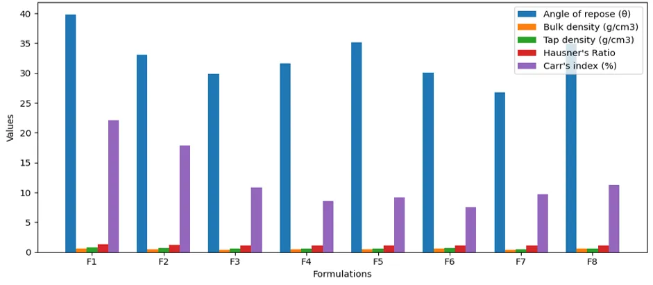
Figure 4.6: Characterization of proniosome powder
The micromeritic properties of proniosomal powder formulations are presented in Table 4.2. The angle of repose ranged from 26.80° to 39.85°, indicating acceptable to excellent flow properties. Bulk density varied between 0.41 and 0.62 g/cm³, while tapped density ranged from 0.50 to 0.80 g/cm³. Hausner's ratio and Carr's compressibility index ranged from 1.07–1.32 and 7.50–22.10%, respectively.
Among all formulations, F6 exhibited the lowest Hausner's ratio (1.07) and Carr's index (7.50%), indicating excellent flowability and compressibility. Although F7 showed the lowest angle of repose (26.80°), the overall micromeritic evaluation suggested that F6 possessed the most favourable powder flow characteristics. The acceptable micromeritic properties observed for all formulations indicate their suitability for pharmaceutical processing and handling.
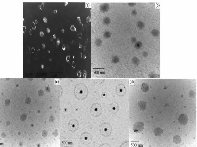
Figure 4.7: TEM Image of Formulation F3
Table 4.3: In vitro drug release of Nepeta cataria proniosome formulation F3 & pure drug
|
Dissolution medium |
Sr. No. |
Time (hr) |
Drug Release of control gel of pure Drug (%) |
Drug Release of Formulation F3 (%) |
|
0.1 N HCl solution |
1 |
0 |
0 |
0 |
|
2 |
0.25 |
15.842 ± 0.712 |
8.964 ± 0.845 |
|
|
3 |
0.5 |
34.921 ± 1.102 |
13.584 ± 1.041 |
|
|
4 |
1 |
73.418 ± 0.932 |
18.209 ± 1.354 |
|
|
5 |
2 |
95.873 ± 1.084 |
24.986 ± 1.102 |
|
|
Phosphate buffer, pH 6.8 |
6 |
3 |
– |
31.204 ± 1.102 |
|
7 |
4 |
– |
36.118 ± 1.041 |
|
|
8 |
6 |
– |
43.875 ± 1.102 |
|
|
9 |
8 |
– |
50.216 ± 0.732 |
|
|
10 |
10 |
– |
58.904 ± 1.621 |
|
|
11 |
12 |
– |
67.118 ± 0.732 |
|
|
12 |
24 |
– |
92.847 ± 1.102 |
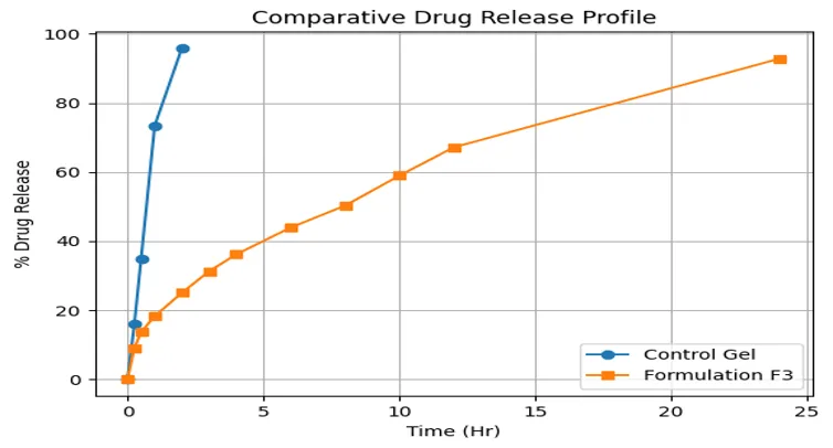
Figure 4.8: Percentage drug release of Nepeta cataria proniosome formulation F3 &Gel
Discussion: The in-vitro drug release data of the pure drug (control gel) and the proniosomal formulation (F3) of Nepeta cataria demonstrate a clear distinction in their release behaviour across different dissolution media.
In 0.1 N HCl (acidic medium), the control gel exhibited a rapid and immediate drug release pattern. Approximately 15.842 ± 0.712% drug was released within 0.25 h, which increased sharply to 95.873 ± 1.084% within 2 hours, indicating that the pure drug lacks any release-retarding mechanism and is readily available for dissolution in gastric conditions.
In contrast, the proniosomal formulation F3 showed a significantly slower release in the same medium. Only 8.964 ± 0.845% drug was released at 0.25 h, gradually increasing to 24.986 ± 1.102% at 2 h. This reduced release in acidic pH can be attributed to the encapsulation of the drug within proniosomal vesicles composed of surfactant and cholesterol. These vesicles act as a barrier, restricting drug diffusion and protecting the drug from immediate release in the gastric environment.
4.8 Drug release kinetic studies
4.8.1 Zero order kinetics
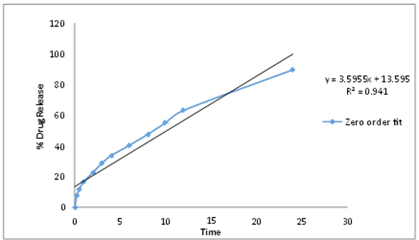
Figure 4.9: Zero order graph of formulation F3
4.8.2 First order kinetics
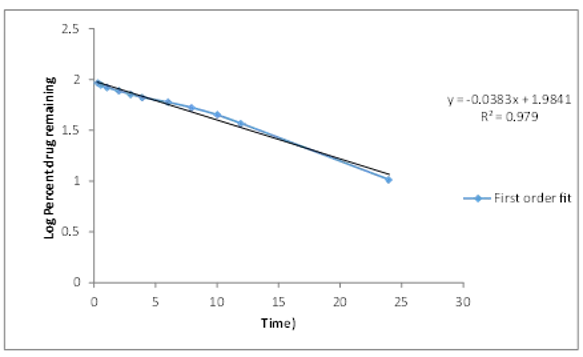
Figure 4.10: First order graph of formulation F3
4.8.3. Higuchi’s Model

Figure 4.11: Higuchi order graph of formulation F3
4.8.4 Korsmeyer-Peppas Model

Figure 4.12: Korsmeyer-Peppas order graph of formulation F3
Table 4.4: Kinetic equation parameter of formulation F3
|
Formulation Code |
Zero order |
First order |
Higuchi |
K. Peppas |
||||
|
K0 |
R2 |
K0 |
R2 |
K0 |
R2 |
K0 |
R2 |
|
|
F3 |
3.595 |
0.941 |
-0.038 |
0.979 |
18.288 |
0.997 |
0.583 |
0.998 |
Discussion: The regression analysis revealed that the Korsmeyer–Peppas model showed the highest correlation coefficient (R² = 0.998), indicating that it is the best-fit model for the drug release data. The Higuchi model (R² = 0.997) also exhibited excellent linearity, suggesting that the drug release is predominantly governed by diffusion.
The comparatively lower R² values for Zero-order (0.941) and First-order (0.979) models indicate that the release does not follow a constant rate or simple concentration-dependent mechanism.
The results suggest that the drug release from formulation F3 follows a non-Fickian (anomalous) diffusion mechanism, which involves a combination of drug diffusion and vesicular erosion. This type of release behavior is typical for proniosomal drug delivery systems, where the vesicular structure controls the release rate.
REFERENCES



Aaryan Sharma, Mona Piplani, Pankaj Bhateja, Saloni Bhatti, Development and Characterization of Proniosomal Formulation of Nepeta cataria L. Extract for Enhanced Antiallergic Activity, Int. J. of Pharm. Sci., 2026, Vol 4, Issue 8, 1839-1855. https://doi.org/10.5281/zenodo.21891155
 10.5281/zenodo.21891155