

We use cookies to ensure our website works properly and to personalise your experience. Cookies policy
1,4Department of Pharmacy, Madan Bhandari Academy of Health Sciences, Hetauda, Nepal.
2,3,5,6,7Department of Pharmacy, Asian College for Advance Studies, Satdobato, Lalitpur, Nepal.
Background: Febuxostat is a non-purine xanthine oxidase inhibitor used for chronic hyperuricaemia in gout. It is practically insoluble in water and behaves as a BCS class II compound, so its dissolution from conventional tablets can be slow and variable. Objective: To develop a liquid self-emulsifying drug delivery system (SEDDS) of febuxostat from lipidic excipients and to compare it’s in vitro dissolution with commercially available febuxostat tablets. Methods: Oils, surfactants and co-surfactants were screened by drug solubility and percentage transmittance. Seven liquid SEDDS (F1–F7) were prepared with Caproyl (propylene glycol monocaprylate) as oil, Acrysol K 160 as surfactant and Transcutol P as co-surfactant, and one comparator (O1) with oleic acid as oil. All contained 40 mg febuxostat. The formulations were evaluated for self-emulsification time, robustness to dilution, transmittance, drug content, FT-IR compatibility and dissolution in 900 mL phosphate buffer (pH 6.0). Profiles were compared with four marketed 40 mg tablets (A–D) using dissolution efficiency (DE??), the difference factor (Fd) and the similarity factor (Fs). Results: A UV method at 314 nm was linear from 2 to 12 µg/mL (R² = 0.9963) and precise (RSD 0.77%). Febuxostat was most soluble in Acrysol K 160 (100 mg/mL), Caproyl (66.7 mg/mL) and Transcutol P (66.7 mg/mL). All formulations emulsified within 21 s and showed no phase separation at 1:50 to 1:500 dilutions in water, pH 6.0 buffer or 0.1 N HCl. F7 (Caproyl:Acrysol K 160:Transcutol P, 2.1:2:1 w/w) emulsified fastest (13 s), had the highest transmittance (98.7%) and a drug content of 98.95%, and released 97% of the drug at 60 min. The marketed tablets released 32.5–77.1% over the same period. F7 had the highest DE?? (61.8%), and its Fs values against the four tablets (17.0–43.6) confirmed that its profile was different from, and higher than, the commercial products.
Gout is the commonest inflammatory arthritis in adults. It follows long-standing hyperuricaemia, when monosodium urate crystals deposit in and around joints, most typically the first metatarsophalangeal joint, and later form tophi [1,2]. Its prevalence is usually put at 1–4% of the general population, and it mostly affects middle-aged and older men [1]. Acute flares are treated with colchicine, NSAIDs or corticosteroids, but long-term control depends on lowering serum urate, and xanthine oxidase inhibitors are the mainstay of that strategy [2,3].
Allopurinol remains the recommended first-line urate-lowering drug [3]. Its use is limited in some patients by hypersensitivity reactions, which can be severe, and by the need for cautious dosing in renal impairment [2,4]. Febuxostat, a 2-arylthiazole derivative without a purine ring, is a potent and selective non-purine inhibitor of xanthine oxidase [4]. It needs no dose change in mild to moderate renal or hepatic impairment and lowers serum urate more effectively than fixed-dose allopurinol in several trials [4]. Its cardiovascular safety has been debated. The CARES trial reported higher cardiovascular and all-cause mortality with febuxostat than with allopurinol [5], while the larger FAST trial found it non-inferior [6]. Febuxostat is therefore now positioned mainly for patients who cannot take or do not respond to allopurinol [3].
Chemically, febuxostat is 2-[3-cyano-4-(2-methylpropoxy) phenyl]-4-methyl-1,3-thiazole-5-carboxylic acid (C₁₆H₁₆N₂O₃S, MW 316.4 g/mol). It is a white, non-hygroscopic crystalline powder melting at about 238–239 °C. It is freely soluble in dimethylformamide, sparingly soluble in ethanol and practically insoluble in water. After oral dosing it is well absorbed, reaches peak plasma levels in about 1–1.5 h, is about 99% protein bound and is cleared mainly by glucuronidation, with a half-life of 5–8 h [4,7].
Because febuxostat combines high permeability with very low aqueous solubility, it is generally treated as a BCS class II drug. For such compounds, the rate at which the drug dissolves in gastrointestinal fluids, rather than its permeation across the gut wall, tends to control the onset and consistency of absorption [8]. Dissolution of febuxostat from conventional tablets may also depend on the medium and on product quality, so formulation approaches that present the drug in a pre-dissolved state are worth exploring [9].
Lipid-based formulations are one of the most practical ways to deal with this problem [10,11]. Self-emulsifying drug delivery systems (SEDDS) are isotropic mixtures of oil, surfactant and co-surfactant (or co-solvent) in which the drug is dissolved. On gentle agitation in aqueous media, such as the stomach contents, they disperse spontaneously into fine oil-in-water emulsions [12,13]. The drug therefore reaches the absorptive surface already in solution, spread over a very large oil–water interface, and the dissolution step that limits conventional tablets is largely bypassed [12,14]. Surfactants in these systems may also increase membrane permeability, and lipids can promote lymphatic transport of very lipophilic drugs [11,13]. SEDDS can be filled into soft or hard capsules, or converted into solid forms by adsorption, spray drying or melt granulation [15–17].
The approach has improved the dissolution and, in several cases, the bioavailability of many BCS class II drugs, including fenofibrate, nevirapine, valsartan, rosuvastatin, glipizide, nateglinide and cefpodoxime proxetil [18–24]. For febuxostat, Patel et al. reported a self-microemulsifying system based on Capmul PG-8, Acrysol K 160 and Transcutol P that released more than 99% of the drug within 40 min [9]. Comparisons between such systems and the tablets actually sold in a given market are still uncommon, however, and few studies have examined how the oil phase itself changes performance.
The aim of this study was to formulate liquid SEDDS of febuxostat using lipidic excipients selected by solubility and transmittance screening, to characterise them in vitro, and to compare their dissolution with four febuxostat 40 mg tablets marketed in Nepal. A formulation based on oleic acid was included to show how the choice of oil affects performance.
MATERIALS AND METHODS
2.1 Materials
Febuxostat was a gift from Asian Pharmaceuticals Pvt. Ltd., Nepal. Caproyl (propylene glycol monocaprylate), Transcutol P (diethylene glycol monoethyl ether) were gifts from Corel Pharma Chem. Acrysol K160 (polyoxyl 60-hydrogenated castor oil; from Corel Pharma) was used as the surfactant. Oleic acid, sesame oil, castor oil, Tween 20, Span 20, Span 80, PEG 400, propylene glycol, methanol, potassium dihydrogen phosphate, sodium hydroxide and hydrochloric acid were of analytical grade and supplied by Asian College for Advance Studies, Satdobato, Nepal. Four local (Nepali) brands of febuxostat 40 mg tablets (coded A–D) were bought from local pharmacies.
Equipment used: analytical balance (Ohaus, India), double-beam UV–visible spectrophotometer (T80+, PG Instruments, UK), dissolution apparatus (Labindia, India), vortex mixer (CM 101 Plus, Remi, India) and FT-IR spectrophotometer (Agilent Technologies, USA).
2.2 Analytical method
A stock solution of febuxostat (100 µg/mL) was prepared by dissolving 100 mg in 1000 mL methanol. A dilution scanned from 200 to 400 nm gave the absorption maximum. Working standards of 2–12 µg/mL were prepared from the stock in methanol and read at 314 nm. The method was checked for linearity, precision and specificity following ICH Q2 (R1) [25–27]. Precision was taken as the relative standard deviation (RSD) of six replicate 5 µg/mL solutions. For specificity, methanolic solutions of the excipients at the working concentration were read at 314 nm. The limits of detection (LOD) and quantification (LOQ) were calculated as 3.3σ/S and 10σ/S, where σ is the residual standard deviation of the regression and S its slope.
2.3 Solubility screening
The solubility of febuxostat was estimated in four oils (Caproyl, oleic acid, castor oil and sesame oil), four surfactants (Acrysol K 160, Tween 20, Span 80 and Span 20) and three co-surfactants (Transcutol P, PEG 400 and propylene glycol). Febuxostat (100 mg) was placed in a glass vial and the vehicle was added in small portions with vortex mixing until a clear solution was seen. Solubility (mg/mL) was calculated from the volume needed and classified according to pharmacopoeial descriptive terms.
2.4 Transmittance screening of excipients
As a second screen, dilutions of each excipient (5 µg/mL) were prepared and their percentage transmittance was read on the UV spectrophotometer. Vehicles that combined high solubility for the drug with high transmittance were taken forward.
2.5 Preparation of liquid SEDDS
Eight formulations were prepared (Table 1), each containing 40 mg febuxostat. The drug was dissolved in the oil. In a separate beaker the surfactant and co-surfactant were mixed until homogeneous, and this mixture was then added to the oily drug solution with continuous stirring using a glass rod. In F1 and F3–F7 the surfactant: co-surfactant ratio (Smix) was fixed at 2:1, while the oil fraction varied between 27.6% and 57.1% w/w. F2 used an Smix of 1:1. O1 had the same composition as F7, but with oleic acid in place of Caproyl. The preparations were stored in closed glass vials at room temperature until tested.
Table 1: Composition of the Liquid SEDDS (mg per Unit; 40 mg Febuxostat in Each)
| Code | Oil | Oil (mg) | Acrysol K 160 (mg) | Transcutol P (mg) | Oil (% w/w of vehicle) | Oil:Smix |
|---|---|---|---|---|---|---|
| F1 | Caproyl | 80 | 140 | 70 | 27.6 | 1:2.63 |
| F2 | Caproyl | 100 | 120 | 120 | 29.4 | 1:2.40 |
| F3 | Caproyl | 125 | 160 | 80 | 34.2 | 1:1.92 |
| F4 | Caproyl | 125 | 140 | 70 | 37.3 | 1:1.68 |
| F5 | Caproyl | 140 | 140 | 70 | 40.0 | 1:1.50 |
| F6 | Caproyl | 140 | 70 | 35 | 57.1 | 1:0.75 |
| F7 | Caproyl | 150 | 140 | 70 | 41.7 | 1:1.40 |
| O1 | Oleic acid | 150 | 140 | 70 | 41.7 | 1:1.40 |
2.6 Self-emulsification time and dispersibility
One milliliter of each formulation was added dropwise to 500 mL distilled water at 37 °C under gentle stirring with a glass rod. The time to complete dispersion was recorded, and the appearance of the dispersion was graded A–E [24]. Grade A: rapidly forming (within 1 min), clear or slightly bluish Nano-emulsion. Grade B: rapidly forming, slightly less clear, bluish-white emulsion. Grade C: fine milky emulsion formed within 2 min. Grade D: dull, greyish-white, slightly oily emulsion that takes more than 2 min. Grade E: poor or minimal emulsification with large oil globules on the surface.
2.7 Robustness to dilution
Each formulation was diluted 1:50, 1:100 and 1:500 with distilled water, phosphate buffer pH 6.0 and 0.1 N HCl. The dilutions were left for 12 h and inspected for phase separation or drug precipitation.
2.8 Percentage transmittance of the formulations
Each formulation (0.5 mL) was diluted to 50 mL with distilled water, filtered, and its transmittance read on the UV spectrophotometer at 314 nm.
2.9 Drug content
An amount of each formulation equivalent to one unit was extracted with methanol on a vortex mixer for 5 min, filtered, diluted to about 5 µg/mL with methanol and read at 314 nm. Twenty marketed tablets of each brand were weighed and powdered, and an amount of powder equal to 40 mg febuxostat was assayed the same way.
2.10 In vitro dissolution
Dissolution was carried out in a USP apparatus I (basket) at 75 rpm, in 900 mL phosphate buffer pH 6.0 kept at 37 ± 0.5 °C. Each unit contained 40 mg febuxostat (n=6). Samples were withdrawn at 15, 30, 40 and 60 min, (replaced with fresh medium), filtered, suitably diluted and read at 314 nm. The four marketed tablets were tested under the same conditions.
2.11 FT-IR compatibility
FT-IR spectra of pure febuxostat, Caproyl, Acrysol K 160, Transcutol P and F7 (fresh and after one week of storage at room temperature) were recorded from 4000 to 650 cm⁻¹ on an Agilent FT-IR spectrophotometer. The characteristic drug bands were compared across the spectra.
2.12 Comparison of dissolution profiles
Profiles were compared using model-independent methods [28–31]. The difference factor (Fd) and the similarity factor (Fs) were calculated as:


Here n is the number of time points, and Rj and Tj are the cumulative percentages dissolved for the reference (marketed tablet) and the test (F7) at time t. Profiles are considered similar when Fd is 0–15 and Fs is 50–100 [28,29]. Dissolution efficiency up to 60 min (DE₆₀) was calculated as the area under the dissolution curve from 0 to 60 min (trapezoidal rule) expressed as a percentage of the rectangle representing 100% dissolution over the same time [31].
RESULTS
3.1 Analytical method
Febuxostat in methanol showed its absorption maximum at 314 nm. Absorbance was linear from 2 to 12 µg/mL (y = 0.0878x − 0.0141, R² = 0.9963; Figure 1). The six replicate 5 µg/mL solutions gave a mean absorbance of 0.4325 with an RSD of 0.77%, well below the usual 2% limit. The excipients did not absorb at 314 nm at the concentrations used, so the method was specific for the drug. LOD and LOQ, calculated from the regression residuals, were 0.84 and 2.55 µg/mL.
3.2 Excipient screening
Solubility and transmittance results are given in Table 2. Caproyl was the best oil (66.7 mg/mL), followed by castor oil, oleic acid and sesame oil. Among the surfactants Acrysol K 160 dissolved twice as much drug as Tween 20, and Transcutol P was the best co-surfactant. The same three excipients also gave the highest transmittance (93.5–98.5%) and were chosen for formulation.
Table 2: Solubility of Febuxostat in Candidate Vehicles and Transmittance of the Selected Excipients
| Function | Vehicle | Solubility (mg/mL) | Transmittance (%) |
|---|---|---|---|
| Oil | Caproyl | 66.7 | 98.5 |
| Oil | Castor oil | 50.0 | |
| Oil | Oleic acid | 33.0 | |
| Oil | Sesame oil | 11.0 | |
| Surfactant | Acrysol K 160 | 100.0 | 93.5 |
| Surfactant | Tween 20 | 50.0 | |
| Surfactant | Span 80 | 20.0 | |
| Surfactant | Span 20 | 10.0 | |
| Co-surfactant | Transcutol P | 66.7 | 97.1 |
| Co-surfactant | Propylene glycol | 50.0 | |
| Co-surfactant | PEG 400 | 33.3 |
3.3 Characterisation of the liquid SEDDS
All eight formulations dispersed within 13–21 s and gave fine milky emulsions (grade C). None showed phase separation or visible precipitation after 12 h at any dilution (1:50, 1:100, 1:500) in water, pH 6.0 buffer or 0.1 N HCl. F7 emulsified fastest (13 s) and gave the highest transmittance (98.7%). Drug content of the Caproyl systems ranged from 89.6% to 103.9%, whereas O1 assayed at only 73.08% (Table 3).
Table 3: Physicochemical Characteristics and Dissolution Efficiency of the Liquid SEDDS
| Code | Emulsification time (s) | Grade | Transmittance (%) | Drug content (%), n =3 | Released at 60 min (%) | DE₆₀ (%) |
|---|---|---|---|---|---|---|
| F1 | 14 | C | 87.4 | 99.23 ± 0.25 | 80.2 | 59.2 |
| F2 | 14 | C | 90.0 | 89.55± 0.61 | 81.4 | 59.3 |
| F3 | 14 | C | 87.4 | 95.58± 1.58 | 71.0 | 49.4 |
| F4 | 16 | C | 90.1 | 103.85± 0.85 | 59.7 | 48.2 |
| F5 | 19 | C | 83.4 | 92.00± 1.69 | 59.4 | 45.2 |
| F6 | 17 | C | 88.0 | 92.67± 1.53 | 57.3 | 40.2 |
| F7 | 13 | C | 98.7 | 98.95± 0.05 | 97.0 | 61.8 |
| O1 | 21 | C | 89.0 | 73.08± 1.51 | 12.9 | 7.9 |
3.4 In vitro dissolution
F7 released 47% of the drug at 15 min, 67.6% at 30 min, 79% at 40 min and 97% at 60 min (Figure 2). It was the only formulation to approach complete release within the test. F1 and F2, which had the lowest oil content, released about 80% at 60 min. F3–F6 reached only 57–71%. The oleic acid system O1 released less than 13% throughout. Among the tablets, brands A, B and C released 67.0–77.1% at 60 min, and brand D released only 32.5%. Brand A showed a lower value at 40 min (56.1%) than at 30 min (65.6%).
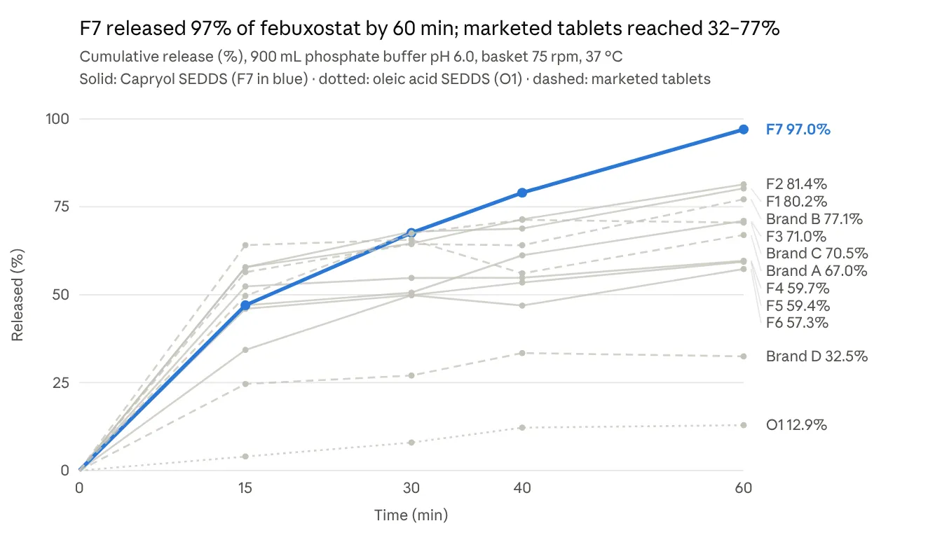
Figure 2: Cumulative in Vitro Release of Febuxostat from the Liquid SEDDS (F1–F7, O1) and Four Marketed 40 mg Tablets (a–D) in 900 ml Phosphate Buffer pH 6.0 (USP Apparatus I, 75 rpm, 37 °C).
| Formulations (n=6) | Brand | |||||||||||
|---|---|---|---|---|---|---|---|---|---|---|---|---|
| F1 | F2 | F3 | F4 | F5 | F6 | F7 | O1 | A | B | C | D | |
| 80.2 ±0.72 | 81.4±1.09 | 71±1.11 | 59.7±1.23 | 59.4±0.74 | 57.3±0.69 | 97±1.19 | 12.9±1.46 | 67±1.10 | 77.1±0.66 | 70.5±1.09 | 32.5±0.94 | |
3.5 Comparison with marketed tablets
F7 released 1.3 to 3.0 times more drug at 60 min than the marketed tablets, and its DE₆₀ (61.8%) exceeded that of every brand (Table 4). The Fs values for F7 against the individual brands ranged from 17.0 to 43.6, all below 50, and Fd ranged from 14.3 to 147.2. The F7 profile was therefore different from all four commercial profiles, the difference being a higher and more complete release.
Table 4: Comparison of F7 with Marketed Febuxostat 40 mg Tablets (F7 as Test, Each Brand as Reference)
| Product | Drug content (%) | Released at 60 min (%) | DE₆₀ (%) | Fd | Fs | Fold release at 60 min (F7/brand) |
|---|---|---|---|---|---|---|
| Brand A | 92.50±1.0 | 67.0 | 54.9 | 28.5 | 34.1 | 1.45 |
| Brand B | 102.50±1.99 | 77.1 | 56.4 | 18.1 | 43.6 | 1.26 |
| Brand C | 95.25±1.14 | 70.5 | 56.0 | 14.3 | 42.8 | 1.38 |
| Brand D | 94.53±1.83 | 32.5 | 25.6 | 147.2 | 17.0 | 2.99 |
3.6 FT-IR compatibility
Pure febuxostat showed its characteristic bands, including a sharp nitrile (C≡N) stretch near 2230 cm⁻¹ and the carboxylic C=O stretch near 1680 cm⁻¹. In the F7 spectrum these bands remained at essentially the same positions, without new bands or marked shifts, alongside the C–H stretching (2850–2950 cm⁻¹) and ester C=O (about 1740 cm⁻¹) bands of the excipients. The spectrum of F7 after one week of storage was superimposable on that of the fresh sample. No chemical interaction between febuxostat and the excipients was therefore evident (Figure 3).
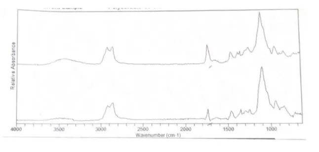
Figure 3: FT-IR Spectrum of Formulated Febuxostat (F7) at the Time of Preparation and After One Week
DISCUSSION
The main finding is that a simple liquid SEDDS made from Caproyl, Acrysol K 160 and Transcutol P (F7) released almost all of its febuxostat within an hour, whereas the four tablets on sale released between a third and three quarters of their dose under the same conditions. Just as important, performance depended strongly on composition. Swapping the oil from Caproyl to oleic acid, with everything else unchanged, reduced release from 97% to under 13%. Changing the proportions of oil and surfactant within the Caproyl series moved release at 60 min between 57% and 97%. These points are discussed in turn below.
4.1 Why these excipients
A SEDDS can only deliver what it holds in solution, so the solubilising power of each component sets the ceiling on drug load and on the margin against precipitation after dispersion [10,32]. Febuxostat dissolved best in Caproyl, a propylene glycol monoester of caprylic acid. Medium-chain mono- and diglycerides and their propylene glycol esters are known to dissolve poorly soluble drugs better than long-chain triglycerides. They are more polar, and their shorter chains pack less tightly around the solute [10,11]. The poor solubility in sesame oil (11 mg/mL), a long-chain triglyceride, fits this pattern. Castor oil did better (50 mg/mL), probably because of the hydroxyl group on ricinoleic acid, but it is viscous and harder to emulsify.
Acrysol K 160, a polyoxyl hydrogenated castor oil, dissolved twice as much drug as Tween 20 and five to ten times as much as the Span surfactants. Its high HLB favours spontaneous formation of oil-in-water emulsions, while its castor oil–derived lipophilic part interacts well with lipophilic drugs [12,33]. The low-HLB Span surfactants are unsuitable as the main emulsifier in a SEDDS whatever their solubilising ability. Transcutol P was the best co-surfactant. It is a strong solvent for many poorly soluble drugs and, by penetrating the surfactant layer, increases interfacial flexibility and aids spontaneous dispersion [14,34]. The same three-component approach was taken by Patel et al. in their febuxostat system, with the closely related Capmul PG-8 as the oil [9], which supports this choice of excipients.
The solubility figures should be read as estimates. They were obtained by adding vehicle until a clear solution formed, not by equilibrium shake-flask measurement, and so they rank the vehicles more reliably than they quantify them.
4.2 The oil phase decides performance
The most striking result is the failure of O1. It differed from F7 only in the oil, yet it released 12.9% instead of 97% and assayed at only 73%. Three explanations are likely, and they reinforce each other. First, febuxostat is about half as soluble in oleic acid (33 mg/mL) as in Caproyl, so at 10% w/w loading part of the dose was probably never fully dissolved in O1. The low assay value supports this, since undissolved or crystallised drug would be unevenly distributed and harder to extract. Second, oleic acid is a long-chain fatty acid. It is harder to emulsify than a medium-chain ester, and it tends to give larger droplets with less interfacial area for drug release [10,33]. Third, at pH 6.0 oleic acid will be partly ionised and may form soap-like structures. The drug could then precipitate as the solvent capacity of the system collapses on dilution [32]. Whatever the relative contribution of each, the comparison shows that solubility screening is not just a preliminary step. An oil that holds less drug produces a formulation that releases less drug.
4.3 Effect of oil and surfactant proportions
Within the Caproyl series a broad trend can be seen. F1 and F2, with the least oil (27.6–29.4%) and the highest ratio of surfactant mixture to oil, gave the fastest early release (about 58% at 15 min) and reached about 80% at 60 min. F6, which had the most oil (57.1%) and the least surfactant, was the slowest at 15 min (34.3%) and released only 57.3% by 60 min. This agrees with the general observation that higher surfactant-to-oil ratios give finer droplets and faster release, while surfactant-poor systems emulsify less completely [33,34]. F2 (Smix 1:1) performed almost exactly like F1 (Smix 2:1), suggesting that within this range Transcutol P can partly take over the role of the surfactant.
F7 does not fit this trend neatly. F4, F5 and F7 share the same surfactant and co-surfactant amounts and differ only in Caproyl (125, 140 and 150 mg). However, F4 and F5 released about 60% at 60 min, while F7 released 97%. A 10 mg increase in oil is unlikely on its own to cause such a jump. F7 also gave the highest transmittance and the shortest emulsification time, which points to genuinely better dispersion. Even so, the size of the difference suggests that small changes near this composition have a large effect, or that run-to-run variability played a part. Replicate preparations and droplet size measurement are needed to settle which explanation is correct, and a statistically designed study (for example, a mixture design) would map this region more reliably than one-factor changes.
Drug load may also matter. Each unit contained 40 mg febuxostat in 360 mg vehicle (about 10% w/w). Taking a weighted average of the individual solubilities, F7 would be expected to dissolve roughly 80 mg/mL, whereas the loading corresponds to roughly 110 mg/mL if a density near 1 g/mL is assumed. This is only an estimate, because solubility in mixtures is rarely additive. Still, it suggests that several formulations were at or above saturation. Such systems can hold the drug in a metastable supersaturated state that precipitates during dispersion or dissolution [32,35]. This would help to explain why most formulations, despite being pre-dissolved, did not reach complete release within 60 min. Measuring equilibrium solubility in the final formulation and following the drug in solution after dispersion would test this directly.
4.4 Shape of the release profile
Even F7 released its drug more gradually (47% at 15 min) than is often reported for SEDDS, which frequently release more than 85% in the first 15 min [18,20]. Patel et al. reported 99.5% at 40 min for their febuxostat system [9]. Several features of our method may account for this. The liquid had to leave its container and pass through the basket mesh before dispersing in the medium, and the basket apparatus at 75 rpm gives relatively gentle mixing for a liquid system. The pH 6.0 buffer is also a demanding medium for a weak acid such as febuxostat, whose solubility rises with pH. A slower profile is not necessarily a disadvantage, but it should be confirmed in a paddle apparatus and in media spanning the gastrointestinal pH range.
4.5 Marketed tablets
The four tablets differed widely. Brands A, B and C released 67–77% at 60 min and brand D released only about a third of its dose, although all four assayed at 92.5–102.5%. Such low release from an immediate-release tablet would usually be a concern. However, the test conditions here were not those of any pharmacopoeial monograph, and only a few units were tested, so no conclusion on product quality should be drawn. The dip seen for brand A at 40 min (56.1% versus 65.6% at 30 min) cannot reflect a real fall in dissolved drug unless precipitation occurred. It more likely points to sampling or analytical error, or to the absence of a correction for medium replaced at each sampling. The between-brand spread nevertheless supports the case for a formulation whose performance depends less on the tablet's solid-state and manufacturing variables.
4.6 Interpretation of Fd, Fs and DE₆₀
The Fd/Fs approach was designed to show that two profiles are similar, typically a generic and its reference, and it is normally used with 12 units per product and with limits on variability at each time point [28,29]. Here the aim was the opposite: to show that F7 is different, and better. The Fs values below 50 against all four brands confirm a difference, but they say nothing about its direction. For this reason, DE₆₀ and release at 60 min were reported alongside them. F7 had the highest DE₆₀ (61.8%), only slightly above brands B and C (about 56%), because F7 started more slowly and then overtook them. Most of its advantage therefore lies in the second half of the test, where the tablets level off at 67–77% and F7 continues to rise to 97%. This plateau of the tablets suggests that their dissolution is limited by the solubility of the drug in the medium, a limit that the SEDDS largely avoids by keeping the drug solubilised in emulsion droplets.
4.7 Comparison with earlier work
Our findings agree with those of other SEDDS studies on BCS class II drugs. Patel and Vavia reported complete release of fenofibrate from a SMEDDS within 15 min, against much lower release from the plain drug [18]. Kulkarni et al. found about four-fold higher release of rosuvastatin from a self-nanoemulsifying system than from the pure drug [21], and Dixit et al. made similar observations for valsartan [20]. Comparable improvements over conventional dosage forms have been reported for ranolazine with an oleic acid–Cremophor EL–Transcutol P system [36], for nevirapine [19] and for glipizide [22]. One difference from the ranolazine work is worth noting. Oleic acid served well there, but it failed here, which shows that the best oil depends on the drug and cannot be carried over from one formulation to another.
4.8 What the results mean for bioavailability
It is tempting to read faster dissolution as higher bioavailability, but some caution is needed. Febuxostat is already well absorbed after oral dosing [4,7], so any in vivo benefit of a SEDDS is more likely to be faster and more consistent absorption than a large rise in the extent of absorption. In the gut, a lipid formulation is also digested by pancreatic lipase, which can change its solubilising capacity and sometimes trigger precipitation [10,32]. Neither digestion nor the effect of food was simulated here. In vitro lipolysis testing followed by a pharmacokinetic study would be needed before any bioavailability claim is made.
4.9 Limitations
The study has several limitations that should be addressed in further work. Droplet size, polydispersity and zeta potential were not measured, and these are standard for characterizing SEDDS. The number of replicates and their variability were not reported for the dissolution data, and pure drug dissolution was not tested. Formulation transmittance was measured after filtration, which may remove large droplets and overstate clarity. Stability was followed for only one week and by FT-IR alone. Finally, F7 contains about 39% w/w surfactant, and gastrointestinal tolerability at that level would need to be considered for chronic use.
CONCLUSION
A liquid SEDDS of febuxostat containing Caproyl, Acrysol K 160 and Transcutol P (2.1:2:1 w/w) emulsified within seconds, withstood dilution in media of different pH, and released 97% of its drug in 60 min. Over the same period, four marketed tablets released 32–77%. The choice of oil was decisive: replacing Caproyl with oleic acid almost abolished release. The proportions of oil and surfactant also had a marked effect. These results support SEDDS as a practical way to overcome the dissolution limits of febuxostat. The next steps are droplet size and zeta potential measurement, longer stability studies, conversion to a solid dosage form, in vitro lipolysis testing and a pharmacokinetic study.
Declarations
Acknowledgements:
The authors thank Asian Pharmaceuticals Pvt. Ltd. for the gift of febuxostat, Funding: No funding
Conflict of interest: None
Ethics approval:
Not applicable. The study involved no human participants or animals.
REFERENCES

Sailendra Chaudhary, Sumit Chandra Shrestha, Chandrika Adhikari Mahat, Kiran Bhatt, Binu Prajapati, Nisha Gupta, Ruja Dhaubanjar, Febuxostat Self-Emulsifying Drug Delivery System: Formulation and In-Vitro Characterization, Int. J. of Pharm. Sci., 2026, Vol 4, Issue 11, 805-816. https://doi.org/10.5281/zenodo.23940139
 10.5281/zenodo.23940139