

We use cookies to ensure our website works properly and to personalise your experience. Cookies policy
1,2,3Doctor of Pharmacy, Department of Pharmacy Practice, SET’S College of Pharmacy, Dharwad, S R Nagar 580002, Karnataka, India.
4Department of Clinical pharmacology (PHARM-D), Clinical Pharmacologist at HCG-Suchirayu Hospital, Hubballi, Karnataka India.
Heparin-induced thrombocytopenia (HIT) is a potentially fatal drug reaction, caused by the formation of IgG antibodies, usually following treatment with heparin. We report the case of a 77-year-old hypertensive man with a history of deep vein thrombosis of the right lower limb who was treated with anticoagulation therapy with heparin and acenocoumarol. Four days later, his platelet count had reduced from 154 × 10³/µL to 55 × 10³/µL, a 64% reduction. The patient had several complications which included acute kidney injury, atrial fibrillation, and a right frontal acute cerebral infarct. The patient had a left inferior vena cava filter placed, and subsequently underwent catheter directed thrombolysis and thrombectomy. One week later, he had recurrent epistaxis and bleeding from the gums. He was severely anemic (Hb 6.8 g/dL) and had deranged coagulation. The patient had advanced kidney failure, was anemic and was 77 years old, which increased his risk of bleeding. The 4 Ts score indicated a high clinical suspicion of HIT, however, heparin/PF4 antibody testing and functional assays were not performed, and therefore, a definitive diagnosis of HIT was not made. The patient was managed with fresh frozen plasma, packed red blood cells, and tranexamic acid. The patient improved clinically. This case illustrates the need for prompt assessment of declining platelet counts in the presence of heparin therapy and consideration of HIT.
Heparin-induced Thrombocytopenia (HIT) is an adverse drug reaction to heparin that increases the risk of thrombosis and results in thrombocytopenia.1,2 The incidence of major bleeding is high in patients with HIT.5 If HIT is not treated, patients may develop life-threatening thrombotic complications at a rate of approximately 6.1% per day.5,3 The classification of HIT is based on its mechanism and clinical findings.1,4 HIT Type 1 is a non-immune mediated, transient, and reversible fall in platelet count, which occurs in the first 2 days of heparin therapy. HIT type 1 usually resolves with the discontinuation of heparin.1,4 Type 2 HIT is an immune mediated thrombocytopenia, in which patients have previous exposure to heparin, and often occurs 5 days or more after the commencement of heparin therapy and is defined as a percentage fall in platelet count of more than 30% from the baseline.1,2,3
CASE PRESENTATION
A 77-year-old male with left lower limb pain and swelling for 3 days and bleeding from the nose and mouth since the day of admission was referred to the general medicine ward.
The patient was apparently healthy until 3 days ago, when he developed pain in his left lower limb that was followed by swelling. He was diagnosed at an outside hospital with subacute deep vein thrombosis (DVT) of the left lower limb, and is currently post-IVC filter placement, post-mechanical thrombectomy, and has acute kidney injury (AKI) due to the left lower limb DVT. His kidney function is stable on rivaroxaban 15 mg BID. The patient has had progressive, epistaxis and oral bleeding, for the last 24 hours. The bleeding was not controlled with nasal packing and clots were present. There was no history of fever, vomiting, headache, giddiness or loose stools. The patient is hypertensive for the last 8 years, on Atenolol 25 mg OD. The patient has no history of alcohol or tobacco use.
On presentation, the patient was alert and oriented. His blood pressure was 130/90 mmHg, pulse rate was 92/min, and respiratory rate was 16/min. His SpO2 was 98% on room air. His blood glucose level was 147 mg/dL. On systemic exam, his heart sounds were normal, with no murmurs. There was no focal neurological deficit. His lungs were clear and equal on both sides. His abdomen was soft and non-tender, with no organomegaly.
Table 1: Timeline of Key Events
| Days | Setting | Key events |
|---|---|---|
| 0 | Hospital A, outpatient | Right leg pain and swelling. Doppler: acute right femoropopliteal DVT. heparin and Acenocoumarol started. |
| 1-8 | Hospital A, inpatient |
Heparin. AKI (creatinine peak 4.78 mg/dL). Acute right frontal infarct (Day 2). Atrial fibrillation. Platelet nadir 55,000/µL (Day 4). Discharged on Acenocoumarol. |
| 12 | Hospital A, review |
INR 7.33, Hb 9.9 g/dL. Referred to Hospital B. |
| 12-16 | Hospital B | IVC filter, catheter-directed thrombolysis and mechanical thrombectomy. Rivaroxaban started. Hb 7.1 g/dL, INR 2.53 at discharge. |
| 18 | Hospital C casualty | Left lower-limb pain and swelling, Epistaxis and haematemesis. Hb 6.8 g/dL. Rivaroxaban stopped. |
| 19-20 | Hospital C inpatient | Melaena. Vitamin K, tranexamic acid, FFP, 2 units red cells. Bleeding resolved. |
Abbreviations: Deep vein thrombosis (DVT), inferior vena cava (IVC), Acute kidney injury (AKI), Fresh frozen plasma (FFP), Red Blood Cell (RBC),
Table 2: Laboratory Trends Across Care Settings
| Days | Hb (12-16 g/dL) |
TLC (4000-11000/µL) | Platelets (125-350 ×10³/µL) | Creatinine (0.6-1.6 mg/dL) | Urea (15-45mg/dL) | PT (13.5s) / INR | aPTT (22.2-37.9 s) |
|---|---|---|---|---|---|---|---|
| 0 | 11.6 | 4,700 | 154 | 1.83 | 50.3 | - | - |
| 1 | 13.6 | 12,700 | 119 | 3.89 | 97.6 | 42.7/3.35 | - |
| 2 | 12.8 | 11,400 | 82 | 4.78 | 120.6 | 22.5/1.71 | 43.0 |
| 3 | 11.0 | 14,000 | 59 | 4.30 | 214.1 | 15.3/1.14 | 44.1 |
| 4 | 11.5 | 17,000 | 55 | 3.78 | 202.3 | 14.1/1.04 | 38.2 |
| 5 | 12.4 | 15,500 | 58 | 3.01 | 194.5 | - | 31.9 |
| 6 | 12.1 | 15,200 | 62 | 2.54 | 176.5 | - | - |
| 7 | 12.7 | 17,100 | 84 | 1.41 | 145.6 | 17.3/1.29 | 33.3 |
| 8 | 11.2 | 17,300 | 96 | 1.26 | 106.0 | - | - |
| 12 | 9.9 | 8,800, | 271 | - | - | >90/7.33 | 41.6 |
| 16 | 7.1 | - | - | 1.2 | - | 31.4/2.53 | - |
| 18-19 | 6.8-7.2 | 5,400-2,910 | 286-234 | 1.2 | 34.4 | 27.1/2.53 | 62.6-63 |
Additional investigations performed during admission showed an elevated D-dimer level of 672 ng/mL and an erythrocyte sedimentation rate (ESR) of 53 mm/hr. Serological screening for hepatitis B surface antigen (HBsAg) and HIV-1/2 was negative and non-reactive, respectively. Serial laboratory trends, including hemoglobin, platelet count, renal function parameters, and coagulation profile,
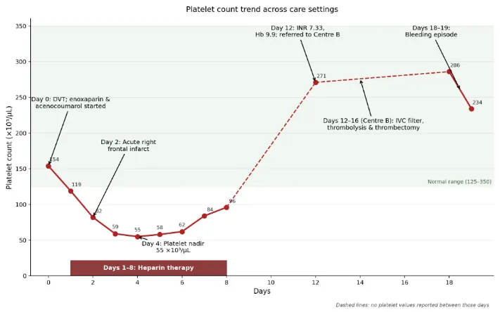
Figure 1: Trend of Platelet Count in Patient During Heparin Therapy and After Discontinuation of Heparin Therapy
Table 3: 4Ts Score for Heparin-Induced Thrombocytopenia
| Component | Clinical findings from case | Score (0–2) | Remarks |
|---|---|---|---|
| Thrombocytopenia (magnitude of platelet fall) | Platelet count fell from 154 × 10³/µL (Day 0) to 55 × 10³/µL (Day 4) — a 64% fall, nadir > 20 × 10³/µL | 2 | Significant fall consistent with HIT |
| Timing of platelet fall | Fall began within 1–4 days of heparin exposure; patient had recent prior heparin exposure (Centre A, inpatient) | 2 | Rapid-onset HIT possible due to prior exposure |
| Thrombosis or other sequelae | Documented acute right femoropopliteal DVT and acute right frontal infarct during heparin therapy | 2 | New thrombosis supports HIT |
| Other causes for thrombocytopenia | No strong alternative cause; AKI and infection possible but not sufficient to explain platelet trend | 1 | Possible but not definite alternative cause |
| Total score | 7 / 8 | High probability of HIT | |
The platelet count declined by 64% during heparin exposure, from 154 × 10³/µL to 55 × 10³/µL. The concurrent acute cerebral infarction raised concern for a possible thrombotic complication. However, the timing component of the 4Ts score requires verification of previous heparin exposure, and the initial DVT preceded the documented platelet decline. As the PF4/heparin immunoassay and functional testing were not performed, HIT remained a clinical suspicion and could not be confirmed.
TREATMENT
The patient was treated with intravenous injection of vitamin K 10 mg once daily, injection tranexamic acid 1 g intravenously as required for bleeding control, injection ceftriaxone 1 g intravenously twice daily, and injection pantoprazole 40 mg intravenously once daily. Botroclot nasal drops were administered for local control of epistaxis. Owing to significant mucosal bleeding, coagulopathy, and severe anemia, the patient received four units of fresh frozen plasma (FFP) and packed red blood cell transfusion along with supportive care. Antihypertensive therapy with tablet amlodipine 5 mg once daily was continued, and oral anticoagulation with tablet Acitrom 2 mg once daily was reinitiated before discharge with advice for regular PT-INR monitoring. Following treatment, the bleeding resolved, hemoglobin levels stabilized, and the patient's general condition improved.
DISCUSSION
The case demonstrates diagnostic and therapeutic difficulties when there is marked platelet decline, recurrent thrombosis, and major bleeding on anticoagulation for Deep Vein Thrombosis (DVT). The patient’s platelet count was 154 × 10³/µL, which decreased to 55 × 10³/µL, a 64% decrease, during exposure to heparin. The co-occurrence of cerebral infarction and advancing thromboembolic disease raised the possibility of heparin-induced thrombocytopenia (HIT). However, a definitive diagnosis was not possible due to the absence of the PF4/heparin antibody tests and functional assays. 6
A substantial decline in platelet count during heparin therapy with new or worsening thromboembolism should raise the suspicion for HIT. Kumar et al. also reported similar diagnostic and management dilemmas with HIT-complicated thrombosis of a DVT. 7 In the current case, the 4Ts score and other clinical features also suggested HIT. However, definitive tests were not performed. Therefore, HIT is likely to explain the thromboembolic and thrombocytopenic conditions of this case. 6
Thrombotic events, including venous and arterial, occur with HIT. Barcellona et al. described the case of HIT with extensive thrombosis, including cerebral thrombosis. 8 Therefore, the case of our patient is also consistent with the idea of HIT since heparin was the only irritant in the clinical case.
Major bleeding, as evidenced by epistaxis, hematemesis, and melena, and a nadir hemoglobin concentration of 6.8 g/dL, complicated this patient’s condition. Bleeding is not classically associated with HIT; however, multiple factors, including this patient’s age, AKI, pre-existing anemia, supratherapeutic anticoagulation (INR of 7.33), and the multiple other antithrombotic interventions, may have played a role. Bleeding complications occur with therapeutic anticoagulation in the elderly.9
This case illustrates the need to closely monitor platelet and coagulation counts, kidney function and hemoglobin levels in the elderly, when anticoagulant therapy is initiated. If a rapid and significant drop in platelet counts is seen, along with a new thrombosis, the HIT 4 Ts assessment should be performed. If HIT is likely, based on the 4 Ts assessment, and laboratory tests are available, they should be performed to aid in the diagnosis. Thrombosis and bleeding complications can occur in the elderly with multiple comorbidities and if HIT is not identified and anticoagulation is not immediately adjusted, thrombotic and bleeding complications can occur.10
CONCLUSION
The abrupt drop in platelet count with superimposed thrombotic events in this elderly patient with HIT was substantiated by strong evidence in the presence of heparin exposure and absent PF4/Heparin antibody testing and functional assays. The major bleeding event was likely to be multifactorial with the presence of advanced age, AKI, anemia, supratherapeutic anticoagulation with an INR of 7.33 and the presence of multiple antithrombotics. Closely monitoring platelet counts, coagulation and renal function should be emphasized in high-risk patient populations to prevent a decompensation event. The presence of profound thrombocytopenia with heparin should always be evaluated using the 4Ts score. This should be followed by laboratory confirmation of the diagnosis when possible.
ACKNOWLEDGEMENTS:
The authors would like to thank the invaluable contributions of Dr. Aishwarya Suresh Pattanshetti, clinical pharmacologist in the thorough clinical assessment and clerking of the case presented in this case report.
CONFLICT OF INTEREST:
The authors declare that there is no conflict of interest among them regarding the publication of this study.
FUNDING: This case report did not receive any funding from public, commercial, or not- for-profit organizations.
ETHICS STATEMENT
: A single case report is exempt from ethical approval in our institution. Written informed consent was obtained from the patient for the publication of this Report.
AUTHORS’ CONTRIBUTION
: All authors contributed equally to conceptualizing the Case report study, and they collaborated in writing and revising the manuscript. All the authors have read and approved the final version of the manuscript and agreed to be accountable for all aspects of the work.
REFERENCES

Anand Gudnal, Umesha E. Karegowdra, Neha Gupta, Aishwarya Suresh Pattanshetti, Suspected Heparin-Induced Thrombocytopenia with Subsequent Major Bleeding Following Anticoagulation for Deep Vein Thrombosis: A Case Report., Int. J. of Pharm. Sci., 2026, Vol 4, Issue 11, 768-773. https://doi.org/10.5281/zenodo.23939912
 10.5281/zenodo.23939912