

We use cookies to ensure our website works properly and to personalise your experience. Cookies policy
1,2Department of Pharmacognosy, Faculty of Pharmacy, M S Ramaiah University of Applied Sciences, Bengaluru 560054, Karnataka, India.
3Department of Pharmacology, Faculty of Pharmacy, M S Ramaiah University of Applied Sciences, Bengaluru 560054, Karnataka, India.
Ulcerative colitis (UC) is a chronic inflammatory illness characterized by colonic mucosal damage, inflammation, and oxidative stress. Hygrophila schulli seeds contain a variety of bioactive Phytoconstituents with antioxidant and anti-inflammatory activity. The purpose of this work was to create and test a lignin-chitosan Nano suspension loaded with H. schulli seed hydro alcoholic extract (HSSE-LCNs) as a potential UC therapy. Microscopic, histochemical, fluorescence, extraction, and phytochemical studies were conducted on H. schulli seeds. UHPLC-Q-Exactive Plus LC-MS was used for phytochemical profiling. A lignin-rich seed polymer was extracted and analysed utilizing phytochemical assays and ATR-FTIR. HSSE-LCNs were made from lignin, chitosan, and hydro alcoholic seed extract and tested for particle size, polydispersity index (PDI), morphology, FTIR, UV-Vis profile, and entrapment efficiency. Anti-colitic action was assessed in DSS-induced female Wistar rats utilizing the disease activity index, ulcer index, colon weight and length, and oxidative stress markers. HSSE-LCNs showed promising anti-colitic and antioxidant properties in DSS-induced UC, paving the way for further research into this plant-based Nano system as a viable therapeutic method.
As a "barrier disease," inflammatory bowel disease (IBD) is caused by a leaky intestinal lining rather than a malfunctioning immune system. A combination of genetics, an imbalance in gut flora, and a lack of the natural antibiotic proteins that typically shield the intestinal wall are the causes of this leakiness. As a result, treatment takes a step-by-step approach, beginning with simple anti-inflammatory medications and progressing to more sophisticated therapies like biologic antibodies when necessary. It's also crucial to understand that surgery is frequently performed, and medical professionals mostly use camera scopes (endoscopy) for diagnosis and straightforward stool tests to track inflammation.20
Crohn's disease (CD) and ulcerative colitis (UC), two distinct chronic intestine-relapsing inflammatory diseases, are the two main types of inflammatory bowel disease (IBD). From the mouth to the anus, the entire gastrointestinal tract is transmurally affected by Crohn's disease. The mucosal lining of the colon and rectum is impacted by ulcerative colitis (UC). With an increasing number of approved and investigational drugs, the current state of treatment for Inflammatory Bowel Disease (IBD) is dynamic. While stalwarts like immunomodulators, corticosteroids like budesonide, and amino salicylates like Mesalamine remain crucial, the field is increasingly dominated by biologic therapy.
Ulcerative Colitis
The breakdown of the intestinal epithelial barrier, abnormal immunological responses to gut microbiota, and atypical type 2 helper T-cell (Th2) and natural killer T-cell cytokine cascades are the main causes of ulcerative colitis (UC). These molecular mechanisms cause superficial ulceration and persistent mucosal inflammation. Pro-inflammatory signalling: Elevated TNF-α, IL-1β, and IL-6 levels increase tissue damage and attract neutrophils to create crypt abscesses. NF-κB activation: The continuous production of inflammatory mediators is driven by the upregulation of nuclear factor kappa B pathways. Rectal haemorrhage, weight loss, stomach pain, and persistent diarrhoea are among the symptoms.18
Many herbal extracts reduce pro-inflammatory cytokines including TNF-α and IL-6, diminish colon tissue damage and ulceration, and maintain the integrity of the intestinal mucosal barrier. Bioactive compounds like terpenes and flavonoids are primarily responsible for their anti-colitic qualities. Additionally, medicinal plants are appropriate supplemental or alternative medicines for the long-term care of ulcerative colitis because they may reduce the side effects of conventional medications and improve symptoms including diarrhoea, stomach discomfort, and rectal bleeding. Medicinal herbs offer a realistic and safer alternative to conventional medication for the treatment of ulcerative colitis (UC).11
Suppression of signaling pathways that promote inflammation, Chemicals produced from plants reduce the production of inflammatory cytokines such TNF-α, IL-1β, IL-6, and IL-17 by inhibiting the NF-κB, MAPK, and JAK/STAT pathways and decrease in oxidative stress
Numerous Phytoconstituents reduce lipid peroxidation (MDA, TBARS) and ROS/RNS levels in colonic tissues while enhancing endogenous antioxidant defences (by increasing SOD, catalase, and GSH). Herbal remedies reduce myeloperoxidase activity and tissue damage by preventing neutrophils, macrophages, and T lymphocytes from being drawn to and activated in the colonic mucosa. defence of the intestinal epithelium.
Polyphenols are secondary metabolites derived from plants with potent anti-inflammatory, immunomodulatory, and antioxidant properties. Pro-inflammatory cytokines (TNF-α, IL-1β, and IL-6) are suppressed, the NF-κB and NLRP3 inflammasome pathways are altered, and reactive oxygen species (ROS) are scavenged. Polyphenols also enhance the integrity of the intestinal barrier by promoting a healthy gut flora and raising tight junction proteins (ZO-1, occludin, and claudin-1). Important polyphenols with anti-Colitic activity that have demonstrated significant preclinical and clinical efficacy in reducing inflammation, oxidative stress, and disease activity in UC patients include quercetin, Kaempferol, Luteolin, Gallic acid, ellagic acid, Curcumin (2-3 g/day), resveratrol (500 mg/day), Anthocyanins, and flaxseed Lignans .
Strong antioxidant activity is demonstrated by the research of Hygrophila schulli seed, which shields erythrocytes from oxidative haemolysis and lipid peroxidation and DNA against strand scission. By maintaining cellular integrity and lowering inflammation-induced damage at a fundamental level, these characteristics are essential for combating chronic oxidative stress, a major factor in the etiology of inflammatory illnesses like IBD.
The cell walls of lignocellulosic biomass include lignin, a complex, naturally occurring aromatic heteropolymer. p-coumaryl, coniferyl, and sinapyl alcohols are the three major monolignols used in its synthesis. These alcohols undergo oxidative coupling to generate *p*-hydroxyphenyl (H), guaiacyl (G), and syringyl (S) units. Different C-O and C-C bonds, such as β-O-4, β-β, and β-5 connections, connect these units. Depending on the type of plant and even the particular tissue within that plant, lignin's structure and content can differ greatly. For example, the endocarp of the castor plant has a distinct catechyl (C) lignin, which is a homogenous polymer of caffeoyl alcohol with many benzodioxane connections; other tissues, such as the stalk and root, are loaded with G and S units.3,9,19
The largest yields and best selectivity were obtained from endocarp and epicarp lignins, indicating that precise structural understanding permits targeted conversion into useful bio-based compounds.
Hygrophila schulli
Hygrophila schulli seeds contain a diverse range of bioactive chemicals, including flavonoids, phenolics, alkaloids, tannins, and steroids, many of which have shown powerful antioxidant and anti-inflammatory properties in various animal models. Other species in the Hygrophila genus have also been studied for their hepatoprotective and antidiabetic properties, however research on Hygrophila schulli seed extracts for Ulcerative Colitis is limited. This in vivo anti-inflammatory study is based on the idea that phytochemicals found in plant extracts can reduce colonic inflammation by scavenging reactive oxygen species, lowering lipid peroxidation, restoring endogenous antioxidant enzymes (SOD, Catalase), and modulating pro-inflammatory cytokine release, thereby preserving the intestinal epithelial barrier.6-8
MATERIALS AND METHODS
MATERIALS
The dried seeds of Hygrophila schulli used in this study were procured from the Central Ayurveda Research Institute, Bangalore, Karnataka, India. Animals used in these studies were approved by the Institutional Animal Ethics Committee of M. S. Ramaiah University of Applied Sciences (IAEC certificate no: XXXIII/MSRFPH/PC/PG-02/28.03.2026)
Microscopic and Histochemical Studies
A sharp razor blade was used to cut free-hand transverse portions of the seeds. Sections were mounted in glycerin under cover slips for microscopic inspection after being either left unstained or stained with safranin to highlight tissue components. Free-hand sections were treated with particular reagents for histochemical localization: Phloroglucinol + HCl for lignin, ruthenium red for mucilage, iodine for starch, Sudan III for lipids, and 10% FeCl₃ and K₂Cr₂O₃ for phenolic compounds. After being placed in glycerin, stained sections were examined under a microscope.
Fluorescence Analysis
Seed powder (#120 mesh) was mixed with various reagents (1N NaOH, 1N HCl, 50% KOH, 50% H₂SO₄, conc. H₂SO₄, 50% HNO₃, conc. HNO₃) and examined under UV at 254, 365, and 425 nm.
Soxhlet extraction
Was performed on 50 g of powdered seeds using n-hexane, toluene, chloroform, ethyl acetate, ethanol, water, and hydro alcohol in that order. For the purpose of calculating yield, each extract was concentrated, dried by evaporation, and weighed.
Phytochemical Screening
Following standard procedures, the ethyl acetate, aqueous and ethanolic extracts of Hygrophila schulli were subjected to qualitative phytochemical screening to identify the presence of different bioactive constituents, such as proteins, amino acids, flavonoids, alkaloids, carbohydrates, phytosterols, saponins, and tannins.
LC-MS Analysis
The Hydro alcoholic extract was subjected to phytochemical profiling using UHPLC in conjunction with a Q-Exactive Plus Biopharma Mass Spectrometer. Using a gradient mobile phase of acetonitrile and 0.1% formic acid in water, separation was accomplished on a Syncronis C18 column (100 × 2.1 mm, 1.7 µm). Compound Discoverer and Xcalibur software were used to collect and process the data. Accurate mass and fragmentation patterns were used to identify the compounds.
EXTRACTION OF LIGNIN
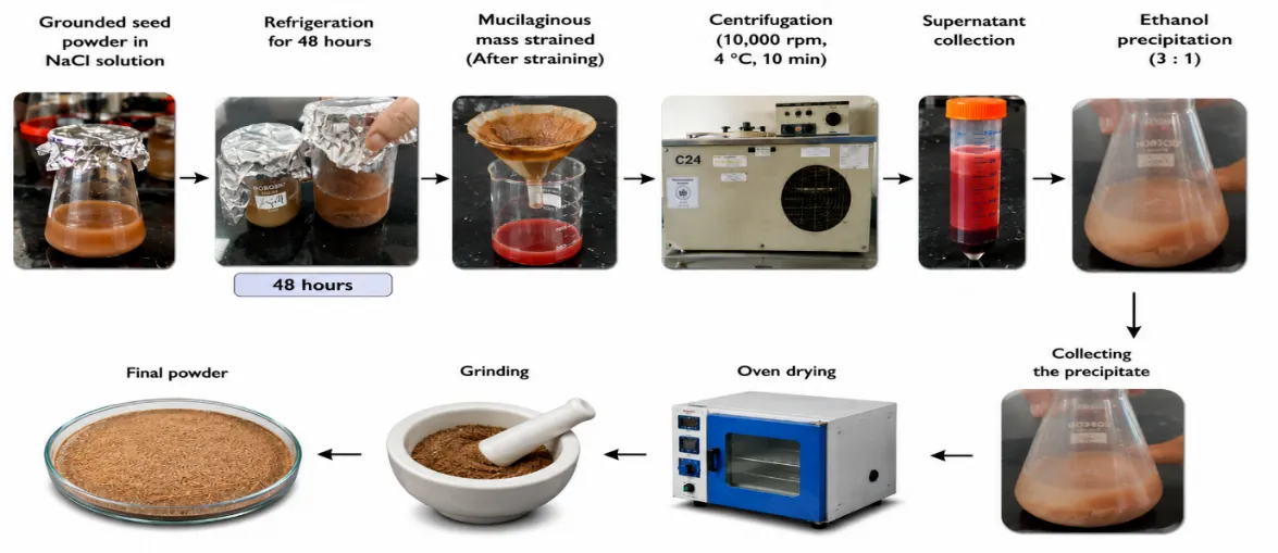
Figure 1: Extraction of Lignin
The dried seeds of Hygrophila schulli were crushed, and 5 g of the powdered material was soaked in 5% sodium chloride solution for 48 hours. The mucilaginous mass was strained, and the collected liquid was centrifuged at 10,000 rpm at 4 °C for 10 minutes. The supernatant was treated with ethanol in a 3:1 ratio to precipitate the mucilage. The precipitate was air-dried to evaporate ethanol, further dried in a hot air oven below 50 °C, and finally ground into a fine powder.
Phytochemical Analysis of Seed Polymer
Standard assays such as Molisch's, Fehling's, Barfoed's, and Benedict's for carbohydrates, ruthenium red for mucilage, Biuret and Xanthoproteic for proteins, and phloroglucinol–HCl for lignin were used to qualitatively screen the separated seed polymer powder for phytoconstituents.
Evaluation Parameters of Seed Polymer
Chloroform, acetone, ethanol, DMSO, and water were among the solvents used to test the isolated polymer's solubility. Gravimetric calculations were used to determine the yield percentage. ATR-FTIR spectroscopy was used for functional group characterization in the 4000–400 cm⁻¹ region.
FORMULATION AND EVALUATION OF HYGROPHILA SCHULLI HYDRO ALCOHOLIC SEED EXTRACT-LOADED LIGNIN-CHITOSAN NANO SUSPENSION
Lignin (0.5 g) was dissolved in 50 mL of aqueous NaOH (0.0625 g per 50 mL), then 20 mg of H. schulli Hydro alcoholic seed extract was added. Separately, 0.5 g of chitosan was dissolved in 2 mL of glacial acetic acid. The chitosan solution was added dropwise to the lignin-extract mixture while continuously stirring at 500 rpm, 30 min. The resultant suspension was centrifuged at 10,000 rpm for 30 minutes, then the pellet was recovered, re-dispersed in distilled water, and probe-sonicated at 40% amplitude (5-second pulses, 10 minutes) to create a homogenous Nano-suspension.
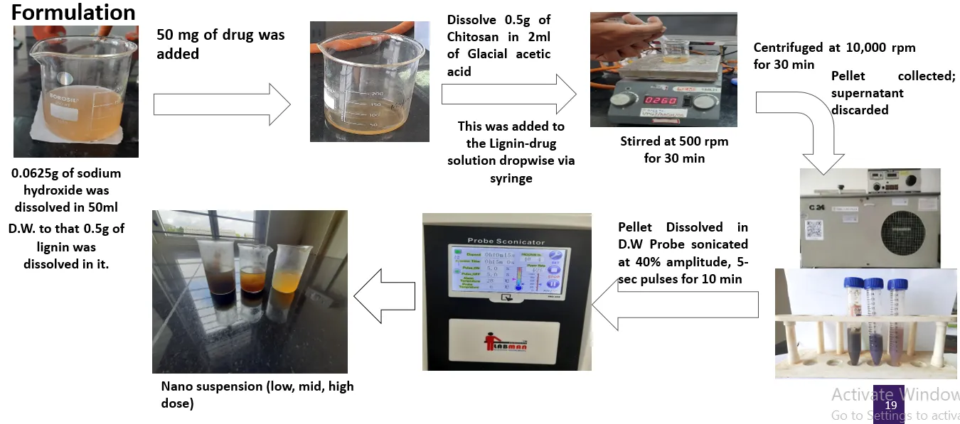
Figure 2: Formulation
EVALUATION OF LIGNIN-CHITOSAN NANO SUSPENSION (HSSE-LCNS)
The particle size and polydispersity index (PDI) of the HSSE-LCNs were measured at 25 °C using dynamic light scattering (Malvern Zetasizer). HR-TEM was used to analyse surface morphology following negative staining with 1% uranyl acetate. Functional groups and intermolecular interactions were evaluated using FTIR spectroscopy (ATR mode, 4000–400 cm⁻¹). By utilizing UV-Vis spectrophotometry to measure the non-encapsulated extract in the supernatant, entrapment efficiency was calculated from this data.
IN VIVO ASSESSMENT OF HSSE-LCNS IN DSS-INDUCED ULCERATIVE COLITIS
Induction, Grouping, and Treatment
Ulcerative colitis was established in female Wistar rats (160-200 g) by administering 6 g/kg DSS orally and 0.5 mL of 30% ethanol intrarectally for 7 days, followed by 7 days of therapy. Rats were placed into six groups (n=6): normal control (0.5% saline), disease control (DSS + ethanol), standard (Sulfasalazine), and three treatment groups that received Nano-suspension orally for 7 days in doses of 100 mg/kg, 200 mg/kg, and 400 mg/kg. After the 14-day study, the animals were slaughtered, and colon tissues were taken for further analysis.
Evaluation Parameters
Disease Activity Index (DAI)
DAI was calculated daily as the average of scores for stool consistency (0-3), faecal blood (0-2), and body weight loss (0-3) using the following formula: DAI = (Stool consistency + Blood score + Weight loss score)/3.
Ulcer Index
Colons were sliced longitudinally and rated (0-2) according to severity: no change (0), mucosal erythema (0.5), mild edema/erythema (1), moderate edema with minor ulcers (1.5), and severe ulceration with necrosis (2).
Colon Weight and Length
Colon length from ileocecal junction to proximal rectum and weight were recorded as indicators of colitis severity.
Antioxidant Studies
Colon homogenates 10% w/v) were prepared in 0.15 M KCl buffer (pH 7.4) and centrifuged at 10,000 rpm for 20 minutes at 4°C. All estimations were performed using the supernatant.
Malondialdehyde (MDA) :
Malondialdehyde (MDA): 500 µL of homogenate was combined with 1 mL TBA:TCA:HCl, heated for 15 minutes, centrifuged, and the absorbance at 532 nm was measured using the molar extinction coefficient and represented as µmol/L of tissue.
Catalase
Catalase activity was measured by adding 100 µL of homogenate to 1.9 mL of phosphate buffer (pH 7.0) and 1 mL of 10 mM H₂O₂. The resulting decrease in absorbance at 240 nm was recorded for one minute. Activity was calculated using the extinction coefficient (43.6 M⁻¹cm⁻¹) and expressed as U/mg of tissue.
Glutathione (GSH)
Glutathione (GSH): 100 µL homogenate and 200 µL Ellman's reagent were combined, incubated for 5 minutes, and absorbance was measured at 412 nm. The GSH concentration was then computed and expressed as nmol/mg of tissue.
RESULTS:
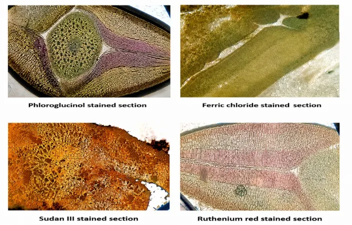
Figure 3: Histochemical Comparison of Hygrophila schulli Seeds
| Table 1: Histochemical comparison of Hygrophila schulli Seeds |
|---|
| Component | Reagent used | Colour | Result |
|---|---|---|---|
| Cellulose | Iodine +60% H₂SO₄ | Blue | - |
| Lignin | Phloroglucinol+ HCl | Pink or Red | + |
| Mucilage | Ruthenium red | Magenta or Red | + |
| Lipids | Sudan III | Orange to reddish orange | + |
| Starch | Iodine | Black | - |
| Tannin | Dil Ferric chloride | Blue Black | + |
Fluorescence Analysis
Tables and figures present the fluorescence analysis findings for Hygrophila schulli seed powders treated with various chemical reagents.
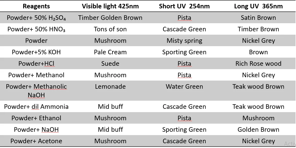
Table 2: Fluorescence Analysis of the Seeds of Hygrophila Schulli Seeds
Note: Colour mentioned in the table is based on Apcolite
LCMS analysis
LC-MS study of the hydro alcoholic extract of Hygrophila schulli seeds revealed 34 compounds, five of which—Ferulic acid, Chlorogenic acid, Genistein, Eriodictyol, and Gluconic acid were screened based on ADMET predictions and documented anti-inflammatory and antioxidant properties.
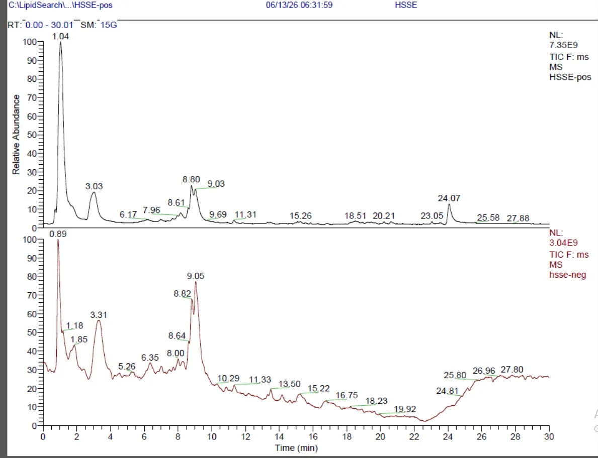
Figure 4: LCMS Chromatogram
Phytochemical analysis for seed polymer
The isolated seed polymer was characterized for yield, solubility, structural nature, and functional group composition. The Phloroglucinol -HCl test confirmed the presence of lignin, as evidenced by an immediate red-to-orange coloration (Figure 5). The extraction yield was 21.5% w/w (0.43 g from 2.0 g seed powder). The polymer was soluble in DMSO (upon sonication), dispersible in water, and readily soluble in alkaline water. Microscopic examination revealed needle-like crystalline structures, indicating a semi-crystalline nature (Figure 6).
The polymer's FT-IR analysis (Figure 7) revealed lignin absorption bands, including a broad O-H stretch at 3282 cm⁻¹, aliphatic C-H stretches at 2925 and 2861 cm⁻¹, a C=O ester band at 1740 cm⁻¹, aromatic C=C at 1642 and 1538 cm⁻¹, and C-O stretches at 1243 and 1037 cm⁻¹, confirming the polymer's lignin-like structure.
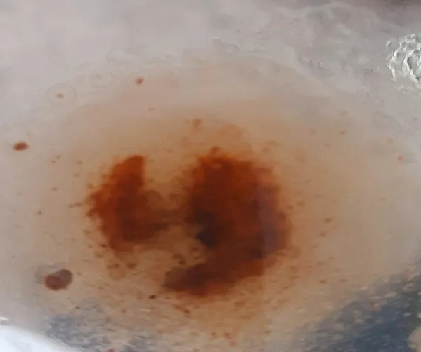
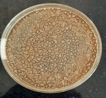
Figure 5: Phloroglucinol -HCl Test for Lignin Figure 6: Semi Crystalline Lignin
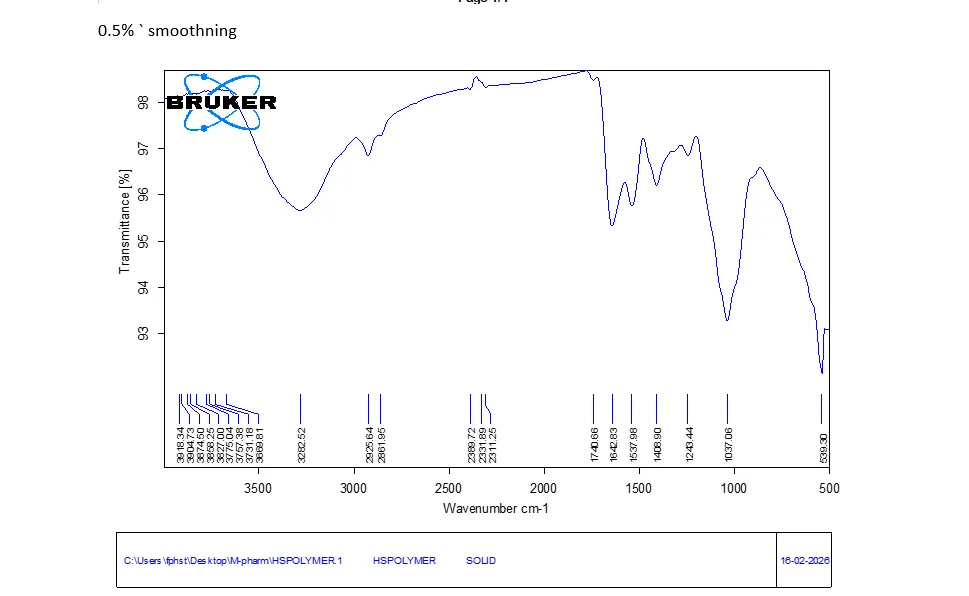
Figure 7: FTIR Spectrum of Lignin
Characterization of HSSE-LCNs
The HSSE-loaded lignin-chitosan Nano suspension (HSSE-LCNs) was characterized for physicochemical properties. DLS analysis showed a particle size of 705.4 d.nm with a PDI of 0.249, indicating uniform size distribution (Figure 8). TEM revealed faceted polyhedral particles forming dendritic agglomerates (1–2 µm) (Figure 9). FT-IR confirmed successful formulation with characteristic bands at 3287.64 cm⁻¹ (O–H/N–H), 1637.54 cm⁻¹ (amide I shift), and 1415.77, 1074.09, 1016.34 cm⁻¹ (aromatic/C–O), along with encapsulation markers at 2121.80, 927.34, and 502.27 cm⁻¹ (Figure 10). UV-Vis showed λmax at 335 nm . Entrapment efficiency was 74.77%.
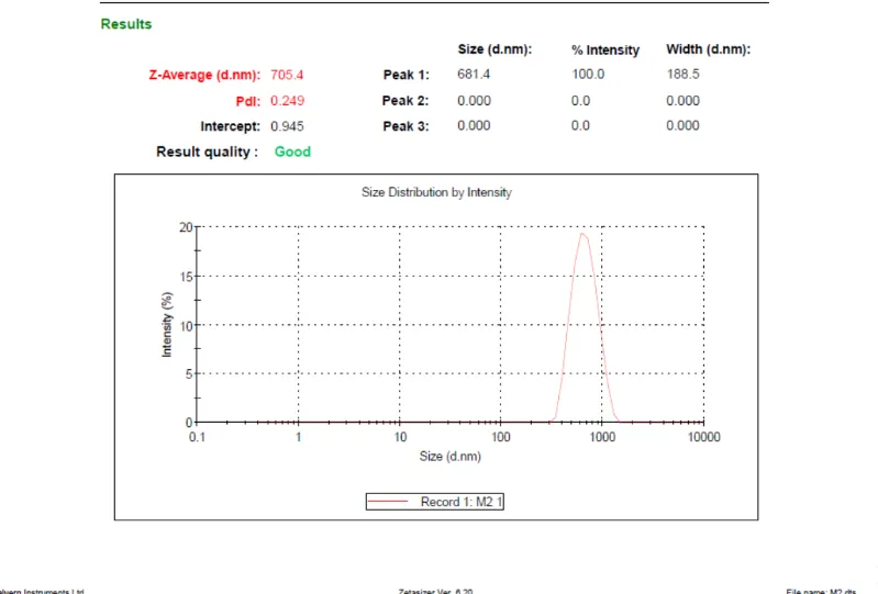
Figure 8: Particle Size of HSSE-LCNs
[Figure - see the formatted PDF]
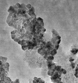
Figure 9: TEM Images Showing Spherical Structures of HSSE-LCNs
[Figure - see the formatted PDF]
Figure 10: FTIR Spectrum of (HSSE-LCNs)
Disease Activity Index (DAI)
Before Treatment: During the 7-day induction phase, the Disease group showed progressive DAI elevation (0.95 ± 0.26), confirming successful colitis induction, while the Normal group remained asymptomatic (0) (Table3).
After Treatment: Over the next 7 days, all treatment groups showed recovery with declining DAI scores, with the High dose (0.67 ± 0.22) showing the most pronounced improvement, nearing baseline by Day 14 (Table3).
Ulcer Index
On Day 14, the Disease group exhibited a significantly elevated ulcer index, while all treatment groups showed scores between 0–0.5, indicating effective mucosal protection against DSS-induced colonic damage at all doses (Figure 11).
Table 3: DAI Before After Treatment
| Groups | Average DAI before treatment | Average DAI after treatment |
|---|---|---|
| Normal | 0 | 0 |
| Disease | 0.95 ± 0.26 | 1.33 ± 0.30 |
| Standard | 0.62 ± 0.19 | 0.81 ± 0.21 |
| Low dose | 0.57 ± 0.19 | 0.90 ± 0.23 |
| Mid dose | 0.38 ± 0.13 | 0.62± 0.19 |
| High dose | 0.47 ± 0.18 | 0.67±0.22 |
Values are expressed as Mean ± SEM; n=6. One-way ANOVA followed by multiple comparison test. ###p<0.01 versus normal control, *p<0.01 versus disease control, **p<0.001 versus disease control, ***p<0.0001 versus disease controlscontrol ***p<0.0001 versus disease control
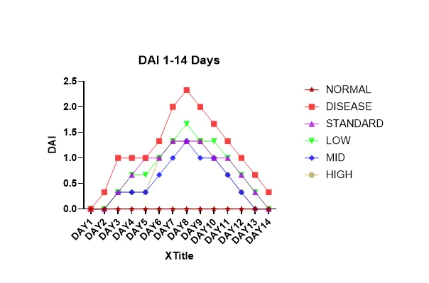
Figure 11: DAI of 14 Days Colitis Study
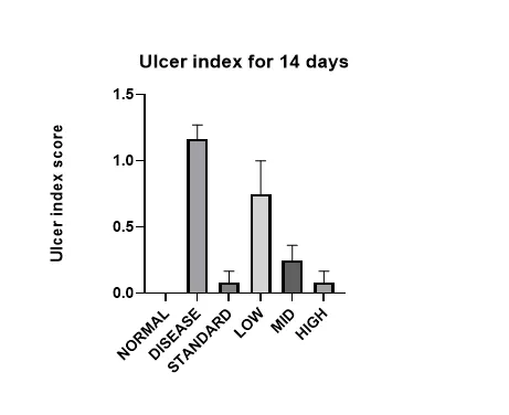
Figure 12: Ulcer Index of 14 Days Colitis Study
Colon Length and Weight
The Disease group showed increased colon weight and decreased length, indicating significant inflammation. Low and mid doses partially reduced colon weight with minimal length restoration, while the high dose normalized both parameters, comparable to the standard drug, demonstrating effective anti-inflammatory activity and tissue protection (Figure 15)
Table 4: Colon Length and Weigh
| Groups | Colon weight (g) | Colon Length (cm) |
|---|---|---|
| Normal | 1.702±0.006 | 16.33±0.189 |
| Disease | 2.372±0.018 | 12.5±0.169 |
| Standard | 2.108±0.007 | 15.683±0.192 |
| Low | 2.137±0.095 | 12.267±0.123 |
| Mid | 1.87±0.131 | 12.8.7±0.154 |
| High | 1.535±0.165 | 13.483±0.174 |
Values are expressed as Mean ± SEM; n=6. Statistical analysis was performed using one-way ANOVA followed by multiple comparison tests. Significance levels are indicated as: ###p<0.01 versus normal control, *p<0.05 versus disease control, **p<0.001 versus disease control, ***p<0.0001 versus disease control.
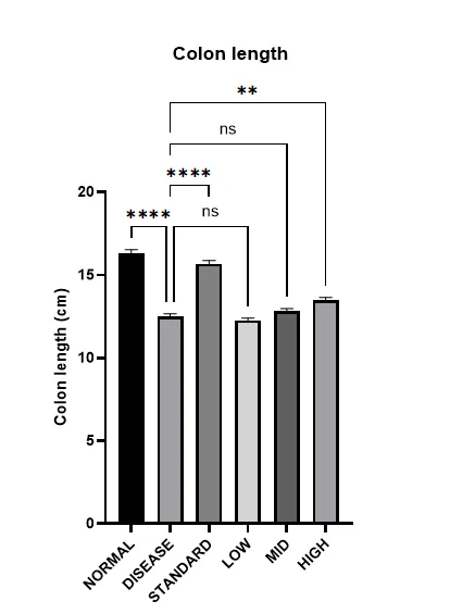
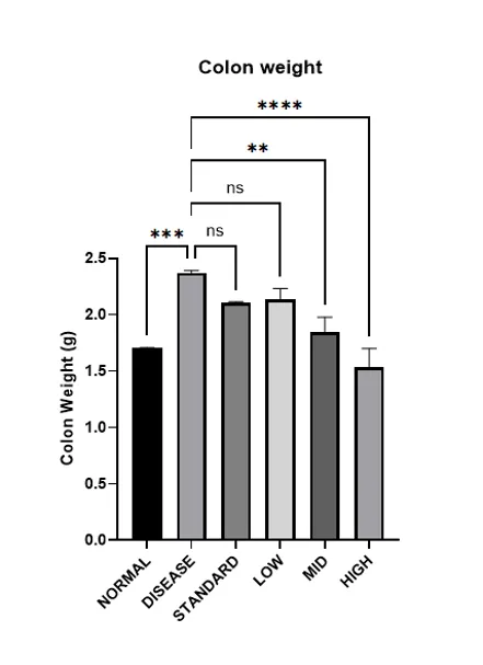
Figure 13: Effect of HSE-LCNPs on Colon Weight Figure 14: Effect of HSE-LCNPs on Colon Length
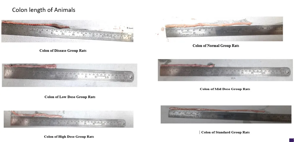
Figure 15: Colon Length of Normal, Standard, Disease, Low, Mid, High Groups
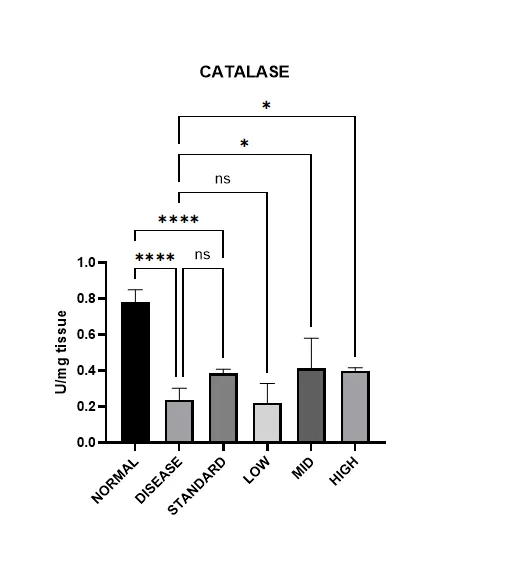
Figure 16: Effect of HSE-LCNPs on Catalase (U/mg)
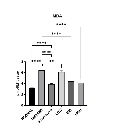
Figure 17: Effect of HSSE-LCNPs on MDA (Μmol/L)
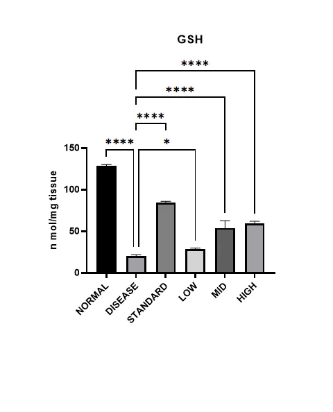
Figure 18: Effect of HSSE-LCNPs on GSH (N mol/mg Tissue)
Table 5: Anti-Oxidant Parameters After 14 Days of Study
| Groups | MDA | GSH | Catalase |
|---|---|---|---|
| Normal | 3.28±0.068 **** | 128.735±1.483 **** | 0.781±0.068 **** |
| Disease | 6.480±0.184 | 20.190±1.738 | 0.240±0.063 |
| Standard | 3.932±.091 **** | 84.55±1.320 **** | 0.385±0.023 **** |
| Low dose | 6.184±0.177 ** | 28.553±1.195 * | 0.222±0.106 |
| Mid dose | 4.406±0.042 **** | 53.829±8.877 **** | 0.413±0.168 * |
| High dose | 4.143±0.123 **** | 59.190±2.729 **** | 0.400±0.017 * |
Values are expressed as Mean ± SN n=6. Statistical analysis was performed using one-way ANOVA followed by multiple comparison tests. Significance levels are indicated as: ###p<0.01 versus normal control, *p<0.05 versus disease control, **p<0.001 versus disease control, *p<0.0001 versus disease control.
Antioxidant Activity
HSE-LCNPs demonstrated dose-dependent antioxidant effects in DSS-induced colitis. The Disease group showed significantly elevated MDA (lipid peroxidation) and reduced GSH and catalase levels, indicating severe oxidative stress. Low-dose treatment showed minimal improvement, while mid and high doses significantly reduced MDA and restored GSH and catalase levels, with the high dose showing effects comparable to the standard drug. These findings confirm the potent antioxidant activity of HSE-LCNPs at mid and high doses (Figure 16-18).
Colon histopathological study
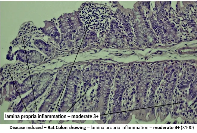
Figure 19: Disease Induced – Rat Colon Showing – Base of Lamina Propria Inflammation with Sub Mucosal Blood Vessels Congestion – Moderate 3+ (X100)
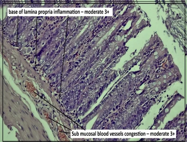
Figure 20: Disease Induced – Rat Colon Showing – Lamina Propria Inflammation – Moderate 3+ (X100)
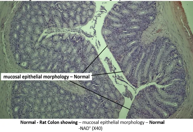
Figure 21: Normal - Rat Colon Showing – Mucosal Epithelial Morphology
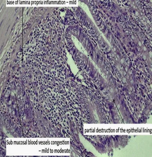
Figure 22: Standard - Rat Colon Showing – Mucosal Epithelial Morphology – Normal, NAD+ (X40)
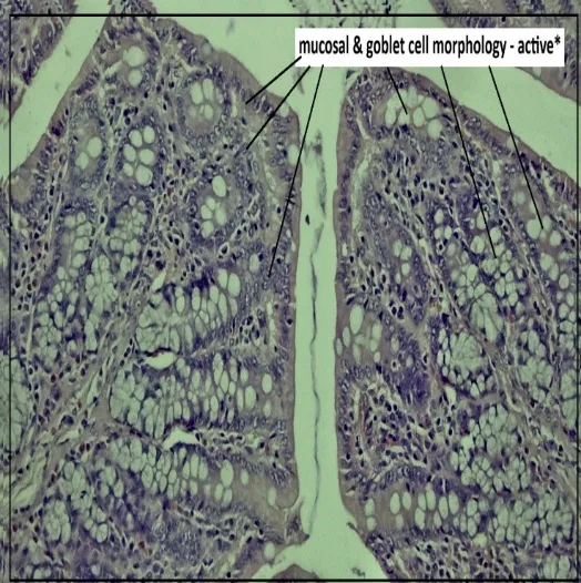
Figure 23: Mid Dose – Rat Colon Showing – Mucosal & Goblet Cell Morphology - Active* - NAD+
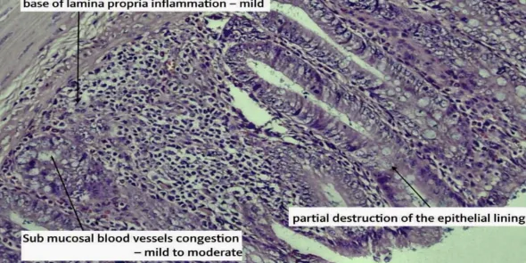
Figure 24: Low Dose - Rat Colon Showing – Mucosal Epithelial Morphology – Normal, NAD+ (X40)
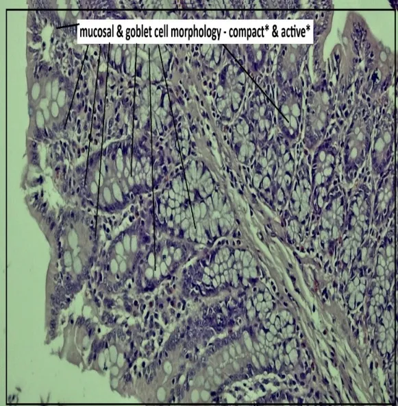
Figure 25: High Dose – Rat Colon Showing – Mucosal & Goblet Cell Morphology - Compact* & Active* - NAD+ (X100)
CONCLUSION:
This research work established the therapeutic potential of Hygrophila schulli seed hydro alcoholic extract-loaded lignin-chitosan Nano suspension (HSSE-LCNs) against DSS-induced ulcerative colitis. LC-MS phytochemical profiling revealed various bioactive compounds, including Ferulic acid, Chlorogenic acid, Genistein, and Eriodictyol, all of which indicate the extract's pharmacological potential. The isolated seed polymer exhibited lignin-associated features, but the formed HSSE-LCNs had appropriate physicochemical parameters, such as a particle size of 705.4 nm, PDI of 0.249, and entrapment efficiency of 74.77%. HSSE-LCNs showed promising anti-colitic and antioxidant actions, improving disease activity, lowering lipid peroxidation, restoring antioxidant defences, and preserving colonic mucosal integrity, especially at higher dosages.
REFERENCES

Meghana S. Nadgir, Ashoka Babu V. L., Gouri Nair, Development of a Hygrophila Schulli (Ham.) Seed-Derived Nanocarrier for Targeted Therapeutic Intervention in DSS-Induced Ulcerative Colitis, Int. J. of Pharm. Sci., 2026, Vol 4, Issue 11, 899-914. https://doi.org/10.5281/zenodo.23971675
 10.5281/zenodo.23971675