

We use cookies to ensure our website works properly and to personalise your experience. Cookies policy
1,2,5,6 Soniya Education Trust’s College of Pharmacy, Dharwad, 580002
3,4 Karnataka Institute of Medical Science, Hubballi, 580029
Background: Stevens–Johnson syndrome (SJS) is a rare but potentially life-threatening mucocutaneous hypersensitivity reaction, most commonly triggered by medications. Early recognition, prompt withdrawal of the offending drug, and appropriate supportive management are essential to reduce morbidity and mortality. Non-steroidal anti-inflammatory drugs (NSAIDs), including aceclofenac, have been implicated as causative agents. Case Presentation: We report the case of a 23-year-old male who developed extensive erythematous targetoid lesions involving the oral mucosa, trunk, upper and lower limbs, palms, soles, and genital region following the administration of aceclofenac, domperidone, and topical medications for fever. Clinical examination and temporal association with drug exposure supported the diagnosis of drug-induced Stevens–Johnson syndrome. Laboratory investigations revealed leukocytosis and mild anemia, while liver and renal function tests were within normal limits. The suspected medications were immediately discontinued, and the patient received supportive care including intravenous fluids, dexamethasone, cyclosporine, topical corticosteroids, oral care, and symptomatic treatment. Progressive clinical improvement was observed, with complete healing of lesions over 18 days, leaving only post-inflammatory hypo- and hyperpigmentation. Conclusion: This case highlights the importance of maintaining a high index of suspicion for Stevens–Johnson syndrome in patients presenting with acute mucocutaneous lesions following recent drug exposure, particularly NSAIDs such as aceclofenac. Early diagnosis, prompt discontinuation of the suspected drug, and multidisciplinary supportive management are crucial for favorable clinical outcomes. Increased awareness among healthcare professionals regarding early recognition and prevention of recurrent exposure is essential to minimize disease-related complications.
Steven Johnson syndrome (SJS) is an IgE mediated hyper sensitivity reaction characterized by erythematous macula’s or fat, atypical targeted lesions with epidermal detachment of <10% body surface area, often complicated by ocular conjunctivitis or uveitis and symblepharon formation 1. It might be called drug-induced Stevens-Johnson syndrome or mycoplasma-induced Stevens-Johnson syndrome. SJS/Toxic Epidermal Necrosis (TEN) is not an inherited condition. However, the genetic changes that increase the risk of developing SJS/TEN can be passed from one generation to the next.
If Reaction is occupied 10-30% of the Body Surface Area (BSA) that is considered as SJS but if more than 30% then it is called as TEN. Incidence range from 1.2 to 6 cases per million per year, the condition is fatal in 5% of treated cases and in 15% of untreated cases2. More cases of SJS occur in females than males. Infections like pneumonia are the most likely cause of SJS in children, whereas medications are the most likely cause of SJS/TEN in adults3. Most patients with Stevens-Johnson syndrome are treated symptomatically. In principle, the symptomatic treatment of patients with this disorder does not differ from the therapy applied to patients with extensive thermal or chemical burns 4.
Due to the high risk of mortality, management of patients with SJS/ TEN requires rapid diagnosis; evaluation of the prognosis using [SCORTEN] Severity-of-Illness Score for Toxic Epidermal Necrolysis, SCORTEN is an illness severity score that has been developed to predict mortality in SJS and TEN cases 5.
Each person’s experience with Stevens-Johnson syndrome can be different. Skin can regret in a matter of weeks, but recovery can take months if symptoms are severe. Some long-term reactions may develop; it may redevelop if one is exposed to the same medication known to have triggered the condition the first time. In such cases, the second episode is usually more severe than the first episode 6.
Antimicrobials are commonly reported group of drugs followed by anticonvulsants, antipyretics, and NSAIDs. Sixteen patients (35.5%) had experienced SJS/TEN due to antimicrobials. The majority of TEN cases (44%) were due to antimicrobials which was in concordance with the studies from (42%) West Germany & (44%) South India 9.
CASE PRESENTATION
The collection of data was done in accordance with moral guidelines.
A 23-year-old male patient attended the SKIN and STD department in Karnataka institute of medical science [KIMS], Hubballi on 22nd February 2023.
Chief complaints were painful red lesions in oral cavity since days, itchy red lesions over upper limb, lower limb, chest, trunk, back, and genital area since days
History of presenting illness:
Patient was apparently normal 3 days back and he had fever since 2 days, and had visited local hospital for the same and was prescribed medications candid oral paints, Aceclofenac and Domperidone.
Patient developed mild itchy red lesions over 2 days; it was insidious in onset and gradually progressive. The lesions increased in number to involve BL, LL, trunk and genital. His temperature was 101ͦ F and Blood Pressure (BP) was 110/70 mmHg
The medication and topical applicants prior to onset of lesions, there was no history of joint pain, abdominal pain, burning maturation and hematuria
Table 1: Affected Body Parts with Causes
|
Affected part |
Cause |
|
Scalp |
No active skin lesions present. |
|
Eyes |
No redness and not watery |
|
Oral cavity |
Multiple well-ill demarcated erythematous erosions with crusting with multiple well-ill demarcated white plaques present over upper and lower lip mucosa, left tonsil and right tonsil bucklemucosa, soft tongue and hard palate and floor of mouth. |
|
Palms and soles |
Multiple well-ill demarcated erythematous maculae’s and patches present. |
|
Nails |
Normal. |
|
Genital |
Few Well-Ill Demarcated Erythematous Erosions Present. |
O/c/e: multiple discrete to coalescing well to ill demarcated erythematous targeted plaques present over upper limb, lower limb, trunk and genitals.
There were no fresh complaints for 4 days but on 6th February patient came uphaving complaints of burning sensation over foot, multiple well-ill demarcated hyper pigmented plaques with multiple ill demarcated erosions over upper and lower limb, trunk, and back.
The patient appetite and sleep decreased, diet was mixed, bowel and bladder regular and not addictive to smoke and alcohol.
Microscopy Test for Pus: 2-3 pus cells were present but infection not found.
Systemic Examination
Table 2: systemic examination
|
Cardio vascular system |
S1, S2 heard no murmur. |
|
Central nervous system |
Higher mental function intact. |
|
Respiratory system |
NVBS heard, no added sounds |
|
Per abdomen |
Soft, non-tender, no organomegaly |
Hematology report
Table 3: Hematology report
|
Items |
Results |
Units |
Alarm |
|
WBC |
11.8 |
10^3/UL |
Increased |
|
LYM% |
17.1 |
% |
Decreased |
|
GRAM% |
73.0 |
% |
Increased |
|
HGB |
10.2 |
g/dl |
Decreased |
|
HCT |
30.0 |
% |
Decreased |
|
MCV |
56.2 |
fL |
Decreased |
|
MCH |
19.1 |
pg |
Decreased |
|
RDW-SD |
27.8 |
fL |
Decreased |
|
PLT |
320 |
10^3/uL |
Increased |
|
PDW |
9.2 |
% |
Decreased |
|
P-LCR |
5.7 |
% |
Decreased |
DAY 1


|
Multiple well-ill demarcated erythematous erosion with crusting with multiple well-ill demarcated white plaques present over upper and lower lip mucosa, left tonsil and right tonsil buckle mucosa, soft tongue and hard palate and floor of mouth. |


|
Multiple well-ill demarcated erythematous maculae’s and patches present on palm, legs and foot. |

|
Few well-ill demarcated erythematous erosions present On Genital area. |
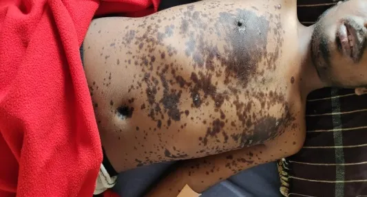

|
Flat or slightly pink blister like structures are appears on both chest and back. |
DAY 6


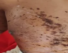

|
The lesions are covered with brown to black colored crust. |
DAY 12


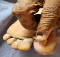

|
The skin has started to exfoliate and painless exfoliate after 11th day |
DAY 18


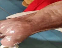

|
The lesions have healed leaving behind post inflammatory hypo and hyper pigmentation |
Figure: 1 Picture of affected area with their Description
Patient was diagnosed with Drug Induced Stevens Johnson Syndrome. He was immediately admitted in emergency ward and given fluid resuscitation with normal saline, The intravenous medicines included corticosteroids (Dexamethasone 2CC I.V), Immunosuppressant (Cyclosporine) and H2 blockers(Ranitidine 50 mg i.v), The oral applicants were povidine iodine gargle, Clotrimazole mouth paint, Mucopain gel, the local applicants like Triamcinolone gel, Soframycine ointment, Calamine lotion. Due to the above adverse effects, medication was withdrawn and the attendant was strictly instructed not to administer the medicine again. His Blood Sugar (Fasting), Liver Function Tests (LFT) and Kidney Function Tests (KFT) were done and found to be normal.
Table 4: Treatment chart
|
Medication |
Frequency |
Route of administration |
|
Injection Dexamethasone |
1-0-0 |
intravenous |
|
Cyclosporine |
1-0-1 |
intravenous |
|
povidine iodine gargle |
1-0-1 |
Oral |
|
Clotrimazole mouth paint |
1-1-1 |
Oral |
|
Mucopain gel |
1-1-1 |
Oral |
|
Triamcinolone gel |
1-1-1 |
local applicant |
|
Soframycine ointment |
1-0-0 |
local applicant |
|
Calamine lotion |
1-0-1 |
local applicant |
|
Fluids (20ml NS and 20ml RL) |
100 ml/hr |
intravenous |
DISCUSSION
Stevens-Johnson syndrome is in most cases, triggered by medications, there’s no way to know – before taking medications – that a person might experience an adverse reaction to the drug. If a medication is identified that has triggered this condition, one can avoid that drug or related drug again 6.
Mortality from SJS depends on the time of reaction onset, the extent of epidermal detachment, the patient’s age, and underlying conditions. From the least to the most severe degree of skin involvement, mortality is almost 10% for patients with SJS, approximately 30% for patients with SJS/TEN overlap, and almost 50% for patients with TEN. It should be noted that SJS and TEN are considered two clinical spectrums of the same disease.
There are gaps that need to be urgently addressed in SJS/TEN research. There is an urgent need for reproducible methods of measuring disease severity that are sensitive to changes induced by therapeutic interventions and that more accurately predict outcomes beyond the acute stage by including the systemic and internal organ effects of SJS/TEN. Potential solutions include consensus on definitions, advances in diagnostic imaging and biomarker assessment, and development of AI platforms for the detection and monitoring status of disease 8.
It is important to suspect the culprit drug by the temporal relationship (2 days to 8 weeks) 10 but the extremely ‘rapid onset’ in this case. The exact aetiopathogenesis being imprecise is thought that some noxious metabolites, inflammatory mediators/modifiers, as well as cytotoxic T-lymphocytes, regulatory T cells and dermaldendrocytes could provoke apoptosis/ necrosis of epithelial cells 8-9.
As the drugs are too small to trigger an immunogenic response, three mechanistic models have been proposed to explain how small molecular synthetic compounds are recognized by T cells in an MHC dependent fashion. These include the hapten/prohapten model, the pei model, and the altered repertoire model.This concept proposes that drugs or drug metabolites can bind to specific MHC molecules, within the pocket of their peptide binding grooves, with exquisite specificity, thus allowing a new repertoire of endogenous self-peptides to be bound and presented 10.
CONCLUSION
By this case report of drug induced SJS underscores the importance that physicians bear in mind that SJS can be developed within only a few days after ingestion of over-the-counter medications, such as Aceclofenac, Anticonvulsants, Antipyretics, and NSAIDs.
As Aceclofenac is a widely used drug, physicians should be aware with this adverse reaction for early detection and intervention. Affected patient and their first-degree relatives should be instructed to avoid any identified drugs or chemicals that may be responsible.
There are gaps that need to be urgently addressed in SJS/TEN research. There is an urgent need for reproducible methods of measuring disease severity that are sensitive to changes induced by therapeutic interventions and that more accurately predict outcomes beyond the acute stage by including the systemic and internal organ effects of SJS/TEN. Potential solutions include consensus on definitions, advances in diagnostic imaging and biomarker assessment, and development of AI platforms for the detection and monitoring of disease.
Evidence-based practice related to the care of patients with SJS is still evolving. Management includes early identification, withdrawal of the suspected drug, and early transfer to a specialized center. Further research and clinical evidence are needed to develop appropriate and cost-effective treatment guidelines for optimal care of these patients.
REFERENCES



Ashwini Angadi, Adarsh G S, Janaki Torvi, Ravi Rathod, Preeti Kulkarni, Venkatrao Kulkani, Drug Induced Steven-Johnson Syndrome: A Case Report, Int. J. of Pharm. Sci., 2026, Vol 4, Issue 6, 7774-7780. https://doi.org/10.5281/zenodo.21071939
 10.5281/zenodo.21071939