

We use cookies to ensure our website works properly and to personalise your experience. Cookies policy
M.Pharm, Department of Pharmacy, M.J.P. Rohilkhand University, Bareilly (243006), Uttar Pradesh, India.
Alzheimer's disease (AD) is a leading cause of dementia, marked by irreversible cognitive decline and behavioral dysfunction due to pathological features like amyloid-? plaques and neurofibrillary tangles. Current diagnostic tools, including cerebrospinal fluid biomarkers and amyloid PET imaging, inadequately detect early molecular changes, highlighting the need for novel biomarkers for early diagnosis and personalized treatment. Advances in multi-omics technologies have unveiled susceptibility genes (e.g., APOE, APP, PSEN1) and emerging proteomic biomarkers related to amyloid metabolism and synaptic integrity. Research shows AD's progression is linked to dysregulated signaling pathways that affect neuronal survival and plasticity. This review encapsulates the state of biomarker discovery, discusses the integration of multi-omics, and emphasizes the potential for improving diagnostics and therapeutic interventions in AD
Alzheimer's disease (AD) is the most prevalent neurodegenerative disorder and the leading cause of dementia worldwide, accounting for approximately 60–70% of all dementia cases [1]. It is characterized by progressive deterioration of memory, cognition, language, executive function, and behavior, ultimately resulting in complete functional dependence and premature mortality. The disease represents a major global public health challenge due to its increasing prevalence in aging populations and the absence of curative therapies. According to the World Health Organization (WHO), more than 55 million people worldwide are currently living with dementia, and this number is expected to exceed 139 million by 2050 owing to increased life expectancy and demographic aging. Alzheimer's disease imposes an enormous socioeconomic burden, affecting not only patients but also caregivers, healthcare systems, and national economies [2]. Therefore, early diagnosis and effective disease-modifying strategies remain among the highest priorities in neuroscience research.
Alzheimer's disease is defined by two key neuropathological features: amyloid-β (Aβ) plaques and neurofibrillary tangles (NFTs) made of hyperphosphorylated tau protein. The dominant amyloid cascade hypothesis explains disease progression via abnormal cleavage of amyloid precursor protein (APP), leading to excess Aβ42 peptide production, which forms plaques and triggers a series of pathological events, including synaptic dysfunction and neuronal apoptosis. Familial Alzheimer's, linked to mutations in APP and related genes, underscores amyloid dysregulation. However, anti-amyloid therapies have shown limited efficacy, suggesting additional mechanisms are involved [3]. The tau hypothesis posits that tau hyperphosphorylation destabilizes microtubules, disrupts axonal transport, and contributes to cognitive decline and neuronal death. Research indicates that tau pathology correlates more strongly with cognitive impairment than amyloid plaque density, positioning tau as a crucial factor in disease severity. In recent years, neuroinflammation has been recognized as a key factor in Alzheimer's disease pathogenesis, with persistent activation of microglia and astrocytes leading to the production of pro-inflammatory cytokines, chemokines, reactive oxygen species (ROS), nitric oxide, and complement proteins, establishing a chronic inflammatory state in the brain [4]. The urgent need for new biomarkers to improve early diagnosis, disease progression prediction, and personalized treatment strategies for Alzheimer's disease. Advances in high-throughput technologies, including genomics, transcriptomics, proteomics, and metabolomics, have led to the discovery of biomarkers such as Transthyretin (TTR), Clusterin (CLU), Complement C3, and Apolipoprotein E (ApoE). These biomarkers provide insights into synaptic integrity, immune activation, and neuronal survival, expanding beyond traditional Aβ and tau measurements. Additionally, understanding dysregulated signaling pathways is critical for deciphering the molecular mechanisms of Alzheimer's [5]. Significant pathways include the Amyloid Cascade, Tau signaling, and the PI3K/Akt/mTOR cascade, which influence processes like amyloid processing, neuroinflammation, and mitochondrial function [6]. The integration of emerging genomic and proteomic biomarkers with these pathways is essential for clarifying disease mechanisms and identifying new therapeutic targets. The review emphasizes the potential of multi-omics approaches to deepen comprehension of Alzheimer's biology, facilitate biomarker discovery, and enhance clinical management with the goal of altering the disease's progression rather than merely relieving symptoms.
2. Pathophysiology of Alzheimer's Disease
Alzheimer's disease (AD) is a multifactorial neurodegenerative disorder characterized by progressive neuronal dysfunction and irreversible cognitive decline [7]. Although extracellular amyloid-β (Aβ) plaques and intracellular neurofibrillary tangles (NFTs) remain the pathological hallmarks of the disease, accumulating evidence indicates that AD results from the interaction of multiple pathological mechanisms, including oxidative stress, mitochondrial dysfunction, chronic neuroinflammation, synaptic degeneration, and blood-brain barrier (BBB) disruption. These pathological events begin several years before the appearance of clinical symptoms and progressively impair neuronal communication, ultimately leading to extensive neuronal loss and dementia [8].
2.1 Amyloid Cascade
The amyloid cascade hypothesis is a well-studied mechanism behind Alzheimer's disease, starting with the abnormal processing of amyloid precursor protein (APP), mainly in neurons. In healthy conditions, APP is cleaved by α-secretase, producing neuroprotective soluble APPα. However, in Alzheimer's, it is cleaved by β-secretase (BACE1) and γ-secretase, leading to the production of amyloid-β peptides, notably Aβ42, which aggregates more readily than Aβ40 [9]. Soluble Aβ oligomers disrupt synaptic function, leading to the formation of plaques that activate microglia, induce oxidative stress, impair mitochondrial function, and trigger neuroinflammation. Mutations in APP, PSEN1, and PSEN2 increase Aβ42 production, hastening disease progression.
2.2 Tau Hyperphosphorylation
Tau is a microtubule-associated protein mainly found in neuronal axons, stabilizing microtubules and aiding axonal transport. Under normal conditions, tau is reversibly phosphorylated, regulating its microtubule interactions. In Alzheimer's disease, abnormal kinase activation leads to hyperphosphorylation of tau, causing it to dissociate from microtubules and destabilize them, impairing transport. Detached tau aggregates into neurofibrillary tangles (NFTs), key pathological features of Alzheimer's [10]. The accumulation of NFTs disrupts neuronal integrity and correlates with cognitive decline, highlighting the critical role of tau in neurodegeneration over amyloid plaque burden.
2.3 Oxidative Stress
Oxidative stress plays a significant role in Alzheimer's disease, arising from an imbalance between reactive oxygen species (ROS) production and antioxidant defenses. The brain's vulnerability is attributed to its high oxygen usage, lipid content, and limited antioxidants. Amyloid-β oligomers exacerbate ROS generation through mechanisms like NADPH oxidase activation and mitochondrial dysfunction, leading to lipid peroxidation, protein oxidation, and DNA damage. This oxidative damage worsens tau phosphorylation and promotes amyloid deposition, creating a cycle of neuronal injury [11]. Concurrently, antioxidant responses mediated by Nrf2 are diminished, further compromising cellular resistance to oxidative damage.
2.4 Mitochondrial Dysfunction
Mitochondrial dysfunction is a key early event in Alzheimer's disease. Mitochondria play crucial roles in ATP production, calcium balance, reactive oxygen species regulation, and programmed cell death initiation [12]. Accumulation of amyloid-β peptides in mitochondria disrupts oxidative phosphorylation, decreasing ATP synthesis. Damaged mitochondria show impaired electron transport, excessive ROS production, DNA mutations, calcium overload, and activate apoptotic pathways. Abnormal mitochondrial dynamics, such as excessive fission and impaired fusion, jeopardize neuronal survival. Ineffective mitophagy leads to the buildup of dysfunctional mitochondria, further compromising neuronal energy metabolism and synaptic transmission, ultimately increasing vulnerability to degeneration [13].
2.5 Synaptic Dysfunction
Synaptic dysfunction is a key factor in cognitive decline associated with Alzheimer's disease. Soluble amyloid-β oligomers impair synaptic plasticity and neurotransmission, while hyperphosphorylated tau disrupts axonal transport and reduces essential synaptic proteins. Chronic microglial activation exacerbates synaptic loss through enhanced pruning [14].
2.6 Blood-Brain Barrier Dysfunction
The blood-brain barrier (BBB) is crucial for regulating molecular transport between the bloodstream and the central nervous system while ensuring cerebral homeostasis. In Alzheimer's disease, BBB integrity declines due to factors like endothelial dysfunction and neuroinflammation, leading to increased influx of immune cells and inflammatory substances into the brain. This exacerbates neuroinflammation and neuronal damage. Additionally, impaired transport proteins reduce amyloid-β clearance, and elevated levels of certain receptors increase amyloid influx. BBB dysfunction also results in cerebral hypoperfusion and oxidative stress, accelerating amyloid deposition and cognitive decline [15].
3. Emerging Genomic Biomarkers in Alzheimer's Disease
Alzheimer's disease (AD) has a significant genetic basis, involving both rare mutations and common variants that influence disease risk. Recent research through genome-wide association studies (GWAS) and sequencing technologies has advanced the understanding of AD's genetic architecture. Key genes associated with early-onset Alzheimer's disease (EOAD) include APP, PSEN1, and PSEN2, while numerous susceptibility loci such as APOE, TREM2, and others are linked to late-onset Alzheimer's disease (LOAD) [16]. These genes are involved in critical mechanisms like amyloid processing, lipid metabolism, and neuroinflammation. The review emphasizes APOE as the strongest genetic risk factor for LOAD and outlines the contributions of other major genes to AD pathology.
3.1 Apolipoprotein E (APOE)
The APOE gene, located on chromosome 19q13.2, is the most influential genetic risk factor for sporadic late-onset Alzheimer's disease. APOE encodes apolipoprotein E, a lipid transport protein synthesized predominantly by astrocytes and microglia in the central nervous system. Besides maintaining cholesterol homeostasis, ApoE regulates neuronal repair, synaptic plasticity, amyloid-β metabolism, and neuroinflammatory responses.
Three major APOE alleles—ε2, ε3, and ε4—produce structurally distinct protein isoforms that differentially influence AD susceptibility [17].
APOE ε2 is considered the protective allele against Alzheimer's disease. Individuals carrying the ε2 allele exhibit enhanced amyloid-β clearance, reduced tau pathology, lower neuroinflammation, and delayed disease onset. The allele also promotes neuronal survival by improving lipid transport and synaptic maintenance.
APOE ε3 is the most common allele worldwide and is generally regarded as the neutral reference genotype. It supports normal lipid metabolism and physiological amyloid clearance without significantly increasing or decreasing Alzheimer's disease risk [18].
APOE ε4 represents the strongest genetic susceptibility allele for sporadic Alzheimer's disease. Possession of one ε4 allele increases disease risk approximately threefold, whereas homozygous ε4 carriers may experience a 10–15-fold higher lifetime risk. ApoE4 impairs amyloid-β clearance, enhances amyloid aggregation, accelerates tau phosphorylation, disrupts blood-brain barrier integrity, promotes oxidative stress, and intensifies microglial activation, thereby contributing to progressive neurodegeneration [19].
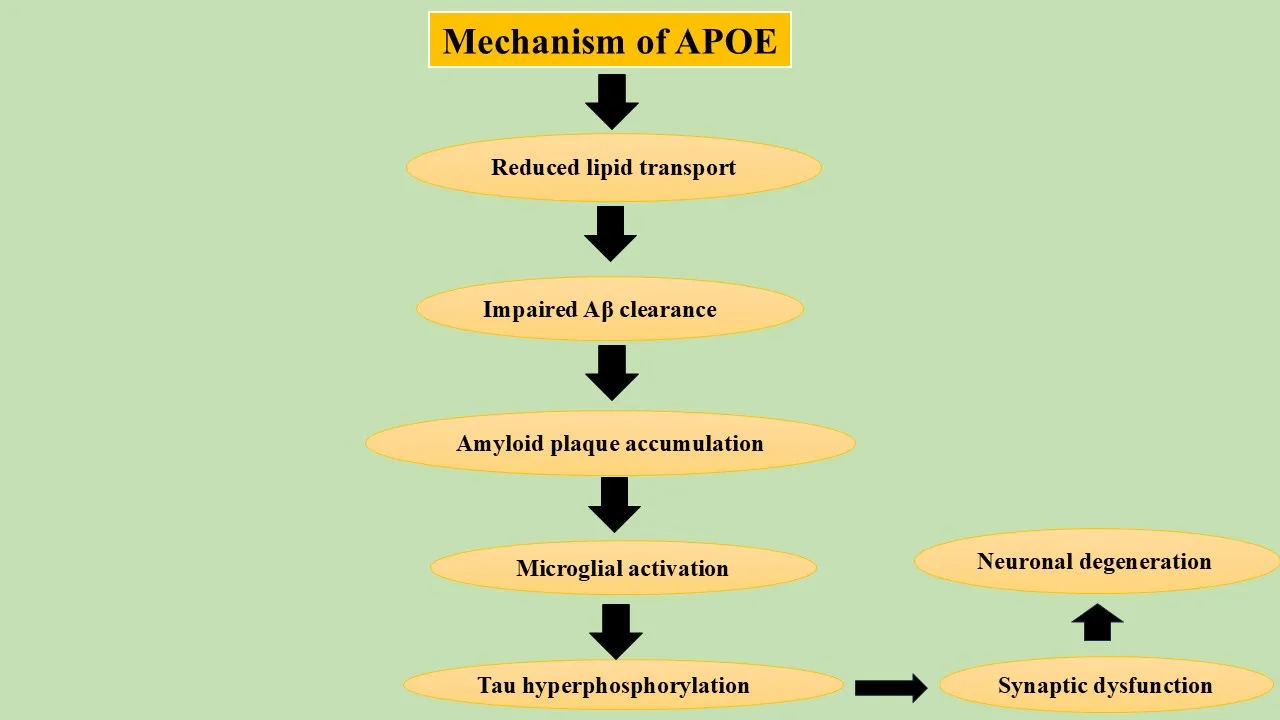
Figure 1:Mechanism of action of APOE
3.2 Amyloid Precursor Protein (APP)
The APP gene on chromosome 21 encodes amyloid precursor protein, essential for neuronal growth and synaptic function. Normally, APP is cleaved by α-secretase, but in Alzheimer's disease, β-secretase (BACE1) and γ-secretase cleave it, leading to the production of amyloid-β peptides, especially Aβ42, which form extracellular plaques. Missense mutations in APP heighten Aβ42 production, contributing to familial early-onset Alzheimer's disease [20].
3.3 Presenilin-1 (PSEN1)
PSEN1, found on chromosome 14, encodes the catalytic part of the γ-secretase complex involved in APP cleavage. Over 300 pathogenic mutations in PSEN1 have been linked to early-onset Alzheimer's disease, increasing the Aβ42/Aβ40 ratio and speeding up amyloid plaque formation and disease progression [21].
3.4 Presenilin-2 (PSEN2)
The PSEN2 gene, found on chromosome 1, encodes a γ-secretase component and can harbor pathogenic variants that elevate Aβ42 production, contributing to familial Alzheimer's disease. Mutations are rarer compared to PSEN1, with later onset and increased phenotypic variability associated with PSEN2-related Alzheimer's disease [22].
3.5 Triggering Receptor Expressed on Myeloid Cells 2 (TREM2)
TREM2, a microglia-specific immune receptor, plays a crucial role in regulating phagocytosis, lipid sensing, inflammatory signaling, and amyloid plaque clearance. Rare variants of TREM2 significantly heighten Alzheimer's disease risk by impairing microglial activation and reducing amyloid clearance. Additionally, dysfunctional TREM2 signaling contributes to chronic neuroinflammation and accelerates tau pathology [23].
3.6 Sortilin-Related Receptor 1 (SORL1)
SORL1 encodes a sorting receptor that manages the intracellular trafficking of APP. In normal conditions, it directs APP towards recycling, which limits amyloidogenic processing. Reduced SORL1 expression increases APP availability for β-secretase cleavage, leading to elevated Aβ production and plaque formation. Genetic variants in SORL1 are linked to both early- and late-onset Alzheimer's disease [24].
3.7 ATP-Binding Cassette Transporter A7 (ABCA7)
ABCA7 plays an important role in lipid transport, phagocytosis, and amyloid clearance. Loss-of-function mutations impair microglial uptake of amyloid-β and reduce cholesterol homeostasis, thereby enhancing amyloid deposition and neuroinflammation. GWAS consistently identify ABCA7 as one of the strongest susceptibility genes for sporadic Alzheimer's disease [25].
3.8 Clusterin (CLU)
The CLU gene encodes clusterin (Apolipoprotein J), an extracellular molecular chaperone involved in complement regulation, lipid transport, and amyloid binding. Clusterin inhibits protein aggregation under physiological conditions but becomes dysregulated in Alzheimer's disease. Genetic variants in CLU influence amyloid clearance, complement activation, astrocyte function, and chronic neuroinflammation, making CLU an important genomic and proteomic biomarker [26].
3.9 Complement Receptor 1 (CR1)
CR1 regulates activation of the classical complement pathway and facilitates immune-mediated clearance of amyloid-β. Alzheimer's disease-associated CR1 polymorphisms alter complement signalling, resulting in excessive synaptic elimination, chronic inflammation, and increased amyloid accumulation. These findings support an important role of innate immunity in Alzheimer's disease progression [27].
3.10 Bridging Integrator 1 (BIN1)
BIN1 is the second strongest genetic risk factor for late-onset Alzheimer's disease after APOE. BIN1 participates in membrane curvature, endocytosis, and cytoskeletal organization. Experimental evidence suggests that BIN1 directly interacts with tau protein and promotes tau aggregation, neurofibrillary tangle formation, and synaptic dysfunction [28].
3.11 Cluster of Differentiation 33 (CD33)
CD33 is an inhibitory receptor expressed on microglia that suppresses innate immune responses. Increased CD33 activity reduces microglial phagocytosis of amyloid-β, thereby facilitating plaque accumulation and chronic neuroinflammation. Genetic variants associated with increased CD33 expression correlate with elevated Alzheimer's disease risk [29].
3.12 Phosphatidylinositol Binding Clathrin Assembly Protein (PICALM)
PICALM regulates clathrin-mediated endocytosis, synaptic vesicle recycling, and intracellular trafficking of APP. Reduced PICALM expression disrupts amyloid-β transport across the blood-brain barrier and impairs neuronal endocytosis, ultimately contributing to amyloid accumulation and synaptic dysfunction. The uploaded review identifies PICALM as a susceptibility locus linked to APP processing, amyloid clearance, endocytosis, and tau pathology [30].
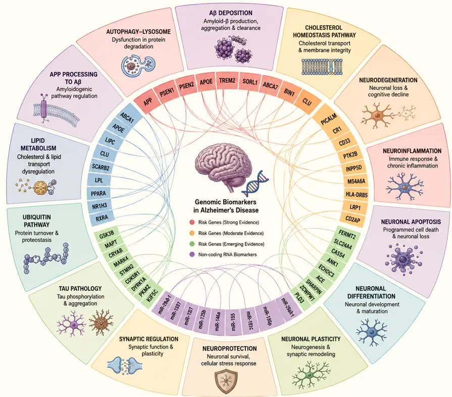
Figure 2. Genomic Biomarkers Associated with Alzheimer's Disease
4. Emerging Proteomic Biomarkers in Alzheimer's Disease
Proteomic biomarkers are crucial for understanding Alzheimer's disease (AD) by enhancing early diagnosis, disease monitoring, and therapeutic assessment. Unlike genomic biomarkers, they reflect dynamic changes in pathology. Techniques such as mass spectrometry and aptamer-based plasma proteomics have identified several proteins linked to amyloid deposition and other pathological aspects. Key proteins like Clusterin, Complement C3, Transthyretin, and others show promise as diagnostic and prognostic markers for AD [31].
Table 1. Proteomic Biomarkers in Alzheimer's Disease [32]
|
Biomarker |
Source |
Major Function |
Alteration in AD |
Diagnostic Significance |
|
Clusterin (CLU) |
Plasma, CSF |
Molecular chaperone |
↑ Increased |
Amyloid burden, neuroinflammation |
|
Complement C3 |
Complement activation |
↑ Increased |
Synaptic loss, inflammation |
|
|
Complement Factor H |
Complement regulation |
Dysregulated |
Complement-mediated neurodegeneration |
|
|
Transthyretin |
CSF |
Aβ binding |
↓ Decreased |
Reduced amyloid clearance |
|
Alpha-2-Macroglobulin |
Plasma |
Protease inhibitor |
↑ Increased |
Aβ sequestration |
|
ApoE |
Plasma, CSF |
Lipid transport |
Isoform-dependent |
Genetic and proteomic risk marker |
|
VGF |
CSF |
Synaptic plasticity |
↓ Decreased |
Cognitive decline |
|
Contactin-2 |
Axonal adhesion |
Synaptic dysfunction |
||
|
NPTX2 |
Synaptic maintenance |
Early cognitive impairment |
||
|
Osteopontin |
Plasma, CSF |
Inflammation |
↑ Increased |
Neuroinflammation |
|
Fibronectin |
Plasma |
Extracellular matrix |
BBB dysfunction |
|
|
Vitronectin |
Complement regulation |
Amyloid-associated inflammation |
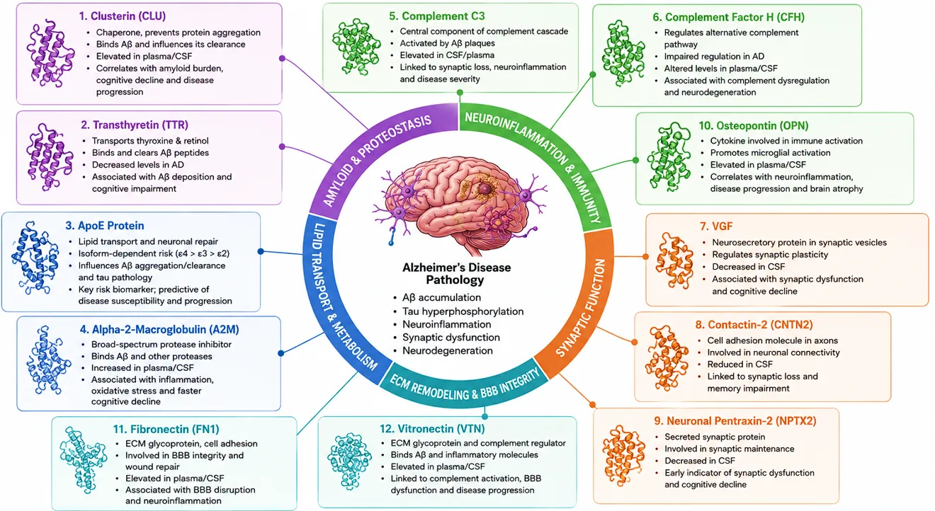
Figure 3. Proteomic Biomarkers and Their Clinical Significance
5. Dysregulated Signalling Pathways in Alzheimer's Disease
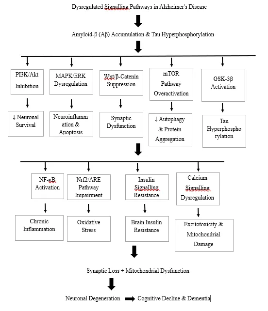
Alzheimer's disease (AD) involves the dysregulation of interconnected signaling pathways leading to amyloid-β accumulation, tau hyperphosphorylation, neuroinflammation, oxidative stress, synaptic dysfunction, and neuronal apoptosis. These pathways, which include the amyloid cascade, tau signaling, and various others like PI3K/Akt/mTOR and NF-κB, interact within a complex regulatory network that drives the disease's onset and progression. Recent advances in genomics and proteomics highlight the significance of these dysregulated pathways for developing disease-modifying therapies and precision medicine strategies [33].
5.1 Amyloid Cascade Pathway
The amyloid cascade hypothesis is the key molecular model for Alzheimer’s disease initiation. It starts with the cleavage of amyloid precursor protein (APP). Normally, APP is cleaved by α-secretase, producing neuroprotective soluble APPα. In contrast, under pathological conditions, APP is processed by β-secretase (BACE1) and γ-secretase, yielding amyloid-β peptides, especially Aβ42, which aggregate more readily than Aβ40. Accumulating soluble Aβ oligomers disrupt synaptic function and lead to extracellular amyloid plaques that activate microglia, stimulate inflammation, and impair neuronal health. The presence of APOE ε4 exacerbates amyloid accumulation, while proteins like Clusterin and Complement C3 influence amyloid clearance and immune responses. This dysregulation creates a vicious cycle leading to significant neuronal damage [34].
5.2 Tau Signalling Pathway
Tau protein is crucial for stabilizing neuronal microtubules and supporting axonal transport. In Alzheimer's disease, excessive phosphorylation of tau, triggered by kinases like GSK-3β, CDK5, ERK, and JNK, leads to its detachment from microtubules and the formation of neurofibrillary tangles (NFTs). These NFTs disrupt axonal transport, impair mitochondrial trafficking, hinder synaptic communication, and activate cell death pathways. Tau pathology spreads in a prion-like manner, correlating more strongly with cognitive decline than amyloid plaques, highlighting tau as a significant therapeutic target [35].
5.3 PI3K/Akt/mTOR Pathway
The phosphatidylinositol-3 kinase (PI3K)/Akt/mTOR pathway regulates neuronal survival, protein synthesis, glucose metabolism, and autophagy. Under physiological conditions, activation of PI3K stimulates Akt phosphorylation, which subsequently activates mammalian target of rapamycin (mTOR), promoting cellular growth and survival. In Alzheimer's disease, chronic activation of mTOR suppresses autophagy, thereby reducing intracellular clearance of amyloid-β peptides and hyperphosphorylated tau. Simultaneously, impaired Akt signalling enhances GSK-3β activity, resulting in increased tau phosphorylation. Dysregulation of this pathway also contributes to insulin resistance, mitochondrial dysfunction, and oxidative stress, making PI3K/Akt/mTOR a central regulator of Alzheimer's disease pathology [36].
5.4 MAPK/ERK Pathway
Mitogen-activated protein kinase (MAPK) signalling regulates neuronal differentiation, synaptic plasticity, cellular proliferation, and stress responses. The extracellular signal-regulated kinase (ERK) cascade is activated by growth factors, inflammatory mediators, and oxidative stress. Persistent MAPK/ERK activation in Alzheimer's disease increases tau phosphorylation, promotes neuronal apoptosis, and stimulates inflammatory cytokine production. Amyloid-β oligomers further activate MAPK signalling, creating a positive feedback loop that accelerates neurodegeneration. Crosstalk between MAPK and NF-κB signalling amplifies inflammatory responses within the brain [37].
5.5 Wnt/β-Catenin Pathway
The Wnt/β-catenin pathway plays a fundamental role in neuronal development, synaptic plasticity, and neurogenesis. Activation of Wnt signalling inhibits GSK-3β, thereby preventing tau hyperphosphorylation and supporting neuronal survival. In Alzheimer's disease, reduced Wnt signalling results in excessive GSK-3β activation, leading to increased tau phosphorylation, impaired synaptic function, and neuronal apoptosis. Loss of β-catenin-mediated transcription also compromises neurogenesis and accelerates cognitive decline [38].
5.6 NF-κB Signalling
Nuclear factor-kappa B (NF-κB) is a master transcription factor regulating inflammatory responses. Amyloid-β oligomers activate Toll-like receptors and pattern-recognition receptors on microglia, resulting in NF-κB nuclear translocation. Activated NF-κB induces transcription of numerous pro-inflammatory mediators including TNF-α, IL-1β, IL-6, cyclooxygenase-2, and inducible nitric oxide synthase. Chronic activation sustains neuroinflammation, enhances oxidative stress, stimulates complement activation, and promotes neuronal apoptosis. Osteopontin, Complement C3, and Complement Factor H are closely associated with NF-κB-mediated inflammatory responses [39].
5.7 TREM2–DAP12 Signalling
TREM2 is a microglial receptor that signals through the adaptor protein DAP12. Activation of TREM2 promotes microglial survival, phagocytosis, lipid metabolism, and clearance of amyloid-β deposits. Mutations in TREM2 impair DAP12 signalling, reducing microglial phagocytic capacity while enhancing chronic inflammation. Consequently, amyloid plaques accumulate, complement activation increases, and tau pathology accelerates [40].
5.8 Complement Cascade
Complement activation represents a critical component of innate immunity in Alzheimer's disease. Amyloid plaques initiate activation of the classical complement pathway through C1q, leading to cleavage of Complement C3 and subsequent activation of C5. Although complement activation initially facilitates amyloid clearance, chronic activation promotes excessive synaptic pruning, microglial activation, and neuronal injury. Clusterin, Complement Factor H, Fibronectin, and Vitronectin regulate multiple components of this pathway [41].
5.9 NLRP3 Inflammasome
The NLRP3 inflammasome is an intracellular multiprotein complex activated by amyloid-β aggregates and oxidative stress.
Activation of NLRP3 stimulates caspase-1, resulting in maturation of IL-1β and IL-18, which amplify neuroinflammation and neuronal damage. Persistent inflammasome activation has been linked to accelerated amyloid deposition and cognitive decline [42].
5.10 JAK/STAT Pathway
The Janus kinase (JAK)/signal transducer and activator of transcription (STAT) pathway mediates cytokine signalling. Increased levels of IL-6 and other inflammatory cytokines activate JAK/STAT signalling in microglia and astrocytes, promoting chronic neuroinflammation and reactive gliosis [43].
5.11 Insulin Signalling Pathway
Brain insulin signalling regulates neuronal glucose uptake, mitochondrial metabolism, and synaptic plasticity. Impaired insulin receptor signalling results in reduced Akt activation and increased GSK-3β activity, promoting tau hyperphosphorylation, amyloid accumulation, and cognitive impairment. This has led to Alzheimer's disease often being described as exhibiting features of "brain insulin resistance." [44]
5.12 Nrf2/Keap1 Pathway
Nrf2 is the principal transcription factor regulating antioxidant defence. Under oxidative stress, Nrf2 dissociates from Keap1 and induces expression of antioxidant enzymes including heme oxygenase-1, superoxide dismutase, catalase, and glutathione-related enzymes.
In Alzheimer's disease, impaired Nrf2 activation reduces antioxidant capacity, thereby enhancing oxidative stress, mitochondrial dysfunction, and neuronal apoptosis [45].
5.13 Autophagy–Lysosomal Pathway
Autophagy–lysosomal degradation is essential for removal of damaged organelles, misfolded proteins, and intracellular protein aggregates. Alzheimer's disease is associated with impaired autophagic flux and lysosomal dysfunction, leading to accumulation of amyloid-β, hyperphosphorylated tau, and dysfunctional mitochondria.
Suppression of autophagy through excessive mTOR activation further accelerates disease progression, highlighting autophagy enhancement as an important therapeutic strategy [46].
6. Multi-Omics Integration in Alzheimer's Disease
Alzheimer's disease (AD) is a complex neurodegenerative disorder influenced by genetic, epigenetic, and environmental factors. Traditional single-biomarker methods, like measuring amyloid-β and tau proteins, are inadequate for fully understanding AD progression. The review advocates for multi-omics integration—combining genomics, transcriptomics, proteomics, and metabolomics—as a strategy to enhance understanding of AD's molecular basis. This approach reveals interactions among molecular layers, aiding in biomarker discovery for diagnosis and treatment, and allows researchers to identify disease-associated molecular networks, leading to novel diagnostic biomarkers and personalized treatment strategies [47].
6.1 Genomics
Genomics investigates inherited genetic variations that influence an individual's susceptibility to Alzheimer's disease. Genome-wide association studies (GWAS), whole-exome sequencing (WES), and whole-genome sequencing (WGS) have identified numerous AD-associated genes. Among these, APOE ε4 remains the strongest genetic risk factor for late-onset Alzheimer's disease, whereas mutations in APP, PSEN1, and PSEN2 are responsible for most cases of autosomal dominant early-onset Alzheimer's disease. Additional susceptibility genes, including TREM2, SORL1, ABCA7, BIN1, PICALM, CLU, and CR1, regulate amyloid processing, lipid metabolism, immune responses, endocytosis, and synaptic maintenance [48]. Although genomic biomarkers identify inherited disease risk, they do not fully explain disease progression because genetic predisposition alone cannot account for the dynamic molecular changes occurring during neurodegeneration. Therefore, genomic information must be integrated with downstream molecular datasets to better understand disease mechanisms [49].
6.2 Transcriptomics
Transcriptomics explores RNA expression profiles to understand gene regulation in Alzheimer's disease. Techniques like bulk RNA sequencing and single-cell RNA sequencing (scRNA-seq) reveal significant changes in mRNAs, miRNAs, lncRNAs, and circRNAs. These studies have shown dysregulation in genes linked to neuroinflammation, synaptic transmission, and neuron survival, among others. Single-cell approaches enhance the classification of microglia, astrocytes, and neuronal subtypes, highlighting cellular diversity in Alzheimer’s. Differential expression of non-coding RNAs impacts APP processing and tau phosphorylation, indicating their potential as biomarkers [50].
6.3 Proteomics
Proteomics bridges the gap between gene expression and cellular function by characterizing protein abundance, post-translational modifications, protein–protein interactions, and signalling networks. Since proteins directly execute biological processes, proteomic biomarkers more accurately reflect ongoing pathological changes than genetic variants alone. Recent advances in liquid chromatography–mass spectrometry (LC-MS/MS), tandem mass tag (TMT) labeling, and aptamer-based proteomics have identified numerous blood- and cerebrospinal fluid-based biomarkers associated with Alzheimer's disease. Proteins such as Clusterin (CLU), Complement C3, Complement Factor H, Transthyretin, Alpha-2-Macroglobulin, ApoE, VGF, Contactin-2, Neuronal Pentraxin-2, Osteopontin, Fibronectin, and Vitronectin reflect multiple pathological mechanisms including amyloid clearance, neuroinflammation, complement activation, synaptic dysfunction, extracellular matrix remodeling, and blood-brain barrier impairment. Integration of proteomic profiles with genomic and transcriptomic data allows identification of disease-specific molecular pathways and improves diagnostic accuracy [51,52].
6.4 Metabolomics
Metabolomics investigates low-molecular-weight metabolites that represent the final products of cellular metabolism. Because metabolites rapidly respond to pathological changes, metabolomic profiling provides a direct reflection of ongoing biochemical alterations within Alzheimer's disease. Numerous metabolomic studies have reported abnormalities in lipid metabolism, amino acid metabolism, glucose utilization, sphingolipid pathways, phospholipid homeostasis, oxidative stress metabolites, and mitochondrial energy production in plasma, cerebrospinal fluid, and brain tissue from Alzheimer's patients. Altered concentrations of phosphatidylcholines, sphingomyelins, branched-chain amino acids, cholesteryl esters, bile acids, and oxidative stress markers have been associated with cognitive decline and disease progression. Integration of metabolomics with proteomics further facilitates identification of dysregulated metabolic pathways contributing to neurodegeneration [53].
6.5 Artificial Intelligence-Based Biomarker Discovery
The enormous volume and complexity of multi-omics datasets require advanced computational methods for meaningful interpretation. Artificial intelligence (AI), machine learning (ML), and deep learning algorithms have become indispensable tools for identifying hidden molecular patterns associated with Alzheimer's disease. These computational approaches integrate genomic, transcriptomic, proteomic, metabolomic, neuroimaging, and clinical datasets to improve disease classification, predict cognitive decline, and identify novel biomarker panels. Machine learning models such as Random Forest, Support Vector Machine (SVM), Gradient Boosting, and Artificial Neural Networks have demonstrated high diagnostic accuracy by simultaneously analyzing hundreds of molecular variables. AI also enables network biology analyses that identify key regulatory genes, signalling pathways, and therapeutic targets, thereby accelerating biomarker discovery and drug development [54].
6.6 Precision Medicine
The integration of multi-omics technologies with artificial intelligence has laid the foundation for precision medicine in Alzheimer's disease. Unlike conventional approaches that apply uniform treatment strategies to all patients, precision medicine aims to classify patients according to their unique molecular profiles and pathological mechanisms. Comprehensive molecular characterization may identify patients with predominant amyloid pathology, tau pathology, neuroinflammatory phenotypes, complement activation, mitochondrial dysfunction, or metabolic abnormalities, enabling personalized therapeutic interventions. Multi-omics integration also facilitates early diagnosis during preclinical stages, predicts disease progression, monitors treatment response, and supports the development of targeted therapies directed against specific signalling pathways. Consequently, precision medicine represents one of the most promising future directions for improving clinical outcomes in Alzheimer's disease [55,56].
7. Clinical Translation of Emerging Biomarkers in Alzheimer's Disease
The translation of emerging biomarkers from laboratory research into clinical practice marks a significant advancement in Alzheimer's disease (AD) management. Traditionally, AD diagnosis relied on neuropsychological assessments and structural neuroimaging, often occurring after considerable neuronal damage. Advancements in genomics, proteomics, metabolomics, and molecular imaging have allowed for the identification of biomarkers that can detect pathological changes during the preclinical stage of AD. These biomarkers are being incorporated into diagnostic frameworks, disease monitoring strategies, and therapeutic decision-making, supporting precision neurology. The review notes that current AD diagnosis uses CSF biomarkers and neuroimaging but highlights the necessity for further biomarkers to enhance early diagnosis and disease progression monitoring. Blood-based biomarkers, due to their minimally invasive nature, are gaining attention for AD screening. Recent advances in ultrasensitive immunoassays and mass spectrometry have enabled accurate quantification of plasma proteins related to AD pathology. Prominent blood biomarkers include phosphorylated tau variants (p-tau181, p-tau217, p-tau231), the amyloid-β42/40 ratio, neurofilament light chain (NfL), and proteins such as glial fibrillary acidic protein (GFAP) [57]. These biomarkers indicate pathological processes such as amyloid deposition and neuroinflammation and demonstrate high diagnostic accuracy for identifying individuals with mild cognitive impairment at risk of developing AD. Cerebrospinal fluid (CSF) is regarded as the gold standard for AD biomarker analysis, providing insights directly from the brain's extracellular environment. The core CSF biomarkers decreased amyloid-β42, increased total tau (t-tau), and elevated phosphorylated tau (p-tau) form the AT(N) classification system. Emerging proteomic biomarkers provide information on synaptic integrity and disease progression, aiding patient stratification and monitoring therapeutic responses in clinical trials. Positron emission tomography (PET) imaging allows in vivo visualization of AD pathology, becoming essential for clinical diagnosis and research [58]. Amyloid PET tracers detect cerebral amyloid deposition, while tau PET imaging assesses neurofibrillary tangles. Additionally, ^18F-fluorodeoxyglucose (FDG)-PET evaluates cerebral glucose metabolism and neuronal activity. Emerging PET tracers improve understanding of neuroinflammatory processes and disease progression, with combined PET imaging and biomarker integration enhancing diagnostic accuracy and facilitating early detection. The integration of genomic, proteomic, metabolomic, and neuroimaging biomarkers supports the development of personalized medicine for AD. Precision medicine aims to tailor treatments based on individual molecular profiles, with differing responses to therapies among patients exhibiting various pathologies. Biomarker-guided treatment selection has the potential to maximize therapeutic efficacy while optimizing clinical trial recruitment and predicting disease progression [59]. Companion diagnostics are increasingly relevant in precision medicine, identifying patients who are likely to benefit from specific therapies. Biomarkers such as plasma phosphorylated tau and APOE genotype are utilized to determine eligibility for targeted treatments. As new disease-modifying drugs are developed, companion diagnostic assays that integrate genomic and proteomic biomarkers will play a crucial role in treatment selection and individualized patient management.
8. Challenges and Future Perspectives
Despite significant advancements in Alzheimer's disease biomarker research, several challenges hinder routine clinical implementation. Most biomarkers need extensive validation, particularly in larger, ethnically diverse cohorts, to ensure reliability across different disease stages. Variability in patient populations, analytical platforms, and sample collection complicate comparisons, highlighting the need for standardized procedures in biomarker analysis. Key issues include the requirement for international standardization of pre-analytical processes and methodological quality control to enhance reproducibility [60]. Although technologies like mass spectrometry and next-generation sequencing offer promising discoveries, their high costs limit accessibility, particularly in low- and middle-income regions. Moreover, artificial intelligence (AI) is proving valuable in integrating omics data with clinical information, aiding in the identification of disease subtypes and optimizing biomarker selection. Future advances are expected to leverage explainable AI to refine precision diagnostics. To address the heterogeneity of Alzheimer's, clinical practice will likely move towards using multi-biomarker panels that combine various types of biomarkers, thereby improving diagnostic accuracy and therapeutic decision-making. Large international collaborations will be vital for validating these biomarkers across diverse populations, ensuring that advances are universally applicable and beneficial.
CONCLUSION
Alzheimer's disease is a complex neurodegenerative disorder influenced by various factors such as genetic predisposition, amyloid deposition, tau hyperphosphorylation, neuroinflammation, oxidative stress, mitochondrial dysfunction, synaptic degeneration, and cellular homeostasis disruption. Recent advancements in genomics, proteomics, metabolomics, and systems biology have elucidated these interconnected mechanisms, leading to the identification of numerous biomarkers with significant potential for diagnosis and therapy. Key genomic biomarkers include APOE, APP, PSEN1, and TREM2, which contribute to insights into disease susceptibility. In proteomics, biomarkers like Clusterin and Complement C3 reflect important pathological processes such as amyloid clearance and neuroinflammation. Dysregulated signaling pathways, including those associated with the amyloid cascade and tau signaling, drive disease progression and are considered promising therapeutic targets. The combination of multi-omics approaches—genomics, transcriptomics, and proteomics—along with artificial intelligence and molecular imaging, paves the way for precision medicine in Alzheimer's disease. Future diagnostic strategies are expected to employ comprehensive biomarker panels for early disease detection, progression prediction, therapeutic monitoring, and individualized treatment guidance. Validation through extensive multi-center studies and the establishment of standard analytical methods will be critical for effective clinical application. In conclusion, the exploration of emerging biomarkers and disrupted signaling pathways not only enhances our understanding of Alzheimer’s disease biology but also offers new avenues for early diagnosis and personalized treatment, aiming to improve patient care and alleviate the dementia burden globally.
REFERENCES Bottom of Form
Zhang H, Therriault J, Kang MS, Ng KP, Pascoal TA, Rosa-Neto P, Gauthier S; Alzheimer’s Disease Neuroimaging Initiative. Cerebrospinal fluid synaptosomal-associated protein 25 is a key player in synaptic degeneration in mild cognitive impairment and Alzheimer's disease. Alzheimers Res Ther. 2018 Aug 16;10(1):80. doi: 10.1186/s13195-018-0407-6. PMID: 30115118; PMCID: PMC6097333.


Shikha Sharma, Kamal Kishore Maheshwari, Prashant Kumar. Emerging Biomarkers & Dysregulated Signaling Pathways in Alzheimer’s Disease, Int. J. of Pharm. Sci., 2026, Vol 4, Issue 8, 3617-3636, https://doi.org/10.5281/zenodo.22055539
 10.5281/zenodo.22055539