

We use cookies to ensure our website works properly and to personalise your experience. Cookies policy
Department Of Pharmaceutics, Mar Dioscorus College of Pharmacy, Sreekariyam, Thiruvananthapuram, Kerala.
Transfersomes are ultra-deformable, lipid-based vesicular carriers first described by Gregor Cevc in the early 1990s as a solution to one of the oldest problems in pharmaceutics: how to move a drug molecule across the skin without a needle. Unlike conventional liposomes, whose rigid phospholipid bilayers cannot squeeze through the fine intercellular channels of the stratum corneum, transfersomes contain an additional class of amphiphile known as an edge activator, which destabilizes the bilayer just enough to make it elastic. This single design change allows the vesicle to deform reversibly and pass through pores five to ten times smaller than its own diameter, largely intact and without significant loss of its payload. This review brings together the composition, mechanism, preparation techniques, and therapeutic applications of transfersomes, and places them in context alongside related vesicular systems such as liposomes and niosomes. We discuss the core formulation variables — phospholipid type, edge activator identity and ratio, and hydration conditions — that govern vesicle size, deformability, and entrapment efficiency, and we walk through the major preparation methods reported in the literature, from thin-film hydration to emerging green techniques such as supercritical CO?-assisted production. The applications section surveys transdermal, nasal, ocular, and oncological uses, alongside newer directions in vaccine delivery and gene/peptide co-delivery via microneedle-transfersome hybrids. We close by examining the practical barriers that still separate transfersomes from routine clinical use — chemical instability, scale-up cost, and the absence of standardized characterization protocols — and suggest that ligand-functionalization, hybrid vesicle design, and quality-by-design optimization represent the most promising paths toward clinical translation.
Getting a drug through the skin has always been a peculiar kind of engineering problem. The stratum corneum exists precisely to keep things out, and it does its job well: intercellular channels in this outermost layer are typically no wider than 30–50 nm, which is narrower than most drug-carrying vesicles that pharmaceutical scientists had built by the late twentieth century. Liposomes, for all their biocompatibility, are essentially rigid soap bubbles their phospholipid bilayers hold a fixed shape, and when pressed against a pore smaller than themselves, they simply do not fit. Niosomes, built from non-ionic surfactants instead of phospholipids, share the same structural limitation even though they gained popularity for their chemical stability and low cost. In 1991, Gregor Cevc proposed a different kind of vesicle, one that could change its own shape under stress and then recover, the way a red blood cell squeezes through a capillary narrower than its resting diameter [1]. He called it a transfersome, from “transfer” and “some” (body), and the defining feature was the inclusion of an edge activator: a single-chain surfactant mixed into the phospholipid bilayer that locally destabilizes it. Instead of resisting deformation, the bilayer becomes soft enough to bend and elongate, letting the whole vesicle pass through pores five to ten times smaller than its own diameter without rupturing or losing its cargo [1]. Cevc described this property as self-optimizing and self-repairing: the vesicle adapts its local composition and curvature to fit the space available and reassembles once through it [2]. This one modification changed what vesicular carriers could be used for. A transfersome is not just a transdermal delivery vehicle in the way a niosome or liposome might occasionally be pressed into service for skin application — deformability is the whole point [3]. Because the mechanism appears to be driven largely by a transepidermal hydration gradient (vesicles move from the relatively dry outer skin surface toward the more hydrated viable epidermis when the formulation is applied non-occlusively), transfersomes do not require harsh chemical penetration enhancers, iontophoresis, or microneedling to achieve meaningful skin permeation, although they are increasingly combined with these physical methods for a synergistic effect [2,4].
Transfersomes sit within a broader family of engineered vesicles that has grown considerably since the 1990s. Liposomes came first, built from phospholipids alone or with cholesterol. Niosomes followed, replacing costly phospholipids with cheaper non-ionic surfactants. Transfersomes then introduced the edge activator concept to phospholipid vesicles specifically for elasticity [5]. Later entries — ethosomes, which use high concentrations of ethanol to fluidize both the vesicle and the skin lipids; transethosomes, which combine ethanol with an edge activator; and spanlastics, which apply the edge-activator concept to Span-based non-ionic surfactant vesicles — can all be read as variations on the same underlying insight, that a rigid bilayer is the enemy of skin penetration [6,5]. As with niosomes, current interest in transfersomes is increasingly shaped by tools that did not exist when Cevc first described them: quality-by-design statistical optimization (Box–Behnken and full factorial designs are now standard in the formulation literature) [7], green solvent-minimizing production methods such as supercritical CO₂ processing [8], and surface functionalization with targeting ligands for site-specific delivery [9]. This review surveys that body of work, organized to parallel the way niosome literature is typically structured, while giving particular attention to the two features that make transfersomes a genuinely distinct technology: their edge-activator-driven deformability and their well-characterized mechanism of skin penetration [3,10].
1.1 Where transfersomes sit within the vesicular carrier family
It is easiest to place transfersomes on a short timeline of related technologies. Liposomes, introduced by Bangham in the 1960s, established the basic architecture: an aqueous core wrapped in a phospholipid bilayer, capable of carrying both hydrophilic and lipophilic drugs [5]. Niosomes emerged in the 1970s as a lower-cost, more chemically stable alternative built from non-ionic surfactants and cholesterol rather than phospholipids [11]. Transfersomes, described roughly two decades later, kept the phospholipid backbone of liposomes but added an edge activator specifically to solve the deformability problem that had limited both liposomes and niosomes to systemic or topical use [1,3]. Since then, ethosomes, transethosomes, invasomes, menthosomes, and spanlastics have each modified the same basic recipe — phospholipid or surfactant plus a fluidizing or penetration-enhancing component — for particular routes or drug classes [6]. Understanding transfersomes, in other words, means understanding the specific trade-off they were designed around: phospholipids for biocompatibility and bilayer-forming ability, and an edge activator for the elasticity that phospholipids alone cannot provide [3,12].
1.2 Classification by size and lamellarity
Reported transfersome preparations vary considerably by preparation method and formulation, but most fall into recognizable size classes. Small unilamellar transfersomes, typically in the range of roughly 50–150 nm, are the most commonly reported form and the ones most consistently associated with efficient skin penetration [13,4]; nano-transfersomes prepared by rotary evaporation-sonication or microfluidic methods commonly fall between about 70 and 300 nm depending on the phospholipid-to-edge-activator ratio and sonication conditions [7,14]. Large unilamellar and multilamellar transfersomes are also described, generally arising from methods such as simple thin-film hydration without further size-reduction steps, and tend to be reserved for applications where very high entrapment efficiency matters more than penetration depth — for example, depot-style topical formulations [15].
2 STRUCTURE AND COMPOSITION OF TRANSFERSOMES
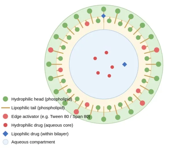
Figure 1: Structure of Transfersome.
2.1 The core components
The vesicle-forming phospholipid:
Soy phosphatidylcholine and egg phosphatidylcholine are the most commonly used amphipathic lipids, chosen for their natural biocompatibility, low immunogenicity, and long history of use in liposomal products [13,16]. Commercial phospholipid mixtures such as Phospholipon® 90G are frequently used in the recent literature because they offer consistent purity and phase behavior [16]. The phospholipid forms the bilayer itself the same basic architecture as a liposome with its hydrophilic head groups facing the aqueous core and the surrounding medium, and its hydrophobic tails packed together in the membrane interior [5].
The edge activator:
This is what distinguishes a transfersome from a plain liposome. Edge activators are single-chain surfactants that insert into the phospholipid bilayer and locally disrupt the tight packing of the lipid tails, lowering the bilayer’s resistance to bending [12,17]. Commonly used edge activators include the polysorbates (Tween 20, Tween 60, Tween 80), the sorbitan esters (Span 60, Span 65, Span 80), and bile-salt-type surfactants such as sodium cholate and sodium deoxycholate; dipotassium glycyrrhizinate has also been reported [18,17]. The choice of edge activator, and its concentration relative to the phospholipid, has a large and fairly predictable effect on the resulting vesicles. Formulations are typically prepared at phospholipid-to-edge-activator weight ratios somewhere between 95:5 and 80:20, with 90:10 and 85:15 being especially common starting points in optimization studies. Because the hydrophilic-lipophilic balance of the edge activator governs how strongly it partitions into the bilayer versus the aqueous phase, higher-HLB surfactants such as Tween 80 (HLB around 15) tend to produce smaller vesicles than lower-HLB surfactants such as Span 80 (HLB around 4.3), which instead tend to associate more strongly with the lipid bilayer itself and produce somewhat larger, though still deformable, structures [8,12]. Deformability and entrapment efficiency do not always move in the same direction: studies comparing edge activators have found that bile-salt-type activators and Span 80 can give higher entrapment efficiency at low activator loading, while Tween 80 tends to confer superior elasticity at higher loading — a trade-off that formulators typically resolve through statistical design-of-experiments approaches rather than intuition alone [12,7].
Ethanol or other short-chain alcohols:
Many transfersome formulations, though not all, include a small amount of ethanol (roughly 3–10% v/v) as a co-solvent. This addition blurs the formal line between “transfersomes” and “transethosomes” in the literature — a point worth noting for anyone trying to reproduce or compare published protocols, since some authors use the terms loosely [6].
The hydrating medium:
Vesicles are almost always rehydrated in water or phosphate-buffered saline, typically at a pH between 6.5 and 7.4, chosen to be compatible both with the phospholipid’s stability and with the drug’s ionization state, since only the unionized fraction of many drugs partitions efficiently into the lipid bilayer [19].
Cholesterol:
A near-universal stabilizing additive in both liposomes and niosomes, is used far less consistently in transfersome formulations. Where it appears, it is generally added in small amounts to improve bilayer rigidity and reduce leakage — but because rigidity is precisely the property transfersomes are designed to minimize, many formulators deliberately omit cholesterol or keep it to a minimum, reserving it for cases where a slower, more depot-like release profile is desired over maximal deformability [5,12].
2.2 Mechanism of penetration: the property that defines the technology
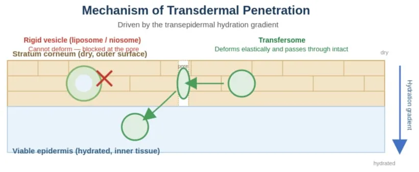
Figure 2: Rigid Vesicles Are Excluded by the Narrow Intercellular Channels of the Stratum Corneum, While Transfersomes Deform to Pass Through, Following Hydration Gradient.
If there is one section of a transfersome review that earns its place, it is this one, because the mechanism of penetration is not incidental — it is the entire reason transfersomes exist as a distinct category rather than simply being called “deformable liposomes” and left at that.
Cevc’s original and still most widely cited explanation is the transepidermal hydration gradient hypothesis [2]. Skin applied non-occlusively — that is, left open to the air rather than covered with an occlusive dressing — naturally loses water from its surface through evaporation, while the deeper, living layers of the epidermis remain comparatively hydrated [2]. This creates a persistent moisture gradient across the stratum corneum. Cevc proposed that transfersomes, being highly deformable and osmotically responsive, are drawn along this gradient from the relatively dry skin surface toward the more hydrated viable tissue beneath, squeezing through the narrow intercellular channels of the stratum corneum in the process [1,2]. Because the driving force is a naturally occurring physiological gradient rather than an externally applied pressure or chemical disruption of the skin barrier, this mechanism is often described as gentler and more physiologically compatible than the barrier-fluidizing approach used by penetration enhancers or high-ethanol systems such as ethosomes [6,20].
The deformability itself is attributed to the edge activator’s effect on the bilayer’s local elastic modulus [12,17]. Where a plain phospholipid bilayer resists curvature and will rupture rather than bend sharply, the edge activator creates zones of higher local curvature tolerance essentially “hinge points” that let the vesicle elongate, narrow, and pass through a constriction, after which the bilayer relaxes back toward its lower-energy spherical shape [17]. Cevc described this as self-optimizing and self-repairing behavior: the local lipid-to-edge-activator ratio within the membrane can redistribute dynamically in response to mechanical stress, which is part of why transfersomes tolerate repeated compression through pores smaller than their resting diameter without the catastrophic rupture seen in rigid vesicles [1,2].
It is worth being honest that this picture, while broadly accepted, is not universally settled. A meaningful strand of the literature debates how much of the observed enhancement in drug delivery comes from genuinely intact vesicles crossing the skin versus a combination of localized drug release near the skin surface and a more conventional penetration-enhancer effect from the free edge activator and lipid components [3,10,21]. Both mechanisms likely contribute in practice, and the balance probably depends on the specific drug, its molecular weight, and the vesicle’s own composition [21]. For a review audience, the fair summary is that there is broad agreement transfersomal formulations enhance drug deposition in deeper skin layers and, in many cases, improve transdermal flux relative to conventional liposomes or a simple drug solution but the precise contribution of “the whole vesicle walked through” versus “the vesicle helped the drug get there” remains an active area of investigation [3,21].
3 METHODS OF PREPARATION OF TRANSFERSOMES
Transfersome preparation methods borrow heavily from liposome technology, which is unsurprising given the shared phospholipid backbone, but each method has been adapted to accommodate the edge activator and, frequently, a size-reduction step aimed at producing the sub-200 nm vesicles most consistently associated with good skin penetration.
3.1 Thin-film hydration (rotary film evaporation) method:
This remains the most widely used approach in the published literature [13,4]. The phospholipid and edge activator are dissolved together in a volatile organic solvent commonly a chloroform-methanol mixture inside a round-bottom flask. The solvent is then removed under reduced pressure using a rotary evaporator, leaving a thin, dry lipid film coating the flask wall. This film is rehydrated with an aqueous buffer at a temperature above the phospholipid’s gel-to-liquid transition temperature, with gentle agitation, which causes the film to swell and spontaneously form multilamellar vesicles [4,22]. A subsequent sonication or extrusion step is usually needed to reduce these to the smaller, more uniform vesicles used in most applications [4].
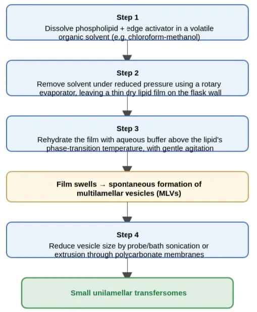
Figure 3: Thin Film Hydration Method.
3.2 Rotary evaporation–sonication method:
Functionally an extension of the thin-film method, this is often reported as a distinct technique in its own right because of how consistently it appears paired with statistical optimization (particularly Box–Behnken designs) in recent formulation studies — for example, in the development of cefepime-loaded nano-transfersomes and sertraline transfersomal gels [23,24]. The defining feature is a deliberate, controlled sonication step (bath or probe) following film hydration, used specifically to drive vesicle size down into the nano range while the formulation is optimized against variables such as phospholipid-to-edge-activator ratio and sonication time [7,23].
3.3 Vortexing sonication method:
Here the phospholipid and edge activator are combined directly in phosphate buffer and mixed by continuous vortexing until a milky suspension forms, without a separate organic-solvent film-forming step. The suspension is then sonicated in a bath sonicator and, in many protocols, subsequently extruded through polycarbonate membranes to narrow the size distribution [19]. This method is comparatively fast and equipment-light, which makes it attractive for early-stage screening work [19].
3.4 Reverse-phase evaporation method:
The phospholipid and edge activator are first dissolved in an organic solvent such as diethyl ether, and the aqueous drug solution is then added to this organic phase [5]. The mixture is sonicated to form a water-in-oil emulsion, after which the organic solvent is slowly removed under reduced pressure. As the solvent is stripped away, the system undergoes a phase inversion and collapses into an aqueous vesicular suspension. This method is generally associated with higher entrapment efficiency than simple hydration methods, particularly for hydrophilic drugs, because the drug is present in the aqueous phase from the earliest stage of vesicle formation rather than being loaded afterward [5].
3.5 Ethanol injection method:
The phospholipid and edge activator are dissolved in an ethanolic solution, which is then injected typically through a fine needle into a warmed aqueous phase under continuous stirring [19,25]. As the ethanol diffuses into the water, the amphiphiles spontaneously self-assemble into vesicles. This method avoids some of the more hazardous chlorinated solvents used in film-hydration approaches and is often described as gentler for heat- or solvent-sensitive drugs, though it introduces the practical complication of residual ethanol that may need to be removed or accounted for in the final formulation [19].
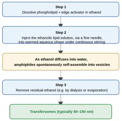
Figure 4: Ethanol Injection Method.
3.6 Freeze–thaw method:
A previously formed vesicle suspension (often prepared first by thin-film hydration) is subjected to repeated cycles of freezing, typically in liquid nitrogen, followed by controlled thawing in a warm water bath [22]. Each cycle disrupts and reforms the bilayer, which tends to reduce lamellarity, redistribute encapsulated material, and can meaningfully improve the encapsulation of larger or more fragile payloads such as proteins and growth factors [22]. Studies specifically examining freeze-thaw cycling on transfersomes have shown measurable, cycle-number-dependent effects on particle size, polydispersity, and encapsulation efficiency, making this as much a post-processing optimization tool as a standalone preparation method [22].
3.7 Other and emerging methods:
High-pressure homogenization and centrifugation-based processes have both been used, mainly to achieve the tighter size distributions required for injectable or ocular formulations [4]. Suspension homogenization is sometimes used as an intermediate step between initial hydration and final sizing [4]. Perhaps the most notable recent development is the supercritical carbon dioxide-assisted process, sometimes marketed under the name “Supersomes,” which uses supercritical CO₂ as a processing medium instead of large volumes of organic solvent [8]. This approach has been used to produce phosphatidylcholine-based transfersomes loaded with antioxidants such as ascorbic acid, with reported encapsulation efficiencies above 95% and the practical advantage of dramatically reduced organic solvent use [8] — an attractive feature for both environmental and regulatory reasons, echoing the “green chemistry” motivations that have also shaped recent niosome research.
4. CHARACTERIZATION OF TRANSFERSOMES
Characterization of transfersomes largely follows the same tests used for liposomes and niosomes — particle size and polydispersity index by dynamic light scattering, zeta potential as an indicator of colloidal stability, morphology by transmission or scanning electron microscopy, entrapment efficiency, and in vitro release [7,14] but one test is essentially unique to this class of vesicle and deserves particular mention.
Deformability index: Because elasticity is the defining functional property of a transfersome, formulators routinely measure it directly by forcing the vesicle suspension through a filter membrane of defined, sub-vesicle pore size under controlled pressure, and comparing the flux and any change in vesicle size before and after passage to that of a rigid liposome control processed the same way [2,3]. A higher deformability index indicates a vesicle that passes through the pore more readily and with less size change, and this measurement is frequently used alongside entrapment efficiency as a primary output variable in formulation optimization studies [7,12].
Ex vivo skin permeation studies, typically performed using excised animal or, less commonly, human skin mounted in a Franz diffusion cell, remain the standard way of connecting these physicochemical measurements to a functional prediction of transdermal performance, and are reported in nearly every applied transfersome study cited in this review [18,15].
5. ADVANTAGES OF TRANSFERSOMES
The central advantage of transfersomes, is their ultra-deformability: the capacity to squeeze through pores substantially smaller than their own resting diameter without significant loss of structure or payload, which allows them to achieve meaningful transdermal and transmucosal drug delivery where rigid vesicles largely cannot [1,2,21]. This one property carries several practical consequences. Because they can cross the skin non-invasively, transfersomes offer a genuinely needle-free route for drugs that would otherwise require injection, including proteins, peptides, and vaccine antigens [11,26]. Transdermal and transmucosal delivery via transfersomes also avoids hepatic first-pass metabolism and the variability of gastrointestinal absorption, which can translate into more predictable bioavailability for drugs that are otherwise poorly or erratically absorbed orally [20,27].
Transfersomes retain the core advantages of phospholipid-based vesicles more broadly: they are built from natural, biodegradable, and generally well-tolerated lipids, they can encapsulate both hydrophilic drugs (in the aqueous core) and lipophilic drugs (within the bilayer itself), and their self-optimizing, self-repairing bilayer behavior under mechanical stress gives them a degree of structural resilience that most other nanocarriers lack [1,5]. In several head-to-head studies, transfersomal formulations have shown higher entrapment efficiency and superior skin permeation compared with conventional liposomes carrying the same drug [28,29], and because the whole system is administered as a non-invasive topical or transmucosal formulation, patient compliance is generally reported to be favorable relative to injectable alternatives [20].
6. DISADVANTAGES AND CHALLENGES
The same phospholipid backbone that gives transfersomes their biocompatibility also gives them their principal weakness: chemical instability. Phospholipids are prone to oxidative and hydrolytic degradation over time, particularly the unsaturated fatty acid chains common in soy and egg phosphatidylcholine, which limits shelf life relative to the chemically hardier non-ionic surfactants used in niosomes [10,13]. This instability, along with a tendency toward vesicle fusion and aggregation on long-term storage, is one of the most consistently cited barriers to commercial transfersome products [10,21].
Production cost is a second, related concern: purified phospholipids are substantially more expensive than the non-ionic surfactants used in niosome manufacture, and natural phospholipid sources can show meaningful batch-to-batch variability in composition and purity, complicating both formulation reproducibility and regulatory characterization [3,10]. Scale-up from laboratory to industrial production remains comparatively underexplored in the published literature, most of which describes small-batch preparation, and this gap contributes to the broader uncertainty around good manufacturing practice (GMP) compliance for transfersome-based products [3,10].
7. TRANSFERSOMES COMPARED WITH OTHER VESICULAR CARRIERS
| Parameter | Transfersomes | Liposomes | Niosomes |
|---|---|---|---|
| Core forming component | Phospholipid + edge activator | Phospholipid (± cholesterol) | Non-ionic surfactant + cholesterol |
| Deformability | Very high (defining feature) | Low (rigid bilayer) | Low to moderate |
| Skin/pore penetration | Can cross pores smaller than vesicle size. | Generally blocked by intact skin pores. | Generally blocked by intact skin pores. |
| Chemical stability | Moderate (phospholipid prone to oxidation) | Low | High |
| Typical production cost | High | High | Low |
| Entrapment efficiency (lipophilic drugs) | High | High | Moderate–high |
| Primary route of use | Transdermal, transmucosal, nasal, ocular | Parenteral, oral, topical | Oral, transdermal, ocular, parenteral |
8. APPLICATIONS OF TRANSFERSOMES IN DIFFERENT FIELDS
8.1 Transdermal drug delivery
Transdermal delivery remains the founding and still the most extensively documented application of transfersome technology, and it is the setting in which the largest and most varied body of evidence exists [1,20]. Non-steroidal anti-inflammatory drugs, corticosteroids, local anesthetics, hormones, and cardiovascular agents have all been formulated as transfersomes with the consistent goal of improving skin penetration relative to conventional topical formulations while avoiding systemic first-pass metabolism [20,18]. Studies of drugs ranging from ketorolac to sertraline to felodipine have used transfersomal gels or dispersions to demonstrate enhanced drug deposition in and permeation through excised skin [24,30], generally attributing the improvement to the combination of high entrapment efficiency and the deformability-driven penetration mechanism discussed earlier [1,2].
8.2 Protein, peptide, and vaccine delivery
Because transfersomes can carry molecules across a wide range of molecular weights including proteins and peptides that would otherwise require injection — they have attracted particular interest as a platform for non-invasive vaccination [11]. Antigen-loaded transfersomes applied to intact skin have been explored as a way of eliciting both humoral and cell-mediated immune responses without a needle, capitalizing on the skin’s own dense population of antigen-presenting Langerhans cells [11]. This application connects transfersomes to the same production-method literature that also covers liposomes and niosomes for vaccine delivery, and represents one of the clearest points of overlap between the “next-generation” ambitions of all three vesicular platforms [11].
8.3 Cancer therapy
Transfersome-based delivery of anticancer agents spans both topical and more systemically oriented approaches. For skin cancers — melanoma, basal cell carcinoma, and squamous cell carcinoma — transfersomal formulations of chemotherapeutics and photosensitizing agents aim to concentrate drug delivery at the tumor site while limiting systemic toxicity [31], and comparative studies have shown transfersomes outperforming both liposomes and niosomes carrying the same drug in terms of skin retention and cytotoxic effect against cancer cell lines [28,29]. Beyond purely topical use, more elaborate combination strategies have also been described, including transpapillary iontophoresis-assisted delivery of co-loaded chemotherapeutic transfersomes for breast cancer [32], and surface modification with cell-penetrating peptides to improve both skin and tumor tissue penetration for agents such as paclitaxel [9].
8.4 Nasal and nose-to-brain delivery
The nasal mucosa shares some of the same barrier-crossing challenges as the skin, and transfersomes have been adapted for intranasal administration as a route that can bypass hepatic first-pass metabolism and, via the olfactory and trigeminal nerve pathways, potentially reach the brain directly without needing to cross the blood-brain barrier through the bloodstream [27]. This has made transfersomes of particular interest for neurodegenerative disease therapeutics — for example, rasagiline-loaded transfersomes incorporated into a thermosensitive in situ gel have been investigated for nose-to-brain delivery in Parkinson’s disease, where the in situ gel component helps counteract the rapid mucociliary clearance that otherwise limits nasal residence time [27].
8.5 Ocular delivery
Topical ocular drug delivery faces its own persistent barrier problem: short precorneal residence time and limited transocular membrane permeability mean that a large fraction of any conventional eye drop is lost before it can act [33]. Transfersomes have been formulated as eye drops and as electrolyte-triggered sol-to-gel systems — for instance, natamycin-loaded transfersomal gels for fungal keratitis [33] — with the goal of extending ocular surface residence time and improving sustained drug release, and comparative studies evaluating transfersomes alongside liposomes, niosomes, and transniosomes for conditions such as glaucoma have generally found transfersomes competitive on both bioavailability and controlled-release performance [34].
8.6 Anti-infective and wound-healing applications
Transfersomes have also been explored for localized anti-infective therapy, including antifungal and antibacterial applications, and notably for cutaneous leishmaniasis, where miltefosine-polyphenol co-loaded second-generation nano-transfersomes have been developed specifically to improve topical treatment of this parasitic skin infection [35]. In wound care, fusidic acid-loaded transfersomes have been formulated to address burn wound infection, taking advantage of both the antimicrobial payload and the vesicle’s favorable skin retention properties [36].
8.7 Gene and peptide co-delivery via hybrid platforms
A newer and still-developing direction combines transfersomes with physical enhancement technologies, most notably dissolving microneedles, to co-deliver peptide biologics alongside gene-modulating agents [26]. These hybrid microneedle-transfersome platforms aim to combine the microneedle’s ability to physically bypass the stratum corneum with the transfersome’s capacity for sustained, controlled release once past that initial barrier, and represent an active area of translational research aimed particularly at biologics that are otherwise very difficult to deliver non-invasively [26].
8.8 Oral and other routes: a genuine gap relative to niosomes
It is worth noting explicitly, in contrast to the niosome literature, that oral delivery is comparatively underexplored for transfersomes [3,4]. The overwhelming majority of published transfersome research addresses transdermal, transmucosal, nasal, or ocular routes, where the deformability advantage is directly relevant; the gastrointestinal environment does not present the same kind of narrow-pore barrier that transfersomes were designed to overcome, and few studies have systematically evaluated whether transfersome encapsulation offers a meaningful advantage for oral bioavailability enhancement compared with niosomes or other established oral nanocarrier strategies [4].
9. CURRENT DEVELOPMENTS AND INNOVATIONS IN TRANSFERSOME TECHNOLOGY
Several converging trends characterize the more recent transfersome literature. Ligand-conjugated and surface-modified transfersomes — functionalized with folic acid, transferrin, or cell-penetrating peptides such as stearylated R8H3 — are being used to add active targeting on top of the vesicle’s inherent penetration advantage, particularly in oncology applications where directing the carrier preferentially toward tumor tissue can meaningfully improve the therapeutic index [9]. Hybrid vesicle systems, especially transethosomes that combine an edge activator with a meaningful ethanol content, are increasingly popular as a way of capturing benefits from both the transfersome and ethosome mechanisms simultaneously [6].
Stimuli-responsive combination formulations, such as thermosensitive in situ gels built around Pluronic or pectin-based systems and loaded with transfersomes, are being used to address the residence-time limitations of nasal and ocular delivery specifically, converting a free-flowing liquid formulation into a more viscous, retained gel once it contacts the physiological environment [33,27]. On the manufacturing side, green and solvent-minimizing techniques such as supercritical CO₂-assisted production are gaining attention both for their environmental profile and for the very high encapsulation efficiencies reported in early studies [8]. Physical enhancement technologies, particularly microneedle arrays and iontophoresis, are increasingly paired with transfersomal formulations rather than treated as competing approaches, reflecting a broader shift toward combination strategies rather than any single technology solving the transdermal delivery problem alone [26,32]. Finally, much like the niosome literature, transfersome formulation optimization has moved decisively toward statistical, quality-by-design methods — Box–Behnken and full factorial designs are now the norm rather than the exception in recently published work [7], systematically mapping how variables such as phospholipid-to-edge-activator ratio, sonication time, and hydration conditions jointly determine vesicle size, deformability, and entrapment efficiency, rather than relying on one-variable-at-a-time optimization [7,14].
10. REGULATORY STATUS, SCALE-UP, AND CLINICAL TRANSLATION
Despite more than three decades of academic study, transfersome-based products have not yet achieved the same regulatory and commercial maturity as conventional liposomal drugs [3,10]. Several practical obstacles help explain this gap. Characterization methods, particularly the deformability index, are not yet standardized across laboratories, which makes it difficult to compare results or establish consistent specifications for regulatory submission [3]. Sourcing pharmaceutical-grade phospholipid at the purity and consistency required for GMP manufacturing, at commercial scale, remains more complex and costly than sourcing the non-ionic surfactants used in niosome production [10]. Long-term, real-time stability data — as opposed to accelerated or short-duration studies — remain relatively sparse in the published literature [10,21], and this gap is compounded by the still-unresolved mechanistic questions around intact-vesicle versus penetration-enhancer-mediated drug delivery discussed earlier, which complicate the kind of clear in vitro–in vivo correlation that regulators generally expect [3,21]. None of these obstacles are unique to transfersomes, and none appear insurmountable, but together they explain why clinical translation has lagged behind the substantial body of promising preclinical and formulation-stage research [3,10].
CONCLUSION
Transfersomes occupy a distinct and well-justified place within the broader family of vesicular drug carriers. Where niosomes earned their reputation on chemical stability and low cost, and liposomes on biocompatibility and versatility, transfersomes earned theirs on a single engineered property — deformability — that opened up genuinely non-invasive delivery across the skin, nasal mucosa, and ocular surface in a way that rigid vesicles simply cannot achieve. That property has proven applicable across an unusually wide range of therapeutic contexts, from small-molecule anti-inflammatories and anesthetics to proteins, vaccine antigens, and anticancer agents, and recent work extending transfersomes into ligand-targeted, stimuli-responsive, and microneedle-hybrid formats suggests the technology still has considerable room to develop. At the same time, the field’s persistent challenges — phospholipid instability, production cost, scale-up complexity, and an incompletely resolved mechanism of action — are the same issues that have kept transfersomes largely in the preclinical and early formulation-development stage rather than routine clinical use. Closing that gap will likely depend on the same combination of tools now reshaping niosome research: quality-by-design formulation optimization, standardized characterization protocols, and a clearer, more mechanistically settled account of exactly how these remarkable, self-optimizing vesicles make their way through the body’s tightest barriers.
REFERENCES

S. Karthik, Transfersomes as Next-Generation Nanocarriers for Stable, Controlled and Targeted Drug Delivery: a Comprehensive Review, Int. J. of Pharm. Sci., 2026, Vol 4, Issue 10, 205-217. https://doi.org/10.5281/zenodo.23090152
 10.5281/zenodo.23090152