

We use cookies to ensure our website works properly and to personalise your experience. Cookies policy
St. Joseph's College of Pharmacy, Dharmagiri College Campus, Naipunnya Road, Cherthala, Keralam 688524, India.
The stratum corneum is the principal barrier limiting topical and transdermal drug delivery, particularly when the therapeutic target lies beyond the dermis. Transethosomes are ultra-deformable phospholipid vesicles that combine ethanol-mediated skin-lipid modification with edge-activator-induced membrane flexibility. This hybrid design may enhance cutaneous transport, drug deposition and local pharmacological activity. Preclinical studies involving analgesic and anti-inflammatory agents have reported favourable vesicle characteristics, improved skin permeation and, in selected models, increased local tissue exposure. However, enhanced skin permeation cannot by itself establish therapeutically meaningful delivery to deep anatomical targets. This review critically examines the composition, biophysical mechanisms and formulation variables governing transethosome performance and evaluates evidence from analgesic, inflammatory and musculoskeletal applications. Particular emphasis is placed on the distinction between skin permeation, tissue deposition and target-tissue exposure. The review also considers the translational relevance of transethosomes to spine-related leg pain and identifies the experimental evidence required to demonstrate reproducible deep-tissue drug delivery. Current evidence supports transethosomes as a promising research platform for enhanced local delivery, while direct evidence of therapeutically relevant exposure in deep spinal or neural tissues remains limited.
Topical and transdermal delivery offer important advantages, including avoidance of gastrointestinal exposure and first-pass metabolism, ease of administration and the potential for local drug action. However, the stratum corneum remains a major barrier to drug transport, and delivery becomes considerably more challenging when the therapeutic target lies beyond the dermis, requiring drug distribution through subcutaneous and deeper tissues.1,2
Ultra-deformable vesicles (UDVs) were developed to overcome limitations of conventional lipid vesicles by providing highly flexible membranes capable of deformation during skin transport. Transfersomes demonstrated the importance of membrane elasticity and hydration gradients in facilitating skin penetration, while ethosomes introduced ethanol-mediated modification of the skin barrier and increased membrane fluidity.3,4 Transethosomes subsequently combined these principles by incorporating ethanol with edge activators, providing a formulation platform that integrates skin-barrier modification with enhanced vesicle deformability.5,6
Although transethosomes have demonstrated improved skin permeation, drug deposition and pharmacodynamic activity in several preclinical studies, enhanced skin permeation should not be equated with deep-tissue delivery. Evidence from conventional topical drugs shows that drug can reach underlying tissues such as muscle, tendon and synovial structures, but the extent of exposure depends on drug properties, formulation and anatomical site.7,8 For advanced vesicular systems, the more relevant question is therefore whether improved cutaneous transport can translate into therapeutically meaningful drug concentrations at the intended deeper target.
This distinction is particularly important for inflammatory, musculoskeletal and pain-associated disorders involving tissues beyond the dermis. Current evidence does not establish that intact ultra-deformable vesicles routinely reach deep anatomical targets; rather, their potential lies in enhancing skin interaction, drug deposition and subsequent tissue distribution.5,9 Transethosomes therefore represent a promising platform for investigating delivery beyond the skin, while the extent to which they can achieve reproducible and therapeutically relevant deep-tissue exposure remains an important translational question.
This issue is particularly relevant to disorders involving deeper musculoskeletal and neural structures. Sciatica, more precisely described within the broader terminology of spine-related leg pain, represents one such clinical condition. Spine-related leg pain encompasses heterogeneous conditions, including radicular pain and radiculopathy, commonly involving the lumbosacral nerve roots that contribute to the sciatic nerve 10,11. The structures involved in radicular pain and radiculopathy are located deep within the body and are protected by the lumbar vertebrae, paraspinal muscles, and connective tissue layers 11,12. Important pathological sites include the herniated or degenerated intervertebral disc, spinal nerve roots within the neural foramina, the dorsal root ganglion (DRG), and adjacent paraspinal muscles and facet joints 11,13.
At the site of nerve root compression or irritation, inflammatory processes involving chemical neuritis, oedema, and microvascular ischaemia can develop 12,14. This provides a rationale for delivering therapeutic agents locally to the paraspinal region rather than relying solely on systemic distribution. However, local delivery is challenging because the target tissues lie at considerable depth beneath the skin. A drug applied to the lower back must therefore pass through several anatomical barriers and tissue planes before reaching the deeper structures 12.
Conventional topical formulations generally have limited ability to cross these barriers because of their restricted penetration and lack of mechanisms for overcoming the skin barrier. Consequently, drugs may remain within superficial skin layers or undergo clearance before reaching deeper paraspinal tissues 12,14,15.
For pain associated with chemical radiculopathy, may involve mechanical compression, inflammation, nerve sensitisation, neuropathic mechanisms, or combinations of these, requiring an adequate drug concentration at the local site of inflammation is important. NSAIDs and other analgesics exert their effects through mechanisms such as inhibition of cyclooxygenase (COX) enzymes and modulation of inflammatory and nociceptive pathways 12,16. Local delivery may produce higher drug concentrations in tissues near the application site while limiting systemic exposure compared with oral administration 12,14. Thus, an improved local-to-systemic drug exposure profile may enhance therapeutic efficacy while reducing systemic adverse effects 12,17.
It is important to distinguish the terms topical, dermal, transdermal, and deep tissue drug delivery. Topical delivery refers broadly to application of a formulation to the skin, usually with the aim of producing a local effect at or near the skin surface 12,18. Dermal delivery involves transport of the drug into the viable skin layers, particularly the epidermis and dermis, to achieve local tissue concentrations while limiting systemic exposure 12,18. In contrast, transdermal delivery involves passage of the drug across the skin with subsequent entry into the systemic circulation, generally for systemic therapeutic effects 12,18.Systemic delivery following transdermal absorption occurs when a drug crosses the skin, enters cutaneous blood vessels, and is subsequently distributed through the systemic circulation 12,18. Deep tissue drug delivery, in the context of sciatica, refers to the targeted transport of therapeutic molecules beyond the superficial skin layers and subcutaneous tissues towards deeper structures such as paraspinal muscles, joints, ligaments, and neural tissues. The objective is to increase drug availability at relevant pain-generating sites while minimising unnecessary systemic exposure 14,19.
2. SKIN AS A BARRIER TO TOPICAL AND TRANSDERMAL DRUG DELIVERY
The skin is the largest organ of the human body and serves as an essential protective barrier against chemicals, microorganisms, ultraviolet radiation, and excessive transepidermal water loss (TEWL) 20,21. It consists of three main layers: the epidermis, dermis, and hypodermis (subcutaneous tissue) 21. The epidermis is the outermost and avascular layer, while the underlying dermis is highly vascularised and contains collagen and elastic fibres that provide strength and elasticity 12,21. The deepest layer, the hypodermis, is mainly composed of adipose tissue along with fibroblasts and macrophages and provides structural and vascular connections between the skin and deeper tissues 21.
The stratum corneum (SC), the outermost non-viable layer of the epidermis, is the principal barrier to percutaneous drug penetration and represents the major rate-limiting step in transdermal delivery 20,21. It has a characteristic “brick-and-mortar” structure, consisting of approximately 10–15 layers of flattened, keratin-filled corneocytes embedded within an extracellular lipid matrix 12,20. The lipid matrix contains predominantly ceramides, cholesterol, and free fatty acids, forming a highly organised and relatively impermeable barrier 12,15. This structure considerably restricts the passive penetration of many exogenous compounds, particularly hydrophilic and larger molecules 12,20.
Drug molecules can cross the SC mainly through three pathways: the intercellular, transcellular, and appendageal routes 15,20,21. The intercellular pathway involves diffusion through the lipid matrix surrounding the corneocytes and is considered the major route for many lipophilic and moderately lipophilic molecules. The transcellular pathway requires repeated partitioning through the hydrophilic corneocyte interiors and surrounding lipids, creating substantial resistance to diffusion. The appendageal pathway involves passage through hair follicles, sebaceous glands, and sweat glands, thereby partially bypassing the SC; however, its overall contribution is limited by the relatively small surface area occupied by these appendages 15,20.
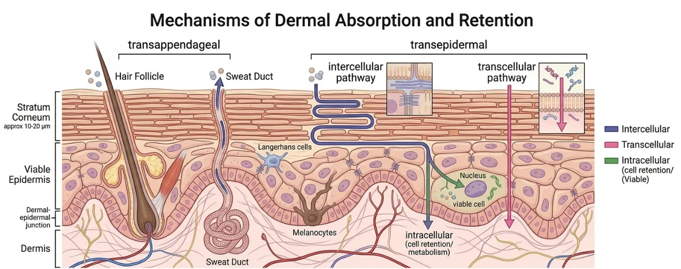
Figure 1: Mechanism of Dermal Absorption and Retention
Passive skin permeation is influenced by the physicochemical properties of the drug. Factors such as molecular size, lipophilicity, aqueous and lipid solubility, and ionisation state can markedly affect its ability to cross the SC 12,15. Percutaneous absorption is also influenced by skin hydration, temperature, anatomical site, age, and skin integrity 12,21. These factors are particularly relevant for delivery across the lumbar region, where variations in skin and underlying tissue characteristics can affect drug penetration.
Conventional topical formulations, such as creams, ointments, emulsions, and hydrogels, generally depend on passive diffusion and therefore provide limited control over transport beyond the superficial skin layers 14,19. Following application, the drug must first partition into and traverse the SC before reaching deeper tissues. The strong barrier properties of the SC can substantially restrict this process, while drug that enters the dermal circulation may undergo systemic absorption rather than reaching therapeutically relevant concentrations in deeper paraspinal tissues 12,14.
These limitations have encouraged the development of strategies to enhance cutaneous and deeper tissue penetration. Physical approaches include iontophoresis, sonophoresis, and microneedles, whereas chemical approaches employ penetration enhancers that modify the barrier properties of the SC 15,20,22. Vesicular and other advanced delivery systems have also been investigated to improve drug transport across the skin and potentially increase drug availability within deeper tissues.
3. VESICULAR SYSTEMS FOR DERMAL AND TRANSDERMAL DRUG DELIVERY
The development of vesicular carriers has progressively expanded the possibilities of transdermal and dermal drug delivery, with newer systems designed to overcome the limitations of earlier vesicles 15,23. Conventional liposomes, introduced in the late 1960s, are spherical vesicles composed of one or more phospholipid bilayers surrounding an aqueous core 15,16. Although they are generally biocompatible and well tolerated, their relatively rigid membranes limit their ability to deform and penetrate the compact structure of the stratum corneum. As a result, conventional liposomes tend to remain within the superficial skin layers, limiting their ability to deliver drugs to deeper tissues 15,18.
Niosomes were subsequently developed as vesicular carriers based on non-ionic surfactants, commonly stabilised with cholesterol 15,20. They offer advantages such as improved chemical stability and lower cost compared with phospholipid-based systems 15,20. However, their relatively rigid membrane structure can similarly restrict their penetration beyond the superficial skin layers, limiting their potential for deep tissue delivery 15,18.
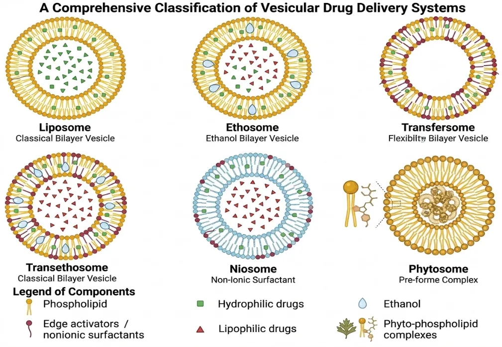
Figure 2: A Comprehensive Classification of Vesicular Drug Delivery Systems
A significant advancement came with the introduction of transfersomes by Cevc and Blume in 1992 15,23. These highly deformable vesicles consist primarily of phospholipids and an edge activator, such as Tween 80 or Span 80 24,25. The edge activator increases membrane flexibility, allowing the vesicles to deform under hydration-driven forces and pass through narrow pathways within the skin 15,25. This exceptional deformability distinguishes transfersomes from conventional liposomes. However, their penetration is strongly influenced by hydration gradients, and the relatively high surfactant content of some formulations may contribute to skin irritation 15.
Ethosomes, introduced by Touitou and colleagues in 1997, incorporated high concentrations of ethanol into phospholipid vesicles 13,15. Ethanol can enhance skin penetration by interacting with and increasing the fluidity of stratum corneum lipids, thereby facilitating drug transport across the skin 13,15. The presence of ethanol also increases the fluidity of the vesicular membrane, allowing ethosomes to interact more effectively with the skin barrier. However, conventional ethosomes do not typically contain an edge activator and therefore do not possess the same degree of membrane elasticity associated with transfersomes 15.
Transethosomes represent a hybrid vesicular approach that combines key features of ethosomes and transfersomes 15,23. They generally contain phospholipids, ethanol, and an edge activator, integrating the penetration-enhancing effect of ethanol with the increased flexibility provided by the surfactant 15,23. Their proposed mechanism therefore involves both modification of the skin barrier by ethanol and increased vesicle deformability. This combination may facilitate transport through the skin and improve drug deposition beyond the superficial layers compared with less deformable vesicular systems 15,23.
Table 1: A Comprehensive Classification of Vesicular Drug Delivery Systems
| Feature | Liposomes | Niosomes | Ethosomes | Transfersomes | Transethosomes |
|---|---|---|---|---|---|
| Principal structural component | Phospholipid | Non-ionic surfactant | Phospholipid + ethanol | Phospholipid + edge activator | Phospholipid + ethanol + edge activator |
| Main permeation principle | Conventional vesicular delivery | Vesicular delivery | Ethanol-mediated fluidisation | Deformability/hydration-driven transport | Combined mechanisms |
| Deformability | Low–moderate | Low–moderate | Moderate | High | High, formulation-dependent |
| Typical evidence | Dermal/local | Dermal/local | Enhanced skin permeation | Enhanced penetration | Enhanced skin permeation/local delivery |
| Deep-target evidence | Limited | Limited | Limited | Limited | Insufficient; requires validation |
| Major limitation | Limited penetration | Stability/rigidity | Ethanol dependence | Surfactant irritation | Mechanistic/stability/scale-up uncertainty |
Overall, the progression from conventional liposomes and niosomes to transfersomes, ethosomes, and transethosomes reflects a gradual shift from relatively rigid vesicles towards systems specifically engineered to enhance skin interaction, deformability, and drug penetration.
4. TRANSETHOSOMES: COMPOSITION AND MECHANISM
Ultra-deformable vesicles, also referred to as elastic or flexible lipid vesicles, were introduced to overcome the limited deformability of conventional liposomes and improve drug transport across the skin 15,23.
The flexibility of the vesicular membrane is largely determined by its lipid composition. In addition to phospholipids, ultra-deformable vesicles contain surfactants known as edge activators 24,25. These molecules reduce membrane rigidity and facilitate redistribution of membrane components when the vesicle encounters a constricted pathway. Consequently, the vesicle can deform and adapt its shape rather than undergoing immediate rupture 15,25. Phospholipids such as phosphatidylcholine provide the principal structural framework of the vesicle bilayer 15,25.
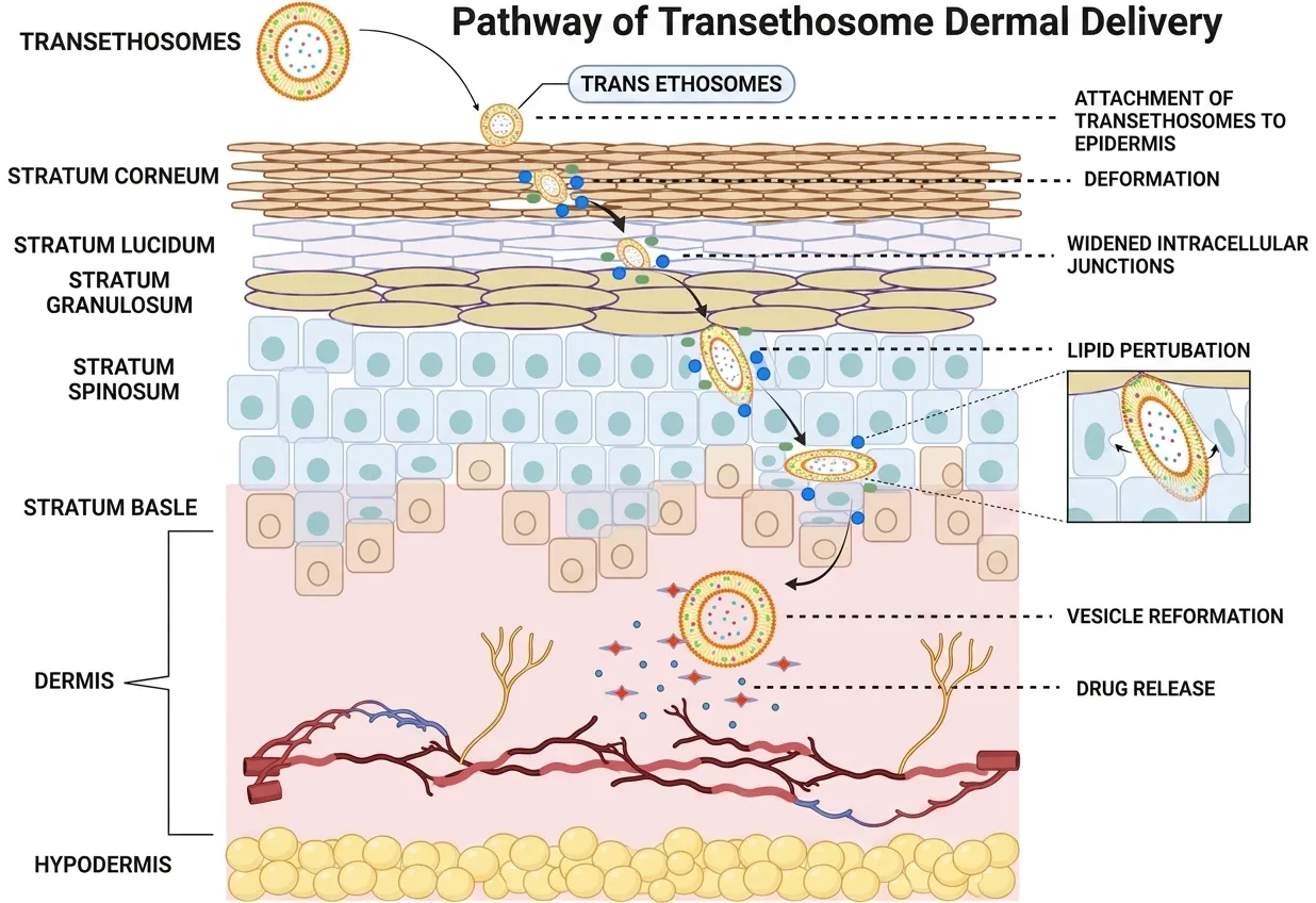
Figure 3: Pathway of Transethosome Dermal Delivery
Transethosomes (TEs) are hybrid lipid-based vesicular systems that combine key features of ethosomes and transfersomes 15,23. They incorporate ethanol to enhance skin permeation and an edge activator to increase vesicle flexibility. This combination is intended to improve vesicle–skin interaction and facilitate drug transport across the stratum corneum 15,23.
Transethosomes generally contain four principal components 15,23. Phospholipids, commonly soya phosphatidylcholine or related phosphatidylcholine sources, form the structural bilayer and provide biocompatibility 15,25. Ethanol acts as both a vesicle-fluidising agent and a skin penetration enhancer by increasing lipid mobility within the vesicle and modifying the organisation of stratum corneum lipids 13,15. The edge activator, which may include surfactants such as Tween 80 or Span 80, reduces membrane rigidity and improves vesicle deformability 15,24,25. The aqueous phase hydrates the lipid components and provides the continuous phase of the vesicular dispersion 15,25. The exact concentrations of these components vary considerably with the drug, preparation method, and desired vesicle characteristics and should therefore be optimised experimentally rather than treated as fixed ranges.
Because of their amphiphilic bilayer structure, transethosomes can accommodate drugs with different physicochemical properties. Hydrophilic compounds are preferentially associated with the aqueous interior, whereas lipophilic compounds can partition into the lipid bilayer 15,25. This structural versatility makes transethosomes suitable for the delivery of a broad range of therapeutic agents.
Ethanol plays an important role in systems such as ethosomes and transethosomes 13,15. At the vesicular membrane, ethanol can increase lipid mobility and reduce membrane packing, thereby contributing to greater membrane fluidity. At the skin surface, ethanol can also interact with the lipid components of the stratum corneum and increase their fluidity, thereby reducing the barrier to drug permeation 13,15. The combined effects of membrane fluidisation and modification of the skin barrier contribute to the enhanced penetration observed with ethanol-containing vesicular systems.
Edge activators are surfactants incorporated into the phospholipid bilayer to increase vesicle flexibility 15,24,25. Examples include Tween 20, Tween 80, Span 80, and sodium deoxycholate. By reducing membrane rigidity and allowing redistribution of membrane components during deformation, edge activators help vesicles withstand mechanical stress and pass through constricted pathways more readily than conventional liposomes 15,25.
Vesicle elasticity can be assessed using the deformability index (DI), which is determined by measuring the amount of vesicular dispersion that can pass through a membrane with defined pore size under controlled pressure 15,25. A higher DI indicates greater membrane flexibility. Transfersomes and transethosomes generally demonstrate greater deformability than conventional liposomes, although the magnitude of this difference depends on vesicle composition and experimental conditions 15,25.
The interaction between ultra-deformable vesicles and the stratum corneum is considered to involve several overlapping processes. Initially, vesicles deposit on and interact with the skin surface. Ethanol and other formulation components may then modify the organisation of the SC lipids, while the flexible vesicles undergo deformation and move through available intercellular pathways 15,25. These processes can enhance the transfer of the encapsulated drug into deeper skin layers.
The proposed mechanism of transethosomal permeation can be considered in several stages. First, stratum corneum modification occurs as ethanol interacts with the intercellular lipid domains and increases their fluidity 13,15. Second, vesicle deformation and penetration are facilitated by the flexible membrane produced by the edge activator, allowing the vesicles to adapt to constricted pathways within the skin 15,25. Third, following penetration into deeper skin layers, the vesicular system may act as a local drug reservoir from which the drug is released and subsequently diffuses into surrounding tissues 12,14,15.
For scientific interpretation, it is important to distinguish established observations from proposed mechanisms. However, the precise fate of intact vesicles after entering deeper skin layers remains incompletely understood. In particular, claims that intact vesicles routinely travel from the skin directly into deep paraspinal tissues or systemic capillaries are not sufficiently established. A more cautious interpretation is that ultra-deformable vesicles enhance drug transport across the skin and may contribute to increased drug deposition in deeper tissues, where the vesicular components can subsequently release the encapsulated drug 12,14,15.
Thus, the enhanced performance of ultra-deformable vesicles is best understood as the combined effect of membrane deformability, skin-lipid interaction, formulation-dependent penetration enhancement, and hydration-driven transport, rather than a single mechanism operating independently.
Transethosomes are therefore mechanistically distinct from conventional ethosomes and transfersomes. Ethosomes rely primarily on the penetration-enhancing and membrane-fluidising effects of ethanol, whereas transfersomes depend predominantly on membrane flexibility imparted by edge activators and hydration-driven transport 15,25. The enhanced skin-delivery behaviour of transethosomes is likely to arise from an interplay of ethanol-mediated barrier perturbation, membrane fluidisation, surfactant-induced deformability, vesicle–skin interactions and formulation-specific drug partitioning.
5. FORMULATION AND PROCESS VARIABLES AFFECTING TRANSETHOSOME PERFORMANCE
The physicochemical properties and performance of transethosomes are influenced by both formulation composition and processing conditions. Careful optimisation of these variables is therefore essential to obtain vesicles with suitable size, stability, deformability, drug loading, and skin permeation characteristics 15,23,25.
5.1 Vesicle Size and Polydispersity Index (PDI)
Vesicle size can influence skin interaction, drug release, and permeation behaviour 16. Smaller vesicles generally provide a larger surface area and may interact more effectively with the skin barrier. Although sub-200 nm vesicles are frequently reported in optimisation studies, vesicle diameter alone is not a validated surrogate for transdermal or deep-tissue performance. 15,25.
The polydispersity index (PDI) describes the breadth of the vesicle size distribution. A PDI below 0.3 is generally considered indicative of a relatively narrow and homogeneous population, whereas higher values indicate greater heterogeneity and may be associated with reduced physical stability and less predictable formulation behaviour 15,25. Therefore, both mean vesicle size and PDI should be considered during formulation optimisation.
5.2 Zeta Potential and Colloidal Stability
Zeta potential provides an indication of the surface charge and electrostatic environment of vesicles and is commonly used to assess colloidal stability 15,25. Higher absolute zeta-potential values can increase electrostatic repulsion between vesicles and thereby reduce aggregation. A value above approximately ±30 mV is often used as a general indicator of good electrostatic stability; however, stability is also influenced by steric effects, lipid composition, ionic strength, and the surrounding medium 15,25.
The surface charge of phospholipid vesicles depends on lipid composition and formulation conditions. Where required, charge-inducing components such as stearylamine or dicetyl phosphate may be incorporated to modify vesicle surface charge and influence stability and skin interaction 15,25.
5.3 Entrapment Efficiency and Loading Capacity
Entrapment efficiency (EE) represents the proportion of the initial drug incorporated into the vesicular system 15,25:
EE (%) = [(A₀ − Aᵤ) / A₀] × 100
where A₀ is the total amount of drug initially added and Aᵤ is the amount of unentrapped drug remaining in the external phase after separation 15,25.
Loading capacity (LC) expresses the amount of drug associated with a given mass of lipid or vesicular carrier. Its calculation should be defined according to the basis used in the study, as different reports may express loading relative to lipid mass, total vesicle mass, or total formulation mass. Both EE and LC are important indicators of formulation efficiency and should be interpreted together with vesicle size and drug solubility 15,25.
5.4 Membrane Deformability and Elasticity
Deformability is one of the key characteristics distinguishing ultra-deformable vesicles from conventional liposomes 15,23. It describes the ability of vesicles to undergo reversible shape changes and pass through pores smaller than their original diameter without extensive structural disruption 15,25.
Deformability is commonly expressed using the deformability index (DI), determined by extruding vesicles through a membrane with a defined pore size under controlled pressure. A commonly used relationship is:
DI = Jᵥ (rᵥ/rₚ)²
where Jᵥ is the volume of vesicular dispersion passing through the membrane during a specified period, rᵥ is the vesicle radius after extrusion, and rₚ is the membrane pore radius 25. Higher DI values generally indicate greater membrane flexibility and deformability, although results should be compared only under standardised experimental conditions.
5.5 In Vitro Drug Release and Skin Permeation
Drug release from transethosomal systems may involve an initial release phase followed by a slower phase as the entrapped drug is progressively released from the vesicular structure 15,25. The extent of any initial burst depends on factors such as the amount of free or surface-associated drug, vesicle composition, and preparation method. Subsequent release is influenced by drug partitioning within the vesicle, membrane characteristics, and the properties of the release medium 15,25.
Skin permeation studies provide complementary information by determining how effectively the formulation delivers drug across or into the skin. Parameters such as cumulative drug permeated, steady-state flux, permeability coefficient, and drug deposition within different skin layers may be used to evaluate performance.
5.6 Formulation and Process Variables
Table 2: Major Variables Influencing Critical Quality Attributes of Transethosomes
| Independent variable | Major effect on CQAs | Biophysical basis |
|---|---|---|
| Phospholipid concentration | Influences vesicle size, EE, and viscosity | Provides the structural bilayer; excessive lipid may favour larger or multilamellar vesicles 15,25 |
| Ethanol concentration | Alters vesicle size, membrane fluidity, and skin permeation | Increases lipid fluidity and modifies stratum corneum lipid organisation 13,15 |
| Edge activator type and concentration | Influences deformability, DI, size, and stability | Reduces membrane rigidity and facilitates reversible deformation 15,24,25 |
| Stirring/homogenisation | Influences vesicle formation and PDI | Controls mixing, dispersion, and vesicle self-assembly 15,25 |
| Sonication conditions | Reduces vesicle size and may improve size uniformity | Acoustic cavitation disrupts larger vesicular structures; excessive energy may cause thermal or chemical stress 15,22 |
| Storage temperature and conditions | Affect size, aggregation, drug leakage, and ethanol retention | Temperature and storage environment influence bilayer mobility and formulation stability 15,22,25 |
Overall, transethosome performance is governed by the interaction between lipid composition, ethanol content, edge-activator concentration, vesicle characteristics, and processing conditions. Because these variables are interdependent, optimisation should focus on their combined influence on critical quality attributes rather than on individual parameters in isolation.
6. EVIDENCE FOR TRANSETHOSOMES IN ANALGESIC, ANTI-INFLAMMATORY AND MUSCULOSKELETAL CONDITIONS
Direct evidence for transethosomes in spine-related leg pain or lumbar radiculopathy remains limited. The available literature is predominantly derived from gout, rheumatoid arthritis, osteoarthritis, acute pain and other inflammatory models. These studies provide evidence for enhanced cutaneous delivery and pharmacodynamic activity, but their findings should be interpreted as indirect evidence for deeper tissue delivery rather than disease-specific validation.26–36
6.1 Colchicine-loaded transethosomes for gout
Abdulbaqi et al. (2018); developed colchicine-loaded transethosomes using the cold method and sequential factorial optimisation, followed by incorporation into Carbopol 940® gel.26 Selected formulations showed vesicle sizes of 87.5–104.0 nm, dispersity values of 0.085–0.138, zeta potentials of −17.0 to −20.9 mV and entrapment efficiencies of 69.37–79.75%. TET 2-0.3 showed 91.0 ± 3.6 nm, PDI 0.111 ± 0.015, −19.86 ± 0.77 mV and EE 78.84 ± 0.65%, while TENa 12-0.3 showed 87.6 ± 1.4 nm, PDI 0.109 ± 0.009, −20.86 ± 1.09 mV and EE 79.75 ± 1.04%.26 Increasing ethanol concentration initially reduced vesicle size, whereas concentrations above the optimal range promoted vesicle enlargement and possible structural disruption. The transethosomal gel also produced greater ex vivo colchicine permeation than the corresponding non-ethosomal gel.26 These findings support enhanced cutaneous delivery but do not demonstrate exposure at deeper pathological tissues.
6.2 Sinomenine hydrochloride-loaded transethosomes for rheumatoid arthritis
Song et al. (2019); investigated sinomenine hydrochloride-loaded transethosomes using sodium deoxycholate, Tween 80 or oleic acid as edge activators.27 The sodium-deoxycholate formulation, TE1, showed a size of 88.7 ± 9.6 nm, PDI 0.152 ± 0.036, zeta potential −20.6 ± 3.2 mV and EE 60.2 ± 6.3%. The antioxidant-surface transethosome (AS-TE) showed 93.2 ± 7.2 nm, PDI 0.168 ± 0.023, zeta potential −17.6 ± 3.6 mV and EE 59.9 ± 4.5%.27 At 0.5 MPa, TE1 demonstrated approximately 5.2-fold greater deformability than the reference ethosome. Ex vivo studies showed greater permeation, while synovial-fluid microdialysis demonstrated increased local drug concentration after AS-TE administration. The formulation also reduced joint swelling in the arthritis model.27 This provides evidence of increased local tissue exposure, although the target remained a peripheral joint rather than a deep spinal structure.
6.3 Baicalin-loaded transethosomes
Adin et al. (2023); prepared baicalin-loaded transethosomes using thin-film hydration and Box–Behnken optimisation with phospholipon 90G, ethanol and sodium cholate.28 The optimised formulation contained 60 mg phospholipon 90G, 30% ethanol and 20 mg sodium cholate and exhibited a size of 141.2 ± 2.98 nm, PDI 0.1135 ± 0.009 and EE 76.39 ± 2.09%.28 After incorporation into Carbopol 934P gel, drug release reached 88.32% over 24 h, with an ex vivo flux of 4.67 µg/cm²/h. CLSM demonstrated penetration to approximately 52 µm, while the formulation produced a of 7.94 ± 0.51 µg/mL and of 42.92 ± 7.90 µg·h/mL compared with 3.94 ± 1.91 µg/mL and 24.96 ± 9.76 µg·h/mL following oral administration.28 The formulation also improved radiographic and histopathological outcomes in experimental arthritis. However, the reported 52 µm penetration remains within the skin and should not be interpreted as deep paraspinal delivery.28
6.4 Mangiferin-loaded transethosomes
Adin et al. (2023); prepared mangiferin-loaded transethosomes by thin-film hydration and employed a Box–Behnken design using phospholipon 90G, ethanol and sodium cholate.29 The optimised vesicles measured 146.8 ± 2.98 nm, with PDI 0.1139 ± 0.009, zeta potential −38.62 mV and EE 74.23 ± 2.09%.29 The transethosomal gel showed 65.32% in vitro release, while transdermal administration produced a of Cmax of 6.94 ± 0.51 µg/mL and an AUC₀–₂₄h of 43.92 ± 7.90 µg·h/mL compared with 3.74 ± 1.91 µg/mL and 22.96 ± 9.76 µg·h/mL following oral administration.29 Greater skin penetration and improved anti-arthritic responses were reported. These findings demonstrate improved transdermal exposure but do not establish preferential drug accumulation at a deep pathological target.29
6.5 Ginger-extract transethosomes
Hassan et al. (2023); investigated ginger-extract-loaded transethosomes using Span 80, Tween 80 and sodium deoxycholate as edge activators.30 The sodium-deoxycholate formulation showed the most favourable characteristics, with a size of 188.3 ± 7.66 nm, PDI 0.276 ± 0.021, zeta potential −38.6 ± 0.08 mV and EE 91.0 ± 0.24%, compared with 254.8 ± 30.84 nm and PDI 0.45 ± 0.02 for Span 80 and 286.23 ± 10.54 nm and PDI 0.327 ± 0.012 for Tween 80.30 After 24 h, the HPMC transethosomal gel produced 436.55 ± 19.79 µg/cm² permeation compared with 377.80 ± 10.06 µg/cm² for the non-transethosomal gel and 179.28 ± 14.16 µg/cm² for the free-drug solution. The formulation also demonstrated anti-inflammatory activity in the carrageenan paw-oedema model.30 The study illustrates the strong influence of edge-activator selection on transethosome performance.
6.6 Ketoprofen-loaded transethosomes for osteoarthritis
Mammella et al. (2024); developed ketoprofen-loaded transethosomes using QbD-based Taguchi and central composite designs and incorporated the optimised vesicles into a hyaluronic-acid/poloxamer 407 gel.31 The formulation exhibited a size of 110.0 ± 1.70 nm, PDI 0.103 ± 0.01, zeta potential −6.08 ± 0.27 mV and conductivity 0.049 ± 0.0001 mS/cm, with drug content of 90.08 ± 1.25% in the final gel.31 In a monosodium iodoacetate-induced osteoarthritis model, the formulation preserved joint architecture and reduced inflammatory markers including IL-1β, IL-6, IL-22 and TNF-α.31 This study is particularly relevant to NSAID delivery, but the peripheral joint target remains anatomically distinct from lumbar nerve roots.
6.7 Caryophyllene-oxide transethosomes
Natsheh et al. (2025); evaluated caryophyllene oxide-loaded transethosomes in porcine skin and an acute analgesic mouse model.32 The optimised formulation showed a mean vesicle size of 450.7 ± 55.03 nm; PDI, zeta potential and EE were not reported in the accessible primary record.32 Skin permeation reached 40.3 ± 0.881 µg/cm² compared with 29.5 ± 10.5 µg/cm² for the conventional emulsion, while skin extraction yielded 251.8 ± 76.03 µg/cm² compared with 13.5 ± 0.6 µg/cm² for the ointment. The formulation produced an 80.5% maximum possible analgesic effect compared with 24.7% for the conventional topical formulation.32 The relatively large vesicle size despite measurable analgesic activity indicates that vesicle diameter alone is insufficient to predict biological performance.
6.8 Methotrexate–baicalin co-loaded nanotransethosomes
Adin et al. (2024); developed co-loaded methotrexate–baicalin nanotransethosomes for rheumatoid arthritis.33 The optimised formulation showed a particle size of 151.3 nm, PDI 0.125 and zeta potential −32.22 mV, with increased penetration of both drugs compared with suspension gel.33 Improved radiographic and histopathological outcomes were reported in vivo, together with modulation of inflammatory responses. Exact EE values were not available in the reported source and therefore should not be inferred.33
6.9 Lornoxicam-loaded transethosomal gel
Muneer et al. (2026); reported lornoxicam-loaded transethosomal gel using Box–Behnken optimisation.34 The optimised formulation showed a size of 150.7 ± 1.2 nm, PDI 0.326 ± 0.04, zeta potential −23.3 ± 0.9 mV and EE 80.9 ± 0.2%.34 Ex vivo Franz-cell experiments demonstrated approximately fourfold greater skin permeation than the conventional formulation, and six-month stability was investigated.34 The PDI value also illustrates why transethosomal performance should be interpreted from the complete formulation profile rather than a single numerical criterion.
6.10 Tacrolimus-loaded transethosomes
Qaiser et al. (2026); evaluated tacrolimus-loaded transethosomal gel for rheumatoid arthritis.35 The optimised vesicles measured 142.0 ± 0.8 nm, with PDI 0.177, EE 87.98% and zeta potential −28.5 ± 2.5 mV.35 The formulation produced a flux of 26.291 µg/cm² and an enhancement ratio of 10.475 compared with conventional tacrolimus gel, together with improved inflammatory, radiological and histopathological outcomes in the arthritis model.35 These findings extend the evidence for anti-inflammatory transethosomal delivery but remain peripheral to the anatomical problem of deep spinal exposure.
6.11 Allopurinol-loaded transethosomes
A recent 2026 study investigated allopurinol-loaded transethosomes for gout using thin-film hydration and Box–Behnken optimisation.36 The optimised formulation showed a Z-average diameter of 155.4 ± 0.2 nm, PDI 0.28 ± 0.01, zeta potential −27.0 ± 0.2 mV and EE 80.7 ± 2.4%.36 The study further supports the application of transethosomes to poorly soluble anti-inflammatory or gout-related therapy while demonstrating continued use of DoE-based optimisation. However, the peripheral target of gout does not provide evidence for delivery to deep spinal or neural tissues.36
6.12 Comparative Interpretation of the Evidence
Collectively, Abdulbaqi et al. (2018), Song et al. (2019), Adin et al. (2023, 2024), Hassan et al. (2023), Mammella et al. (2024), Natsheh et al. (2025), Muneer et al. (2026), Qaiser et al. (2026) and the recent allopurinol study demonstrate that transethosomes can achieve favourable vesicle characteristics, enhanced skin permeation and pharmacodynamic activity across several inflammatory models.26–36
Particle sizes reported across these studies range from approximately 88 nm to 451 nm, while PDI values range from 0.085 to 0.45 and EE values from approximately 60% to 91% where reported.26–36 Importantly, improved permeation does not necessarily correspond to increased exposure at a deeper pathological site.
The strongest evidence comes from studies that extend beyond skin permeation to local tissue exposure, pharmacokinetics or pharmacodynamics. Song et al. (2019), for example, demonstrated increased synovial-fluid drug concentration, while Adin et al. (2023) reported pharmacokinetic enhancement with baicalin and mangiferin systems.27–29 Nevertheless, these models involve peripheral tissues and cannot establish whether comparable exposure can be achieved in deeper spinal or neural structures.
The current evidence therefore supports the capacity of transethosomes to enhance topical and local drug delivery, whereas their ability to generate therapeutically relevant exposure in deep tissues remains unresolved.
7. TRANSETHOSOMES AND MUSCULOSKELETAL CONDITIONS: TRANSLATIONAL RELEVANCE
Preclinical studies of transethosomes in osteoarthritis, rheumatoid arthritis, gout and other inflammatory conditions demonstrate enhanced skin permeation, local deposition and anti-inflammatory or analgesic effects, but these findings cannot be directly equated with efficacy in spine-related leg pain.14,19,26–36
The major limitation is anatomical depth. Peripheral inflammatory tissues are generally more accessible from the skin than lumbar nerve roots, neural foramina and dorsal root ganglia, which are protected by paraspinal muscle, fascia, vertebral structures and connective tissues.10,12,14 For example, the approximately 52 µm penetration reported for baicalin transethosomes represents skin penetration and does not demonstrate delivery to paraspinal muscle or spinal neural structures.28
Table 3: Evidence Status and Translational GAP
| Evidence | Current status | Interpretation |
|---|---|---|
| Skin permeation | Demonstrated in multiple studies | Supports enhanced cutaneous delivery |
| Peripheral inflammatory-tissue exposure | Demonstrated in selected studies | Supports local-delivery potential |
| Analgesic/anti-inflammatory effects | Demonstrated in preclinical models | Pharmacodynamic evidence |
| Lumbar nerve-root exposure | Not demonstrated | Major research gap |
| Paraspinal/deep-tissue exposure | Insufficient evidence | Requires depth-resolved analysis |
| Disease-specific radiculopathy efficacy | Not established | Cannot be inferred from OA/RA/gout models |
Similarly, increased (Cmax) and AUC demonstrate systemic absorption but do not establish preferential target-tissue exposure.28,29 Thus, improved skin permeation or peripheral tissue deposition should not be interpreted as evidence of deep spinal delivery.
The key translational question is therefore not simply whether transethosomes cross the stratum corneum, but whether they produce therapeutically meaningful drug concentrations in deeper target tissues while maintaining acceptable systemic exposure. Future studies should quantify drug distribution across successive tissue layers together with plasma concentrations and disease-relevant pharmacodynamic outcomes.26–36
8. TRANSETHOSOMAL GELS AS A TOPICAL DOSAGE-FORM STRATEGY
Transethosomal dispersions are generally low-viscosity systems and may show limited residence or run-off after topical application. Incorporation into a suitable polymeric gel can improve viscosity, spreadability and skin residence, with Carbopol, poloxamers, chitosan and other hydrophilic polymers commonly investigated.14,15,25
The polymeric matrix may prolong drug release and improve formulation residence; however, excessive viscosity may restrict vesicle mobility and alter drug release or skin permeation. Gel incorporation may also affect vesicle size, deformability and ethanol content. Therefore, polymer concentration and vesicle–polymer compatibility must be optimised experimentally to maintain the desired transethosomal characteristics and skin-delivery performance.14,15,25
Importantly, the gel should be considered an active formulation component rather than merely a viscosity-modifying vehicle, since its properties may influence the overall behaviour of the transethosomal system.14,15,25
9. CURRENT RESEARCH GAPS
Despite encouraging biophysical and permeation findings, several gaps remain in the development of transethosomes for deep-tissue drug delivery. The major challenges include limited disease-specific evidence, inadequate deep-tissue exposure data, poor in vitro–in vivo correlation, formulation variability, and translational limitations.12,14,15
9.1 Limited Disease-Specific and Deep-Tissue Evidence
Most transethosomal studies have focused on dermatological, inflammatory and musculoskeletal conditions rather than spine-related leg pain or lumbar radiculopathy. Moreover, drug permeation and deposition are generally assessed within the skin, providing limited evidence of exposure in deeper tissues such as fascia, muscle or neural structures. Disease-relevant models and quantitative tissue-distribution studies are therefore required.
9.2 Limited In Vitro–In Vivo Correlation
Franz diffusion studies are useful for comparing formulations but cannot fully reproduce blood circulation, lymphatic drainage, metabolism and tissue clearance.15,22 Consequently, increased in vitro permeation may not translate into therapeutically relevant tissue exposure. Correlation between permeation findings and in vivo tissue concentrations should therefore be established.
9.3 Lack of Standardisation and Formulation Reproducibility
Reported deformability values are difficult to compare because experimental conditions vary between studies.15,25 In addition, differences in phospholipid source, ethanol concentration, edge activator and processing conditions can affect vesicle size, PDI, entrapment efficiency, deformability and stability. Standardised methods and well-defined raw-material specifications are required. Deformability measurements are influenced by membrane pore size, applied pressure, extrusion time, vesicle concentration, temperature and calculation method.
9.4 Stability and Manufacturing Challenges
Ethanol-containing vesicles may undergo changes in size, aggregation, drug leakage, lipid oxidation and ethanol content during storage.15,22 Reproducible scale-up also requires control of critical quality attributes, ethanol loss, mixing and size-reduction processes. Long-term stability and repeated-dose safety therefore require further evaluation.
9.5 Regulatory and Translational Challenges
The regulatory pathway for transethosomal products requires product-specific consideration because these systems combine an active drug with a nanoscale vesicular carrier and, potentially, a secondary gel vehicle. Their classification varies with the product, route and intended use.
FDA guidance highlights the need to evaluate physicochemical characteristics, critical quality attributes, manufacturing controls, stability, safety and efficacy. For transethosomal gels, key requirements include comprehensive characterisation, excipient safety, local and systemic exposure, reproducible manufacturing and adequate stability data.
Overall, the major research gap is the lack of a clear relationship between transethosome formulation characteristics, deep-tissue drug exposure and pharmacodynamic response. Addressing this relationship is essential for determining whether enhanced skin permeation can translate into meaningful deep-tissue delivery.
10. PROPOSED TRANSETHOSOMAL DELIVERY FRAMEWORK FOR SPINE-RELATED LEG PAIN
Based on the available evidence, a transethosomal gel containing an analgesic or anti-inflammatory drug may be considered a hypothetical local-delivery strategy for spine-related leg pain; this concept should be clearly distinguished from established clinical efficacy.12,14,19
The proposed sequence is:
Transethosomal gel applied to the lumbar/paraspinal region ↓ Increased formulation residence and skin contact ↓ Ethanol-mediated modification of stratum corneum lipids and increased vesicle membrane fluidity ↓ Edge-activator-mediated vesicle deformability and enhanced passage through skin pathways ↓ Increased drug deposition within deeper skin and subcutaneous tissues ↓ Potential local drug reservoir and subsequent drug release ↓ Diffusion and tissue distribution towards deeper paraspinal structures ↓ Potential local pharmacological effect
This framework is biologically plausible at the level of enhanced skin delivery and local drug deposition; however, several downstream steps remain unverified. In particular, enhanced penetration through the stratum corneum does not demonstrate that intact transethosomes reach lumbar nerve roots, the epidural space or dorsal root ganglia. Similarly, increased local deposition cannot by itself establish therapeutically relevant concentrations at deeper targets.
10.1 Rational Formulation Optimisation and Quality by Design
Future development should increasingly adopt a Quality by Design (QbD) approach rather than relying primarily on trial-and-error experimentation.15,25 Statistical Design of Experiments (DoE), including Box–Behnken and central composite designs, can evaluate the combined effects of phospholipid concentration, ethanol content, edge-activator concentration, drug loading and processing conditions on critical quality attributes such as vesicle size, PDI, zeta potential, entrapment efficiency, deformability, drug release and permeation.15,25
10.2 Advanced Biophysical Characterisation
A more detailed understanding of vesicle structure and membrane behaviour would benefit from Cryo-TEM, SAXS/SANS, NMR and complementary spectroscopic techniques.15,25 These approaches can provide information on morphology, lipid organisation and interactions among ethanol, phospholipids and other formulation components, complementing conventional size, PDI, zeta-potential and deformability measurements.
10.3 Human-Relevant Skin Models and Imaging
Greater use of human ex vivo skin can improve the translational relevance of permeation studies compared with reliance on animal skin alone.15,25 CLSM, Raman spectroscopy and multiphoton microscopy may further characterise formulation–skin interactions and drug distribution. Fluorescent labelling should, however, be interpreted cautiously because dissociation of the label from the vesicle may lead to misleading conclusions regarding intact vesicle penetration.
10.4 Depth-Resolved Drug Distribution
Franz diffusion studies should be complemented by analysis of the stratum corneum, epidermis, dermis, subcutaneous tissue and deeper tissue fractions rather than relying only on receptor-phase permeation.15,25 Quantitative drug extraction and validated analytical methods can distinguish enhanced skin deposition from genuine distribution towards deeper tissues.
10.5 Pharmacokinetic and Pharmacodynamic Validation
Future studies should quantify drug concentrations over time in relevant tissues together with plasma concentrations to establish the relationship between local and systemic exposure.12,14 These data can support PK/PD or PBPK modelling and help determine whether increased tissue exposure produces a corresponding pharmacological response. Importantly, evaluation should focus on drug concentrations rather than assuming that intact transethosomes themselves reach the neural target.
10.6 Disease-Relevant Models of Radicular Pain
Appropriate animal models of radicular pain or nerve inflammation should be used to evaluate local drug concentration, mechanical and thermal hypersensitivity, inflammatory biomarkers and histopathological changes.12,14 The chronic constriction injury (CCI) model should be interpreted cautiously because it primarily represents peripheral neuropathic nerve injury and does not reproduce the complete pathology of human lumbar radiculopathy.
10.7 Safety, Scale-Up and Clinical Translation
Before clinical development, the complete formulation should undergo evaluation for skin irritation, sensitisation, repeated-dose tolerance, systemic exposure, lipid oxidation and ethanol retention. Scale-up should maintain critical quality attributes including vesicle size, PDI, drug content, deformability and ethanol concentration. Reproducible manufacturing, stability, microbiological quality and product-specific regulatory requirements must also be established before clinical investigation.
Ultimately, the proposed translational pathway is:
Formulation optimisation → physicochemical characterisation → human ex vivo skin studies → depth-resolved tissue distribution → disease-relevant animal studies → PK/PD and safety evaluation → clinical investigation.
This staged approach provides progressively stronger evidence while avoiding the assumption that enhanced skin permeation alone represents successful deep-tissue delivery.
CONCLUSION
Sciatica, or spine-related leg pain, presents a complex therapeutic challenge involving mechanical and inflammatory mechanisms. Although systemic pharmacotherapy remains widely used, its clinical limitations and potential adverse effects have encouraged investigation of alternative approaches to local drug delivery. Conventional topical formulations, meanwhile, face substantial resistance from the stratum corneum and may provide limited delivery beyond superficial tissues.
Transethosomes offer an attractive vesicular platform because they combine ethanol-mediated modification of the skin barrier with edge-activator-mediated membrane deformability. These properties can enhance interaction with the skin and, in appropriate formulations, improve drug permeation and local deposition. Evidence from related analgesic, inflammatory, and musculoskeletal models provides a useful proof-of-concept for the broader potential of deformable vesicular systems.
However, the current evidence does not establish transethosomes as a clinically proven treatment for sciatica. In particular, enhanced skin permeation should not be equated with delivery of intact vesicles or therapeutically effective drug concentrations to lumbar nerve roots, the epidural space, or the dorsal root ganglia. Direct evidence in disease-relevant radiculopathy models and well-designed human studies remains limited.
The principal research opportunity therefore lies in determining whether the physicochemical advantages of transethosomes can be translated into meaningful deep-tissue drug exposure and therapeutic benefit. Future work should prioritise rational formulation optimisation, standardised deformability and permeation testing, human-relevant skin models, quantitative tissue-distribution studies, disease-specific animal models, PK/PD evaluation, and rigorous safety assessment.
Thus, transethosomes should currently be regarded as a promising research platform rather than an established therapy for sciatica. Their potential for local analgesic and anti-inflammatory delivery warrants systematic investigation, particularly through studies designed to bridge the gap between enhanced skin permeation and clinically relevant exposure at the deeper tissues involved in spine-related leg pain.
ACKNOWLEDGMENT
None.
CONFLICT OF INTEREST
The authors declare no conflict of interest.
REFERENCES








Nimmi Thankam Biju, Boby Johns G., Fasna Nargees N. H., Gopikrishna S. Pai, Krishnapriya E. K., Athira B. Nair, Athira Balachandran, Rose Mary Kariyil, Emerging Scope and Challenges of Transethosomes for Deep-Tissue Drug Delivery: a Critical Review, Int. J. of Pharm. Sci., 2026, Vol 4, Issue 10, 188-204. https://doi.org/10.5281/zenodo.23089840
 10.5281/zenodo.23089840