

We use cookies to ensure our website works properly and to personalise your experience. Cookies policy
¹Department of Chemistry Education, School of Science, Federal College of Education Technical Potiskum, Yobe State, Nigeria
²Department of Chemistry, Faculty of Chemical Sciences, Gombe State University, Gombe, Nigeria
³Department of Chemistry, Faculty of Science, Gombe State University, Gombe, Nigeria
4Department of Medical Biology, South Ural State University, Chelyabinsk, Russia.
Nanotechnology has emerged as a transformative field, particularly in the synthesis of nanoscale materials with unique physicochemical properties. This study reports the green synthesis and characterization of silver nanoparticles (AgNPs) using the leaf extract of Maerua angolensis, a medicinal plant widely distributed in tropical Africa. The synthesis was carried out by adding 0.01M silver chloride (AgCl) solution to the aqueous leaf extract in a 1:9 ratio, with constant stirring at 60°C for 60 minutes. The formation of AgNPs was confirmed by a color change from yellowish to reddish-brown within the first 15 minutes. Characterization techniques including UV-Visible spectroscopy, Fourier Transform Infrared (FT-IR) spectroscopy, Scanning Electron Microscopy (SEM), and X-Ray Diffraction (XRD) were employed. UV-Vis spectroscopy revealed a characteristic surface plasmon resonance (SPR) peak at 430 nm, confirming nanoparticle formation. FT-IR analysis identified functional groups such as hydroxyl, carbonyl, amine, and aromatic groups responsible for reduction and stabilization of nanoparticles. XRD analysis confirmed the crystalline nature of AgNPs with face-centered cubic (FCC) structure, showing prominent peaks at 2? values of 29.64°, 38.1°, 44.3°, and 64.5° corresponding to (111), (200), (220), and (311) planes, with an average crystallite size of 32 nm. SEM analysis revealed heterogeneous, rough, and compact surface morphology with some aggregation. The antibacterial and antifungal activities were evaluated against Escherichia coli, Pseudomonas aeruginosa, Staphylococcus aureus, Klebsiella pneumoniae, Aspergillus Niger, and Candida species using the agar well diffusion method. The AgNPs exhibited significant antimicrobial activity with zones of inhibition ranging from 8-22 mm for bacteria and 8-25 mm for fungi, showing concentration-dependent activity. The highest antibacterial activity was observed against Pseudomonas aeruginosa (18 mm at 500 µg/ml), while the highest antifungal activity was recorded against Candida (22 mm at 500 µg/ml). The results demonstrated that Maerua angolensis leaf extract is an effective bio-reductant for the green synthesis of AgNPs with promising antimicrobial properties, suggesting their potential application in pharmaceutical and biomedical fields
Nanotechnology has emerged as one of the most transformative fields in modern science, particularly in the synthesis and application of nanoscale materials. Nanoparticles, typically ranging from 1–100 nm in size, exhibit unique physicochemical properties such as high surface area-to-volume ratio, enhanced catalytic activity, and improved optical behavior compared to their bulk counterparts [1]. Among these, noble metal nanoparticles especially silver (Ag) and copper (Cu) have gained considerable attention due to their remarkable antimicrobial, antioxidant, and catalytic properties.
Conventional methods for synthesizing nanoparticles, including chemical reduction and physical approaches, often involve toxic reagents, high energy consumption, and environmentally hazardous by-products. These drawbacks have necessitated the development of eco-friendly and sustainable alternatives. Green synthesis, which utilizes biological entities such as plant extracts, microorganisms, and biomolecules, has emerged as a viable and environmentally benign approach [17]. Plant-mediated synthesis is particularly advantageous due to its simplicity, cost-effectiveness, scalability, and the presence of phytochemicals that act as both reducing and stabilizing agents. Various plants contain bioactive compounds such as flavonoids, phenolics, alkaloids, terpenoids, and proteins, which facilitate the reduction of metal ions into nanoparticles while simultaneously capping them to enhance stability.
1.1 Green Synthesis
Green Chemistry has been one of the most interesting sciences of the past few years as an alternative to traditional chemistry. This is mainly due to the energy crisis and its constraints particularly in developing countries. Therefore, serious consideration given to the development of green chemistry has resulted in significant efforts in the multidisciplinary and interdisciplinary scientific fields [12].
[18] Reported that convergence of Nano-meter-size-scale technologies has created the new field of Nanoscience, including nanotechnology and Nanobiotechnology. This relatively new field focuses on the creation and use of materials at the nanometer scale for advanced biotechnology. Thus nanoscience in green chemistry has opened the door to multiple opportunities by enabling variable numbers of chemical, physical, biochemical, and biophysical transformations in a reliable manner. In this respect, the use of nanoparticles has made the fields of catalysis, synthesis, and enzyme immobilizations as well as molecular interactions, much easier, rapid and controllable.
1.2 Maerua angolensis
Maerua angolensis is a deciduous tree belonging to the Family Capparaceae. The genus name "Maerua" means "drooping" in reference to the drooping foliage. As reported by [4], its common English name is "bead bean". Maerua angolensis is a shrub or small to medium-sized and rounded tree which can grow up to 10 metres high and is widely distributed in continental tropical Africa. The plant materials have been used for numerous ethno medicinal uses across the region. The bark on young stems is purplish to yellowish in colour with light grey corky lenticels, and smooth and grey to rough and dark grey, peeling off in small flakes on older stems. The leaves locally known as 'Leggel bale' in Fulfulde Language are used to treat diabetes in parts of northern Nigeria; and the root and stem bark used as an aphrodisiac, and to cure diarrhoea and epilepsy in Tanzania.
2. MATERIALS AND METHODS
2.1 Materials
2.1.1 Reagents and Chemicals
Methanol, Ciprofloxacin, AgCl, dimethyl sulphoxide (DMSO), Nutrient Agar, sterilized petridish, Hinton sensitivity agar fungal culture plate, Plant extracts (leaf) buffer solution.
2.1.2 Apparatus and Analytical Tools
Conical flask different sizes, Filter paper, Rotary evaporator, Burette, Micropipette, XRD, FTIR (Agilent Technology), SEM.
2.2 Methods
2.2.1 Sample Collection and Authentication
The leaves of Maerua angolensis was collected at Potiskum L.G.A of Yobe State. The plant material was identified and authenticated by a specialist in the herbarium of Department of Botany, given a voucher number GSUH 240, Gombe State University, Gombe, Nigeria.
2.2.2 Sample Preparation
The fresh leaves of Maerua angolensis was collected and washed with distilled water to remove dirty particles and sliced using a stainless-steel knife, and air-dried under a sheath over a period of two weeks and ground using mortar and pestle to obtain fine powder particles used for the analysis [6].
2.2.3 Extract Preparation
The sample preparation was done using a slightly modified literature of [14], where 30 g of the leaf extract was weighed and dispersed into 200 ml distilled water in a 500 ml glass beaker and boiled at 80°C for 30 minutes and allowed to cool and filtered using filter paper. The filtrate was used for the synthesis of nanocomposite.
2.2.4 Preparation of Reagent: AgCl Solution
0.01 M solution of the metal was prepared by dissolving 1.632 g of the salt into 100 ml volumetric flask and distilled water was added to the mark [15].
2.3 Synthesis of Silver Nanoparticles (AgNPs)
By using a slightly modified method of [18], for the synthesis of the metal, leaf extract was prepared. The AgCl solution was added drop wise concordantly into the plant extract in a ratio of 1:9 (that is 10 ml extract and 90 ml metallic precursor) with constant stirring at 60°C for 60 minutes using magnetic stirrer. Within the first 15 minutes the color change was observed which indicated the formation of nanoparticles. The mixture was allowed to settle for 24 hours (one day) after which it was decanted and dried. The collected nanoparticles at 100°C for 2 hours were then ground into powder for further analysis.
2.4 Characterization
Characterization was done on synthesized nanoparticles using various analytical tools such as UV-visible (200 nm to 800 nm), SEM, FT-IR, and XRD.
2.4.1 UV-Visible Spectrophotometry Analysis
UV-Visible spectroscopy analysis was done to confirm the formation of nanoparticles and observe the extract plasmon vibration and excitation. The wavelength was varied at regular wavelength of 200 nm, 300 nm, 400 nm, 500 nm, 600 nm, 700 nm, and 800 nm respectively.
3. RESULTS AND DISCUSSION
3.1 UV-Visible Spectrophotometry Analysis
Table 1: UV-Visible Spectrophotometry Analysis Results for the Leaf Extract
|
Sample |
Absorbance |
|
Maerua angolensis leaf |
450nm |
|
AgNPs |
430nm |
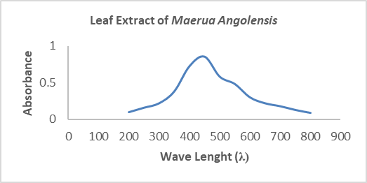
Figure 1: UV-Visible Spectrum of the Maerua angolensis Leaf Extract
The UV-Visible spectrum of the Maerua angolensis leaf extract shows a broad absorption band extending approximately from 250 to 700 nm, with a pronounced maximum (λmax) around 400 nm and decreasing absorbance toward higher wavelengths. This spectral behavior is characteristic of plant-derived extracts rich in bioactive phytochemicals and is highly relevant in the green synthesis and phytochemical analysis [5].
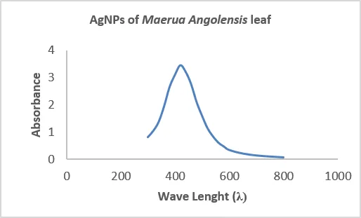
Figure 2: UV-Visible Spectrum of Silver Nanoparticles (AgNPs)
The UV-Visible spectrum of silver nanoparticles (AgNPs) synthesized using Maerua angolensis leaf extract exhibits a distinct and intense absorption peak around 430 nm, which is a hallmark feature of nanoparticle formation. This peak corresponds to the surface plasmon resonance (SPR) phenomenon, arising from the collective oscillation of conduction band electrons on the surface of metallic silver nanoparticles when excited by incident light [16].
3.2 Fourier Transform Infrared (FT-IR) Analysis
FT-IR analysis was done on leaf extract and the synthesized AgNPs in determination of the functional groups present in the sample.
Table 2: FT-IR for Maerua angolensis Leaf Extract
|
FT-IR Peak(cm-1) |
Functional Group |
Phytochemical Class |
|
3226.52 |
O-H stretching (hydroxyl) |
Phenols, alcohols, flavonoids |
|
2980.94 |
C-H stretching Alkanes |
Methyl group |
|
1638.2 |
C=O stretching and C=C stretching |
Proteins, amides, flavonoids, aromatic compounds |
|
1120.85 |
C-O stretching |
Secondary alcohols |
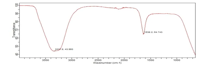
Figure 3: FTIR Spectra of Maerua angolensis Leaf Extract
Fourier Transform Infrared (FT-IR) spectroscopy was employed to identify the functional groups present in the Maerua angolensis leaf extract responsible for the reduction and stabilization of nanoparticles. The FT-IR spectrum showed characteristic absorption bands at approximately 3337.8 cm⁻¹ and 1638.2 cm⁻¹, together with absorption within the fingerprint region below 1200 cm⁻¹. The broad absorption band observed at 3337.8 cm⁻¹ is assigned to O–H stretching vibrations of hydroxyl groups present in phenolic compounds, flavonoids, and alcohols. Hydroxyl-containing phytochemicals are widely recognized as effective reducing agents during the green synthesis of metallic nanoparticles because they donate electrons for the reduction of metal ions while simultaneously acting as capping molecules that stabilize the nanoparticles [1].
Table 3: FT-IR of AgNPs using Maerua angolensis Leaf Extract
|
FT-IR Peak (cm⁻¹) |
Functional Group |
Phytochemical Class |
|
3283.3 |
Hydroxyl(OH)/Amine (N-H) |
Phenols, flavonoids, proteins |
|
2849.5 |
Aldehyde/Alkane (C-H) |
Fatty acids, aldehydes |
|
1720.2 |
Carbonyl (C=O) |
Esters, aldehydes, ketones |
|
1101.4 |
Alcohol/Ether (C-O) |
Glycosides, carbohydrates |
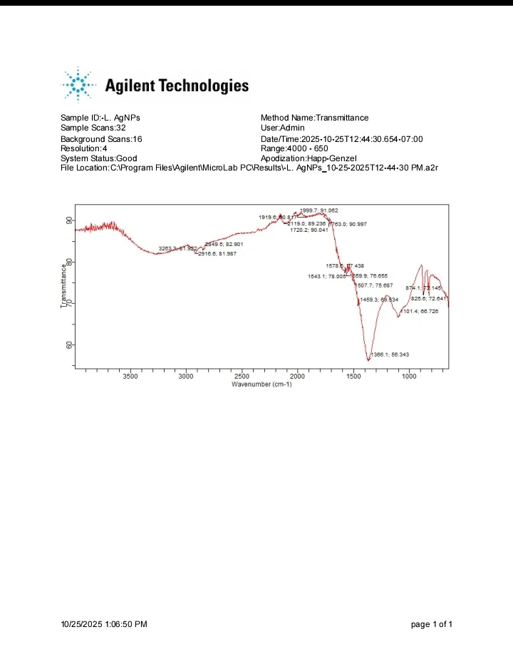
Figure 4: FT-IR Analysis of Silver Nanoparticles (AgNPs)
FT-IR Analysis of Silver Nanoparticles (AgNPs) Synthesized Using Maerua angolensis Leaf. The FT-IR spectrum revealed several characteristic absorption bands, indicating the presence of hydroxyl, carbonyl, amide, aromatic, alkane, and ether functional groups involved in nanoparticle formation. A broad absorption band at 3283.3 cm⁻¹ corresponds to O–H stretching vibrations of hydroxyl groups and may also include N–H stretching of amines or proteins. This peak indicates the presence of phenolic compounds, flavonoids, alcohols, and proteins. These phytochemicals possess strong reducing properties and donate electrons required for the conversion of silver ions (Ag⁺) into metallic silver nanoparticles (Ag⁰). They also function as natural capping agents that stabilize the nanoparticles by preventing aggregation [1].
The absorption bands observed at 2910.0 cm⁻¹ and 2849.5 cm⁻¹ are attributed to C–H stretching vibrations of aliphatic hydrocarbons. These peaks indicate the presence of lipids, terpenoids, and fatty acids, which contribute to the stabilization of the synthesized nanoparticles by forming an organic protective layer around the nanoparticle surface [9].
3.3 Scanning Electron Microscopy Analysis (SEM)
The SEM analysis was done in order to determine the morphology, size and the composition of the elements present in the synthesized AgNPs.
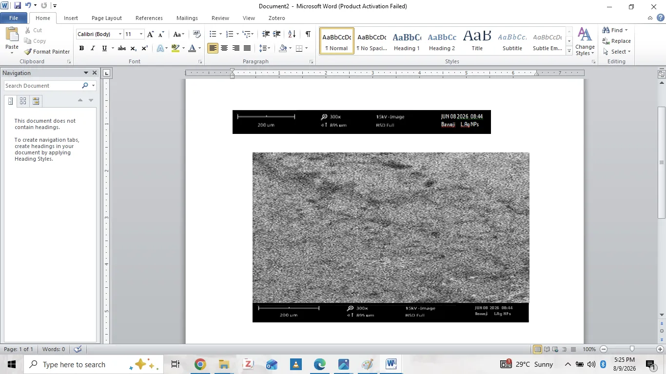
Figure 5: SEM Image of Maerua angolensis Leaf Extract AgNPs
The SEM image of Maerua angolensis leaf extract AgNPs shows a heterogeneous, rough and relatively compact surface with irregularly distributed granular features and some degree of aggregation. The observed aggregation may result from strong inter particle interactions and the high surface energy of nanoparticles, as well as drying during SEM sample preparation. Plant-derived biomolecules can also influence growth, stabilization and the final morphology of AgNPs [2].
3.4 X-Ray Diffraction (XRD) Analysis
XRD analysis was done to find out the average crystalline size. The Debye Scherer equation was used to calculate the average crystalline size:
D=Kλ/βCOSθ
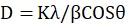
Where: D= Particles size = 0.94
K= Constant volume
λ= X-ray wavelength (0.154nm)
β=Line broadening at half the maximum intensity
θ= Braggs angle (in degree).
Figure 6: The XRD Analysis of the Synthesized Leaf Extract AgNPs
The XRD analysis of the synthesized leaf extract AgNPs revealed prominent diffraction patterns at 2θ values of 29.64°, 38.1°, 44.3° and 64.5° corresponding to the (111), (200), (220), and (311) crystallographic planes of Face Centered Cubic (FCC) structure respectively. The average crystallite size of the AgNPs was found to be 32 nm. The calculated crystallite size of approximately 32 nm is consistent with the size range reported by [14] for silver nanoparticles synthesized using various plant extracts, where crystallite sizes of 45 nm were obtained. [5] Reported crystalline sizes of 40 nm for silver nanoparticles synthesized using Tapidium draba weed extract.
3.5 Antimicrobial Study of the Plant Extract and AgNPs
Table 4: Antibacterial Activity of Maerua angolensis Leaf Extract
Showing the results obtained from the antibacterial activity test for the plant extract Maerua angolensis
Zone of Inhibition in Millimeters (mm)
|
Test organism |
100µg/ml |
200µg/ml |
300µg/ml |
400µg/ml |
500µg/ml |
Control(300µg/ml) |
|
E. Coli |
6 |
7 |
7 |
8 |
10 |
18 |
|
P. Aureg |
7 |
8 |
10 |
13 |
18 |
20 |
|
S.Aureu |
8 |
6 |
8 |
11 |
12 |
22 |
|
K.Pneu |
7 |
11 |
13 |
14 |
19 |
21 |
|
Control = Augmentin E. Coli -= Escherichia Coli, P. Aureg = Pseudomonas Aureginosa, S.Aureu = Staphyloccocus Aureus, K.Pneu = Klebsella Pneumonia, µg/ml = Microgram per mi |
||||||
The antibacterial activity test result for the plant extract showed that Escherichia coli showed an increase in the bacterial growth inhibition with an increase in concentration of the plant extracts. Pseudomonas aeruginosa showed an increase in inhibition of the bacterial growth with an increase in the concentration of the plant extract which is almost closed to that of the control drug. Staphylococcus aureus showed an increase in the inhibition with an increase in the concentration of plant extract. Klebsiella pneumoniae showed an increase in bacterial growth with an increase in concentration of the plant extract which also almost closed to that of the control drug. These results correspond to the literature of [20], which tested two gram-positive and two gram-negative bacteria: E. coli, Pseudomonas aeruginosa (Gram negative), Staphylococcus aureus, Klebsiella pneumoniae (Gram positive) and two fungi: Candida and Aspergillus Niger.
Table 5: Showing the results obtained from the antibacterial activity test for the AgNPs
Zone of Inhibition in Millimeters (mm)
|
Test organism |
100µg/ml |
200µg/ml |
300µg/ml |
400µg/ml |
500µg/ml |
Control(300µg/ml) |
|
E. Coli |
7 |
8 |
8 |
9 |
13 |
19 |
|
P. Aureg |
8 |
8 |
11 |
15 |
19 |
22 |
|
S.Aureu |
9 |
8 |
10 |
13 |
15 |
24 |
|
K.Pneu |
10 |
12 |
14 |
16 |
19 |
22 |
|
Control = Augmentin E. Coli -= Escherichia Coli, P. Aureg = Pseudomonas Aureginosa, S.Aureu = Staphyloccocus Aureus, K.Pneu = Klebsella Pneumonia, µg/ml = Microgram per mi |
||||||
The antibacterial activity test result for the AgNPs showed that, the Escherichia –coli show an increase in the bacterial growth inhibition with an increase in concentration of the AgNPs Pseudomonas aureginosa shows an increase in inhibition of the bacterial growth with an increase in the concentration of the plant extract which is almost closed to that of the control drug. Staphylococcus aureus shows an increase in the inhibition with an increase in the concentration of AgNPs Klebsella pneumonia shows an increase in bacterial growth with an increase in concentration of the AgNPs which also almost closed to that of the control drug. In correspond to the literature of [20]. Which two gram positive and negative bacteria was tested and two fungi was tested are E-coli, pseudomonas aeruginosa. (Gram negative), Staphylococcus aureus, Klebsiella pneumonia (Gram positive) and candida, and Aspergillus Niger (fungi).
Table 6: Antifungal Activity of Plant Extract and AgNPs
|
Zone of Inhibition (mm) |
||||||
|
Test organism |
100µg/ml |
200g/ml |
300µg/ml |
400µg/ml |
500 µg/ml |
Control(500µg/ml |
|
Plant extract A.Niger |
7 |
6 |
7 |
8 |
9 |
17 |
|
Plant extract Candida |
6 |
8 |
7 |
9 |
10 |
18 |
|
AgNPS A.Niger |
8 |
10 |
13 |
11 |
14 |
21 |
|
AgNPS Candida |
10 |
13 |
16 |
19 |
22 |
25 |
|
Control = Fulcin, A.Niger = Aspagillus Niger, Candida = Fungus Candida, µg/ml = Microgram per mil |
||||||
The plant extract against Aspergillus Niger showed an increase in fungal inhibition with an increase in the concentration of the plant extract, showing the high rate of inhibition at 500 µg/ml. The Candida showed an increase in the fungal growth as the concentration increases and the highest inhibition was observed at 500 µg/ml. This shows that as the concentration increases, the zone of inhibition increases. The silver nanoparticles (AgNPs) against Aspergillus Niger showed an increase in fungal growth inhibition with increase in concentration of the synthesized drug (nanoparticles). It showed highest fungi inhibition rate at concentration 300 µg/ml but decreased in concentration 400 µg/ml and 500 µg/ml respectively. This decrease depends on the type of fungi in question. Each fungi has its own activity and resistance to the drug used. In relation to literature of [20].
CONCLUSION
The green synthesis of silver nanoparticles (AgNPs) using aqueous leaf extract of Maerua angolensis has been successfully achieved, demonstrating an eco-friendly, cost-effective, and sustainable approach to nanomaterial synthesis. The formation of AgNPs was confirmed by UV-Visible spectroscopy, showing a characteristic surface plasmon resonance peak at 430 nm, indicative of nanoparticle formation. FT-IR analysis revealed the presence of various functional groups including hydroxyl, carbonyl, amine, and aromatic groups from phytochemicals such as phenols, flavonoids, proteins, and alkaloids, which played dual roles as reducing and stabilizing agents during the synthesis process.
XRD analysis confirmed the crystalline nature of the AgNPs with face-centered cubic (FCC) structure, showing prominent diffraction peaks corresponding to (111), (200), (220), and (311) planes, with an average crystallite size of 32 nm. SEM analysis revealed heterogeneous, rough, and compact surface morphology with some aggregation, which is typical for biologically synthesized nanoparticles.
The antimicrobial studies demonstrated that both the plant extract and synthesized AgNPs exhibited significant antibacterial and antifungal activities against the tested microorganisms. The AgNPs showed enhanced antimicrobial activity compared to the plant extract alone, with concentration-dependent inhibition zones. The highest antibacterial activity was observed against Pseudomonas aeruginosa (18 mm at 500 µg/ml), while the most significant antifungal activity was recorded against Candida (22 mm at 500 µg/ml). The enhanced antimicrobial activity of AgNPs can be attributed to their small size, large surface area-to-volume ratio, and the synergistic effect of silver ions with phytochemicals present on the nanoparticle surface.
This study successfully demonstrates that Maerua angolensis leaf extract serves as an excellent bio-reductant and stabilizing agent for the green synthesis of AgNPs with promising antimicrobial properties. The synthesized nanoparticles showed potential as effective antimicrobial agents against both Gram-positive and Gram-negative bacteria as well as fungal pathogens. The findings suggest that these biosynthesized AgNPs could be developed as alternative therapeutic agents for the treatment of infectious diseases, particularly in the face of increasing antimicrobial resistance. Further studies are recommended to investigate the mechanism of antimicrobial action, cytotoxicity, and in vivo efficacy of these nanoparticles for potential pharmaceutical and biomedical applications.
REFERENCES





Suleiman Mohammed Bawaji, Nasiru Yahaya Pindiga, Buhari Magaji, Umar Muhammad Lawan, Green Synthesis and Characterization of Ag Nanoparticles Using Leaf Extract of Maerua Angolensis for Antimicrobial Studies, Int. J. of Pharm. Sci., 2026, Vol 4, Issue 10, 120-130, https://doi.org/10.5281/zenodo.23074504
 10.5281/zenodo.23074504