
We use cookies to ensure our website works properly and to personalise your experience. Cookies policy
1Department of Pharmaceutics, Department of Pharmaceutical Sciences, RIMSR, Puthuppally
The transition from conventional dosage forms to targeted nanovesicular drug delivery systems has resulted in improved therapeutic efficacy and reduced systemic adverse effects. Ufasomes, a suspension of closed lipid bilayers of unsaturated fatty acids and their ionized species, have proven to be a promising alternative for conventional vesicular carrier systems, which address numerous drawbacks of liposomes, including high production costs, physical instability, and oxidative vulnerabilities. Ufasomes exhibit a fusogenic tendency that promotes deep fusion with the lipid bilayers of the skin due to their distinct pH-dependent stability. Hence, they are considered as an excellent vehicles for topical and transdermal drug delivery systems that provide a localized and sustained release of both lipophilic and hydrophilic drugs. Although switching to continuous microfluidic assembly is still a crucial manufacturing frontier, conventional techniques like thin-film hydration, reverse-phase evaporation, and ethanol injection methods are used for the preparation of ufasomes. This review assesses the structural dynamics, formulation parameters, and current manufacturing bottlenecks of ufasomes, emphasizing the importance of overcoming current systemic delivery limitations, specifically acidic degradation in the stomach, and investigates their emerging therapeutic applications in dermal oncology, dual-drug co-delivery, and AI-optimized precision medicine.
The evolution of the conventional dosage forms to targeted specific nanovesicular drug delivery system signifies a remarkable shift in maximizing therapeutic efficacy and minimizing systemic adverse effects of the drug[1]. The conventional drug delivery system has several drawbacks, including poor drug solubility, rapid degradation in the body, low bioavailability, and an inability to target specific tissues, which leads to significant systemic side effects. These nanocarrier platforms were designed to address these constraints. These nanovescicles were utilized to encapsulate the drug, so as to ensure controlled and sustained release, target specificity, and prevention of premature breakdown[2]. Moreover, by improving the drug's distribution throughout the body, these platforms maximize its effectiveness and reduce its toxicity[3].
The formulation, technology, and systems for delivering a pharmaceutical substance into the body so as need to provide a safe intended therapeutic effects are the main concern of novel drug delivery systems (NDDS)[4]. Various vesicular nanocarriers, such as liposomes, niosomes, transferosomes, ethosomes, cryptosomes, aquasomes, virosomes, erythrosomes, bilosomes, and novasomes, differ in their structural and functional features. The drug combines with the phospholipid structure through chemical bonding within the lipid bilayer which contributes to the enchanced physicochemical parameters and protection of the drug[2]. Liposomes, which are composed of phosphotidylecholine, cholesterol, sphingolipids, steroids, and polymeric materials, were regarded as the traditional and first- studied phospholipid vesicles[4,5]. This offered a number of benefits, but it also had several drawbacks, such as high production costs, drug leakage, and phospholipid susceptibility to oxidation and hydrolysis[4]. Transferomes and ethosomes were designed by incorporating the edge activators and high alcohol concentrations in order to prevail over these limitations. By destabilizing the membrane bilayer, the edge activators give it an elastic and stress-responsive nature, significantly improving its penetration. On the other hand, alcohols improve penetration by fluidizing lipid bilayer and lipid matrix of the skin. Eventually, non-phospholipid substitutes were also developed. Niosomes, which use cholesterol and non-ionic surfactants to address issues including stability, sterilization, and large-scale production [4]. Ufasomes are vesicles of unsaturated fatty acids that naturally improve skin penetration[6].
Ufasomes as a Vesicular Carrier
Ufasomes are closed lipid bilayered vesicles composed of unsaturated fatty acids and their ionized species that serve as a natural permeation enhancer. These vesicles include two forms of amphiphiles: non-ionic neutral form and ionic form, the ratio of which affects the basic of vesicle stability. Compared to phospholipids in liposomes, the primary benefit of ufasome is that the fatty acids are single chain amphilphiles and easily accessible[7]. However, they improve drug retention properties within the cell membrane of skin with time[6].
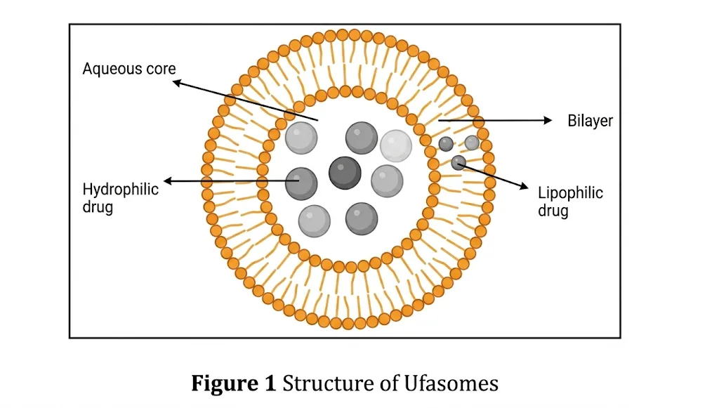
Figure 1: Structure of Ufasome[8]
Advantages[7]
Disadvantages[9]
Generalized Formation Method
Only non-oxidised components, such as 10% oleic and linoleic acid in chloroform treated at 20˚C, are used in the production of ufasomes. Around 0.02ml of the stock solvent is evaporated with a water pump, followed by drying with a nitrogen spray. Using 0.2 ml of 0.1 M Tris-hydroxymethyl aminomethane buffer with a pH of 8–9, the fatty acid layer is broken down in a strong spinning vortex blender, which leads to the production of Ufasomes. The resulting suspension is sonicated using a probe sonicator mounted with a titanium microtip, and the air is expelled by passing a stream of nitrogen through it. Using an ice bath, the consistent temperature can be maintained[6,10].
METHOD OF PREPARATION
Thin Film Hydration Method
This is the most often used method for preparing ufasome. The vesicle components, such as the fatty acids, surfactants, and stabilizers, are taken in a round-bottom flask with an appropriate solvent, often a 1:1 ratio of chloroform : methanol. The solvent is completely evaporated with a rotating vacuum evaporator, resulting in a thin layer that is hydrated with an appropriate phosphate buffer solution[8].
Reverse Phase Evaporation Method
Lipid and surfactants are dissolved in an appropriate organic solvent to produce the organic phase, while water-soluble components are dissolved in water to produce the aqueous phase. A water in oil emulsion developed by combining the two phases and evaporating the solvent at reduced pressure. To achieve a thick, gel-like consistency, an aqueous solution is added. After homogenization, the mixture is vortexed and allowed to stabilize, promoting vesicle formation. The size and uniformity of vesicle can be refined further by sonication[8].
Ethanol Injection Method
The vesicle components, such as fatty acid and stabilizers, were dissolved in ethanol, which constitutes the organic phase, and phosphate buffer of pH 8 was used as the aqueous phase. Both phases were preheated to 65˚C, and the hot lipid phase was injected into the aqueous phase while maintaining the temperature at 65˚C and a constant stirring of 500 rpm. The ethanol was then allowed to evaporate completely by using a rotary evaporator for one hour at 150 rpm to create a suspension of ufasome, which was kept at 2 to 8 ˚C[11].
Autopoetic Process
When aqueous fatty acids are dissolved in a watered buffer solution, fatty acid vesicles develop due to the abrupt pH transition. The vesicles are created when half of the carboxylic acid in the fatty acids is ionised. The hydrocarbon chain forms a bilayer structure that opposes the aqueous compartment, reducing the quantity of water it comes into contact with[6].
KEY ISSUES IN MANUFACTURING OF UFASOMES
Selection of fatty acids
According to research on natural membrane phospholipids and data from pressure region measurements on fatty acid surface films, the 12–22 carbon fatty acid was determined to be the most suitable for stable ufasome preparation. C-18 acids showed highest potential in early studies, thus being the focus of the study. These requirements could only be met by ufasomes of oleic acid and linoleic acid formed membranes. In an oleic acid membrane, stearic acid can be tolerated up to 5% by weight, while palmitic acid can be tolerated up to 33% by weight. Charging the membrane with small amounts of oleic, linoleic, or stearic acid amides had no effect on the preparations. Stability experiments showed that whereas linoleic acid produced significant peroxide within two to three weeks, oleic acid remained free of peroxides for at least six weeks[6,12].
Addition of cholesterol
Cholesterol has the potential to change membrane fluidity, elasticity, and permeability, filling the gap caused by incomplete lipid packing. The ability of vesicles to hold solute rapidly decreases as cholesterol levels rise. Furthermore, membrane impermeability does not increase at any cholesterol level [6,12].
pH
These fatty acid vesicles are developed when half of the carboxylic acids are ionized at pH rangearound 7- 9. Fatty acids form unstructured precipitates below this range and become extremely soluble above it. Micelles, which have a higher ratio of ionized to protonated molecules, are the main aggregate species at higher pH ranges, while oil droplets develop at lower pH levels. At concentrations slightly greater than the critical vesiculation concentration (CVC), the vesicles can be easily distinguished. Colloidal vesicle suspensions are produced when monomers and nonvesicular aggregates form a bilayer structure at the required vesiculation concentration [6,12].
Selection of buffer
The most common buffer for ufasome formation is tris hydroxymethyl aminomethane. The type of buffers to be used depends on the solute to be included. To prepare ufasomes from 1 milligram of fatty acid, 0.1 ml of 0.1 M tris at pH 8 is needed since the right weight of buffer for tris must equal the weight of fatty acid used to form membranes [6].
Electrolyte
Ufasome vesicles are typically prevented by the presence of electrolytes used in the preparation. The spheres may be subjected to phosphate or chloride solutions while keeping the occluded glucose after being stabilized in the required buffer [6].
Peroxidation
Peroxidation alters the usual bilayer structure of fatty acid molecules in ufasome membranes by introducing heavy hydrophilic groups that distort the hydrophobic interior, hence increasing membrane permeability and allowing water-soluble molecules to flow out easier. Linoleic acid oxidizes at a rate of 0.1 percent per minute in air-saturated buffers using more intense preparation techniques like ultrasonic resuspension (30-W irradiations), whereas short periods of hand vortexing do not promote peroxidation. Research by Hicks and Gebicki shows that adding antioxidants such nitroxide radicals, butylated hydroxytoluene (BHT), and alpha-tocopherol can successfully shield the linoleic acid membrane from this peroxidation, even though a maximum 3-minute exposure to ultrasound shouldn't produce significant degradation on its own[12].
Divalent cations
Lipid peroxidation occurs through both enzymatic and non-enzymatic pathways, with the latter relying on metal ion transitions. Since non-variable valence metals like magnesium, calcium, and zinc are unable to take part in redox-coupled homolysis, the single electron valency shift of metals rapidly catalyzes the lipid peroxidation in unsaturated lipids. Calcium has a biphasic effect, activation and inhibition, in ufasome-induced lipid peroxidation. By attaching to negatively charged lipid groups, such as the phosphate groups in lecithin or the carboxyl groups in linolenic acid, Ca2+ activates lipid peroxidation. This raises the concentration of free Fe2+ that can directly accelerate peroxidation by displacing bound Fe2+ ions. Inhibition of Ca2+ interacts with superoxide anion radicals to decrease lipid peroxidation [6].
CHARACTERISATION OF UFASOME
Particle Size and Size Distribution
The average diameter and size distribution of the ufasome suspension were measured using photon correlation spectroscopy at fixed angles of 90˚ and 25˚. After being diluted with phosphate buffer, the suspension was passed over a pH 7.4 polycarbonate membrane to reduce particulate matter interference until sizing[10].
Shape and Surface Morphology
Transmission electron microscopy (TEM) was used to determine the morphological characteristics of sphericity and the accumulation of drug-loaded ufasomal dispersion. The 1% phosphotungstic acid was used to negatively stain the carbon film-covered copper grid to test the selected ufasomal dispersion, after it is being allowed to dry at room temperature for 10mins[10].
Differential Scanning Calorimetry
Differential Scanning Calorimetry was used to determines the physical condition of the compound inside the oleic acid vesicles. The vesicles were placed in a conventional aluminium pan and scanned at a rate of
2°C/min[10].
Entrapment Efficiency
The formulation was ultracentrifuged at 25000 rpm for 3 hours at 4°C, and the supernatant was collected to assess the concentration of unentrapped drug by using UV spectroscopy[10].

In-vitro Drug Release
In vitro drug release determines how quickly the medication leaves the ufasome and with what release kinetics which is carried out using Franz diffsusion cell. The Franz diffusion cell has two compartments, one for the donor and one for the receptor separated by a polycarbonate membrane with 50 nm pores. The ufasome is contained in the donor compartment, while the receptor compartment is filled with a pH 7.4 phosphate buffer that is maintained at 37˚C and continuously stirred at a constant rate using a magnetic stirrer. At predetermined intervals, sample aliquots are collected and replaced with equivalent amounts of fresh PBS[ [10].
CURRENT CHALLENGES
The pH Dependency and Systemic Delivery Barrier
Hashem developed Itraconazole-Loaded Ufasomes for antifungal action against Candida albicans. The partial ionization of fatty acids, which normally requires a narrow, alkaline pH range (generally between pH 7.0 and 9.0), is essential for the structural integrity of ufasomes. The ionized carboxylate group becomes fully protonated due to the acidic environment in the stomach mucosa, disrupting the delicate equilibrium between hydrophobic interactions and electrostatic repulsion. As a result, the ufasomal bilayer rapidly breaks down into micelles or amorphous oil droplets13. This can be addressed through PEGylation, which involves applying PolyEthylene Glycol (PEG) to the surface of the ufasome. This preserves the lipid bilayer by entirely wrapping it in a thick layer of water that shields it from direct contact with stomach acids and gastric enzymes[14].
Active Targeting and Surface Functionalization
The attachment of various active targeting agents, such as peptides or antibodies, to ufasomes has yet to be achieved due to their fragile single chain fatty acid structure, which cannot withstand typical chemical conjugation procedures. Peptide ligands coupled to the surface of ufasomes can be used for functionalized targeting to improve direct cellular absorption and stimuli-responsive targeting, which releases its payload in acidic diseased conditions while remaining stable at a certain pH. Biomimetic cell membrane coatings may prevent immune clearance, and monoclonal antibody-conjugated immuno-ufasomes ensure accurate targeting to skin infections, producing a useful, systematically modified nanovesicle [15].
Transition to Dermal Oncology
The current literature focuses on generic topic applications because ufasomes are fusogenic; they naturally fuse with the skin's lipid layers. This is evident in the studies of Mittal et al. [16] (2013) and Sharma & Arora[17] (2012), which deal with the ufasomal topical drug delivery of dexamethasone and methotrexate, respectively. By encapsulating the potent chemotherapy agents in ufasomes, which can directly target melanoma and non-melanoma skin tumors effectively[18].
Lipid Peroxidation
Ufasomes are composed of unsaturated fatty acids, making their double bonds especially sensitive to autooxidation and lipid peroxidation. During storage, exposure to light, oxygen, or high temperatures can cause oxidative deterioration, which can result in payload leakage, changed vesicle size, and a much shorter shelf life.[13,17]. Co-encapsulation of natural antioxidants can be employed to get around this. The ideal concentration and ratio of antioxidants required in the bilayer can be determined quantitatively using statistical models. The delicate, pH-dependent structure of the ufasome vesicle can be preserved while maximizing oxidative stress by applying Quality by Design (QbD) principles[19].
Reticuloendothelial Clearance Gap
When ufasome is injected intravenously, the lipid vesicles may adhere to the blood proteins, signalling the immune system to rapidly eliminate them, which is a common limitation for all nanocarriers. This can be addressed with PEGylation and biomimetic engineering. The blood plasma protein will be repelled by the barrier created by PEGylation, which coats the surface of the nanocarriers with PEG. On the other hand, with biomimetic engineering, the nanocarrier is encased in a natural cell membrane, which allows the immune system to identify it as a natural protein and a component of the body[15].
FUTURE PERSPECTIVES
Integration of Machine Learning and Quality by Design (QbD)
Ufasomal formulations mostly use iterative, trial-and-error approaches to identify lipid-to-cholesterol ratios. Artificial Intelligence (AI) and Quality by Design (QbD), which are used to accurately estimate vesicle size, long-term stability, and encapsulation efficiency based on the original lipid composition, are essential to the development of ufasomes in the future. These AI models can be used to modify co-encapsulated active chemicals and antioxidants in single-chain ufasomes, significantly lowering formulation bottlenecks [19].
Advanced Co-Delivery and Combination Therapies
In contemporary medicine, combination therapy can be employed to overcome drug resistance and reduce systemic toxicity. Ufasomes have a lipophilic bilayer for hydrophobic drugs and an aqueous core for hydrophilic drugs. This dual-compartment system can be utilized to deliver multiple drugs in future formulations. These kinds of advancements have the potential to transform the treatment strategies of various complicated localized disorders[20].
Transitioning to Microfluidic Scale-Up
For the commercial production of sterile, highly homogeneous vesicles, standard thin-film hydration is inadequate. For unsaturated fatty acid vesicles, future research needs to develop a continuous flow production and microfluidic aseptic assembly. Microfluidics enables the quick, one-step, bottom-up self-assembly of lipidic nanocarriers in a closed, sterile system, in contrast to conventional thin-film hydration. In order to produce highly consistent, repeatable ufasomes at the industrial scale needed for clinical trials and commercial market entrance. The crucial step in implementing ufasomes in clinical practice is to cease this manufacturing gap while concurrently carrying out thorough in vivo pharmacokinetic studies[21].
Regulatory Considerations
Comprehensive preclinical and clinical studies demonstrating the safety, efficacy, and quality of the ufasomal formulation are required for regulatory approval of the formulation. Several regulatory agencies, such as the FDA and EMA, have developed a number of guidelines for the production of ufasomal formulations. These guidelines outline the requirements for pharmacokinetic research, stability testing, characterization, and clinical trial design. Future attempts should focus on simplifying the regulatory procedures, enhancing industry-regulatory authority collaboration, and employing regulatory strategies like rapid approval pathways and adaptive licensing in order to fasten the development and commercialization of the ufasomal formulation[3].
Integration with Other Drug Delivery Platforms
Synergistic effects and better therapeutic outcomes can be achieved by combining ufasomal drug delivery with other platforms, such as nanoparticles, micelles, and polymeric carriers. By using the unique characteristics of several drug delivery systems, combination strategies can be used to get around a variety of platform-specific constraints, including as payload capacity, targeting specificity, and drug release kinetics. Future research must focus on innovative methods to combine ufasomes with complementary drug delivery platforms in order to achieve optimal drug delivery and personalized treatment regimens[3].
Emerging Applications in Personalized Medicine and Theranostics
Theranostics, personalized medicine, and ufasomal drug delivery together provide an efficient means to personalize treatments to the unique characteristics of each patient's disease. Ufasomal formulations can be created to combine medications, diagnostic agents, and imaging probes into multifunctional platforms for the diagnosis and treatment of diseases. Ufasomal theranostic systems combine imaging modalities, biomarkers, and patient-specific data to enable patient classification, treatment optimization, and real-time monitoring of treatment responses. Future research should focus on developing new ufasomal theranostic platforms and applying advancements in precision medicine to improve patient outcomes and healthcare delivery[3].
Table 1: List of drugs that can currently require transformation into ufasomes.
|
Drug Name |
Current Marketed Formulation/ Carrier |
Why Transformation to Ufasome is Required |
|
Oleuropein |
Free extracts, conventional dietary capsules |
This is a potent nutraceutical; it suffers from rapid degradation and poor cellular uptake in its free form. Ufasomes imitate the composition of the cell membrane, which allows them to actively transport antioxidants across cell barriers, enhancing biological efficiency. |
|
Febuxostat |
Oral tablets |
This is used for lowering urate in gout treatment; oral administration has various disadvantages like poor aqueous solubility, high first-pass metabolism, and gastrointestinal adverse effects. Ufasomal transdermal gel creates a localized drug reservoir in the skin for controlled and sustained release, bypassing the GIT. |
|
Aceclofenac |
Oral tablets |
This is an anti-inflammatory often given in combination with Febuxostat to treat acute gout flares, which has poor solubility and GI toxicity. This can be formulated into a dual-loaded ufasomal gel of Aceclofenac and Febuxostat for multiple biological effects like urate lowering and anti-inflammatory effects for topical administration. |
|
Caspofungin |
Intravenous (IV) infusion |
This is a potent antifungal that is currently administered as a systemic IV infusion, which reduces patient compliance and has systemic side effects. Formulating topical ufasomal gel of Caspofungin causes deeper dermal penetration and a sustained release profile directly at the site of topical fungal infections, avoiding systemic circulation. |
|
Terbinafine |
Topical creams, oral tablets |
The oral tablets have various hepatic side effects, whereas the conventional creams are difficult to penetrate in deep fungal infections. Ufasomes improve the transdermal permeation due to the presence of the flexible lipid bilayer, which provides a sustained drug release and improves the antifungal efficacy directly at the site of infection without systemic exposure. |
|
Clotrimazole |
Conventional creams, lozenges |
This is widely used for anti-fungal infections, which have a poor water solubility that severely limits their absorption and local effectiveness. Ufasomal formulations offer higher entrapment efficiency (often >80%) and superior penetration across the stratum corneum, maintaining prolonged antifungal activity over several days. |
|
Etodolac |
Oral capsules, standard topical gels |
This is an orally administered Non-Steroidal Anti-Inflammatory Drug (NSAID) that frequently causes gastrointestinal side effects like dyspepsia and bleeding. By formulating Etodolac into ufasomal topical gels that completely bypass the GI tract. The oleic acid vesicles act as permeation enhancers, delivering localized pain and inflammation relief directly to the targeted joint or muscle. |
CONCLUSION
Ufasomes have proven to be a highly cost-effective, efficient, and adaptable alternative to standard vesicular nanocarriers like liposomes and niosomes. Their distinct fusogenic nature makes them an outstanding nanocarrier for transdermal drug delivery, which improves the penetration and sustained release of both hydrophilic and lipophilic compounds. By employing the single-chain unsaturated fatty acid, they avoid a number of constraints, such as manufacturing costs and physical instabilities typical for conventional phospholipid bilayers. However, addressing several significant biophysical and manufacturing challenges is necessary to close the translational gap from laboratory-scale in vitro research to reliable clinical application. Future research should focus on stabilizing the extremely pH-dependent structure of ufasomes for systemic distribution, mapping their in vivo pharmacokinetic behaviour, and shifting from harmful manual production methods to sustainable, microfluidic continuous manufacture. Ufasomes have the potential to overcome current topical limitations and transform into targeted dermal oncology, combination therapy, and personalized theranostic medicine by integrating artificial intelligence, predictive Quality by Design (QbD) optimization, and sophisticated stimuli-responsive engineering.
REFERENCES


Akhila P.*, Arun Raj, Anandhu Dineshan, Nebin Koshy, Escaping the Topical Trap: Reengineering Ufasomes for Smart Systemic Delivery, Int. J. of Pharm. Sci., 2026, Vol 4, Issue 6, 5144- 5154. https://doi.org/ 10.5281/zenodo.20767165
 10.5281/zenodo.20767165