

We use cookies to ensure our website works properly and to personalise your experience. Cookies policy
Department of Pharmacology, Acharya & BM Reddy College of Pharmacy, Soldevanahalli, Bengaluru, Karnataka, India 560107
Background: Autism spectrum disorder (ASD) is a neurodevelopmental disorder characterized by impaired social interaction, communication deficits, repetitive behaviors, oxidative stress, neuroinflammation, and altered stress responses. Rutin hydrate, a naturally occurring flavonoid with antioxidant and anti-inflammatory properties, has shown neuroprotective potential in several neurological disorders. This study evaluated the therapeutic efficacy of rutin hydrate in a valproic acid (VPA)-induced zebrafish model of ASD. Methods: Adult zebrafish were divided into four groups: vehicle control, VPA (100 mg/kg), VPA + rutin hydrate (35 mg/kg), and VPA + rutin hydrate (70 mg/kg). Behavioral assessments included the light/dark preference, shoaling, mirror attack, and social contact tests. Whole-body cortisol, oxidative stress markers (GSH and TBARS), inflammatory cytokines (TNF-? and IL-6), and total brain protein were evaluated. Data were analyzed using one-way ANOVA followed by Dunnett's multiple comparison test. Results: VPA induced significant behavioral deficits, elevated cortisol, TBARS, TNF-?, IL-6, and total brain protein levels, and reduced GSH levels compared with the control group. Rutin hydrate significantly improved behavioral performance and reversed biochemical alterations in a dose-dependent manner, with the 70 mg/kg dose producing greater neuroprotective effects. Conclusion: Rutin hydrate effectively ameliorated VPA-induced ASD-like behavioral and biochemical abnormalities through antioxidant, anti-inflammatory, and stress-regulating mechanisms, suggesting its potential as a promising therapeutic candidate for ASD.
Autism spectrum disorder (ASD) is a complex neurodevelopmental disorder characterized by persistent deficits in social communication and social interaction, accompanied by restricted and repetitive patterns of behaviour, interests, or activities.[1] The disorder manifests during early childhood and persists throughout life, often affecting cognitive, emotional, and functional development. In addition to the core behavioural symptoms, individuals with ASD frequently present with anxiety, sleep disturbances, gastrointestinal abnormalities, aggressive behaviour, hyperactivity, and impaired adaptive functioning, imposing a substantial burden on affected individuals, families, and healthcare systems(Figure 1).[2] Epidemiological studies indicate that the prevalence of ASD has increased considerably over the past two decades, although changes in diagnostic criteria, improved awareness, and enhanced screening strategies have also contributed to the increasing number of reported cases. Current estimates suggest that approximately 1% of the global population is affected by ASD, with a significantly higher prevalence among males than females.[3]
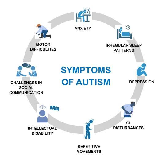
Fig 1: Core & Associated symptoms of ASD
The pathophysiology of ASD is multifactorial and involves a complex interaction between genetic susceptibility, environmental influences, neurodevelopmental abnormalities, immune dysregulation, and oxidative stress.(Figure 2)Genetic alterations affecting synaptic development and neuronal connectivity, including mutations in genes such as CNTNAP2 and SHANK3, have been implicated in ASD.[4] Furthermore, increasing evidence demonstrates that neuroinflammation contributes significantly to disease progression through activation of microglia and elevated production of pro-inflammatory cytokines, including tumour necrosis factor-alpha (TNF-α) and interleukin-6 (IL-6).[5] Oxidative stress also plays an important role in ASD pathology, resulting from an imbalance between reactive oxygen species and endogenous antioxidant defence mechanisms such as glutathione (GSH), catalase, and superoxide dismutase.[6] Excessive oxidative stress promotes lipid peroxidation, protein oxidation, mitochondrial dysfunction, and neuronal injury, ultimately contributing to behavioural and cognitive abnormalities associated with ASD.[7]
Currently available pharmacological interventions for ASD primarily target associated behavioural symptoms such as irritability, hyperactivity, and anxiety rather than the underlying disease mechanisms. Consequently, there is increasing interest in identifying therapeutic agents capable of modulating oxidative stress, neuroinflammation, and neuronal dysfunction. Naturally occurring phytochemicals possessing antioxidant and anti-inflammatory properties have attracted considerable attention because of their favourable safety profile and multitarget mechanisms of action.[8]
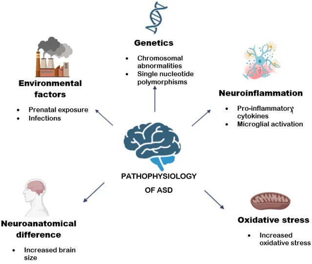
Fig 2: Multifactorial contributors to ASD pathophysiology
Zebrafish (Danio rerio) have emerged as a valuable vertebrate model for investigating neurodevelopmental disorders, including ASD.[9] Their high genetic homology with humans, conserved neurotransmitter systems, rapid development, ease of maintenance, and suitability for high-throughput behavioural screening make zebrafish an attractive experimental model in neuroscience research. Moreover, zebrafish exhibit complex social behaviours, shoaling, aggression, anxiety-like responses, and social preference, which closely resemble behavioural domains affected in ASD.[10] These characteristics have facilitated the development of reliable zebrafish models for evaluating the pathophysiology of ASD and assessing potential therapeutic interventions.[11]
Among the available experimental models, valproic acid (VPA)-induced ASD is one of the most extensively validated models for studying autism-like behavioural and neurochemical abnormalities. Prenatal or experimental exposure to VPA has been associated with impaired social interaction, repetitive behaviours, altered neuronal connectivity, neuroinflammation, oxidative stress, and mitochondrial dysfunction.[12] Experimental studies have demonstrated that VPA disrupts GABAergic neurotransmission, modulates voltage-gated sodium and calcium channels, inhibits histone deacetylases, and alters gene expression involved in neuronal development and synaptic plasticity. These molecular alterations closely resemble several pathological mechanisms reported in individuals with ASD, making VPA-induced zebrafish models suitable for evaluating candidate neuroprotective therapies.[13]
Rutin hydrate (quercetin-3-O-rutinoside) is a naturally occurring flavonoid widely distributed in fruits, vegetables, tea, and medicinal plants.[14] Previous studies have demonstrated that rutin hydrate possesses potent antioxidant, anti-inflammatory, neuroprotective, and free radical scavenging activities. Rutin has been shown to cross the blood-brain barrier and protect neural tissues by reducing reactive oxygen species, enhancing endogenous antioxidant defence systems through increased glutathione levels and antioxidant enzyme activity, and suppressing inflammatory mediators.[15] Experimental studies have further demonstrated its protective effects against oxidative stress-induced neuronal injury, cognitive impairment, and neuroinflammation, suggesting its therapeutic potential in neurological disorders.[16]
Although several studies have investigated the neuroprotective effects of rutin hydrate in different experimental models, limited information is available regarding its therapeutic efficacy against ASD-like behavioural and biochemical alterations induced by valproic acid in adult zebrafish. Therefore, the present study was designed to evaluate the therapeutic potential of rutin hydrate in a valproic acid-induced zebrafish model of autism spectrum disorder. Behavioural assessments, including the light/dark preference test, shoaling behaviour, mirror attack test, and social contact test, were performed to evaluate ASD-like behavioural deficits. In addition, whole-body cortisol, oxidative stress markers (GSH and TBARS), inflammatory cytokines (TNF-α and IL-6), and total brain protein were estimated to investigate the underlying biochemical mechanisms associated with the neuroprotective effects of rutin hydrate.
MATERIALS AND METHODS
Chemicals
Valproic acid (VPA) and rutin hydrate were used in the present study. Rutin hydrate was procured from Sigma-Aldrich, Bengaluru. All chemicals and reagents used throughout the study were of analytical grade.
Experimental Animals
Adult zebrafish (Danio rerio) of either sex, measuring approximately 3–5 cm in length, were used for the study. The fish were acclimatized for 14 days before the initiation of the experiment. Animals were maintained at 28 ± 2°C under a controlled photoperiod of 14 h light and 10 h dark, with water pH maintained between 6.8 and 7.1 and light intensity of approximately 250 lux. Zebrafish were housed in standard tanks equipped with a continuous water circulation and aeration system (36 × 26 × 22 cm), with 10–12 fish per tank. Male and female fish were maintained separately and were fed Tetramin tropical flakes twice daily. [17]All experimental procedures were performed in accordance with the guidelines of the Committee for Control and Supervision of Experiments on Animals (CCSEA), Government of India, and were approved by the Institutional Animal Ethics Committee (IAEC), Acharya & BM Reddy College of Pharmacy (Approval No. IAEC/ABMRCP/2024–2025/25).
Acute Toxicity Study
The acute toxicity study was performed according to OECD Guideline 203. Five concentrations of rutin hydrate were prepared in a geometric progression with a factor not exceeding 2.2. Seven zebrafish were used for each concentration, including the control group (n = 7). Mortality was monitored at 24, 48, 72, and 96 h following exposure to determine the safe dose range for subsequent pharmacological evaluation.[18]
Experimental Design
The zebrafish were randomly divided into four experimental groups (n = 10 per group):
Following treatment, behavioral assessments were performed. At the completion of behavioral studies, the fish were euthanized, brains were dissected, and tissue homogenates were prepared for estimation of oxidative stress markers, inflammatory cytokines, and total brain protein. Whole-body cortisol estimation was also carried out to evaluate stress responses.(Figure 3)
Table 1: Treatment protocol
|
Group |
Treatment protocol (n=10) |
|
I |
Vehicle control |
|
II |
Valproic acid (100 mg/kg) p. o |
|
III |
Valproic acid (100 mg/kg) p. o+ Rutin hydrate (35 mg/kg) p. o |
|
IV |
Valproic acid (100 mg/kg) p.o + Rutin hydrate (70 mg/kg) p.o |
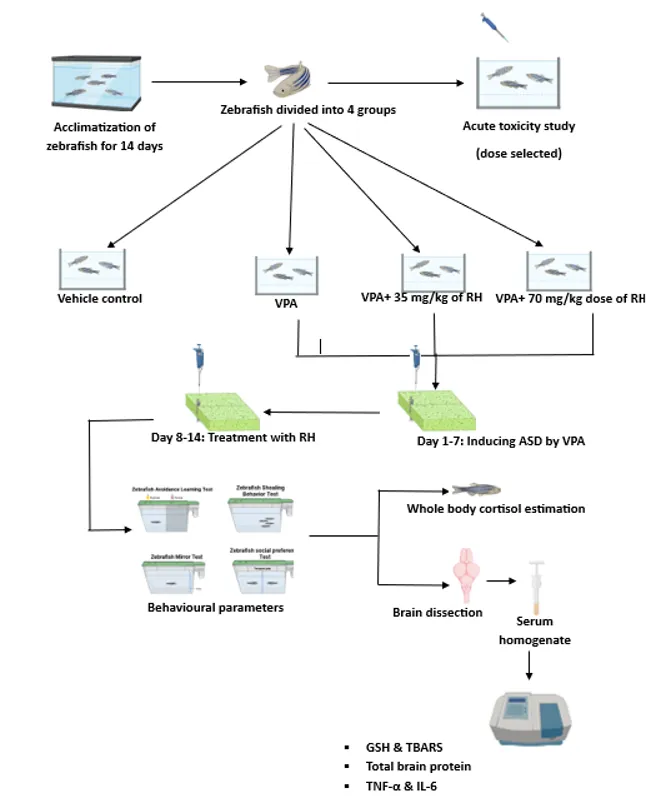
Fig 3: Schematic representation of experimental methodology for valproic acid (VPA)-induced autism spectrum disorder (ASD) in zebrafish and therapeutic evaluation of rutin hydrate treatment
Behavioural Assessment
Behavioral assessments were conducted to evaluate autism spectrum disorder-like phenotypes following VPA administration.
Light/Dark Preference Test
The light/dark preference test was performed using a rectangular tank divided into illuminated and dark compartments without a physical partition, allowing unrestricted movement between compartments. Individual zebrafish were observed for 5 min. The total time spent in each compartment and the total number of transitions between compartments were recorded as indices of anxiety-like behavior and altered exploratory responses.[19]
Shoaling Behavior
Shoaling behavior was assessed in a rectangular tank by placing fish in groups under identical environmental conditions. Following an observation period of 20 min, the average inter-individual distance and nearest-neighbor distance among fish were measured to evaluate social cohesion and group behavior.[20]
Mirror Attack Test
Aggressive behavior was evaluated using the mirror attack test. Individual zebrafish were introduced into the experimental tank and allowed to habituate for 60 s before a mirror was placed adjacent to one side of the tank. Fish behavior was recorded for 5 min. The latency to approach the mirror, frequency of aggressive interactions, and time spent within 1 cm of the mirror were recorded.[20]
Social Contact Test
Social interaction was assessed using the social contact test. Following acclimatization, visual barriers between adjacent tanks were removed, allowing fish to interact visually with conspecifics. Behavioral activity was recorded for 6 min. The average contact time, contact duration, and time spent in the non-social zone were measured as indicators of social preference.[20]
Whole-Body Cortisol Estimation
Whole-body cortisol levels were determined using a commercially available enzyme-linked immunosorbent assay (ELISA) kit according to the manufacturer's instructions. Cortisol estimation was performed to assess the physiological stress response associated with VPA-induced autism-like behavior and the therapeutic effects of rutin hydrate.[21]
Brain Tissue Collection
Following completion of behavioral studies, zebrafish were anesthetized with 0.2% tricaine and euthanized in ice-cold water. Brains were carefully dissected, transferred to microcentrifuge tubes, and stored at −80°C until analysis. Brain tissues were homogenized in phosphate-buffered saline (PBS), and the homogenates were centrifuged at 4000 rpm for 30 min at 4°C. The resulting supernatants were collected and used for biochemical analyses. [22]
Estimation of Oxidative Stress Markers
Reduced Glutathione (GSH)
Reduced glutathione levels were determined using the DTNB (5,5′-dithiobis-(2-nitrobenzoic acid)) colorimetric method. Brain homogenate supernatant was mixed with 10% trichloroacetic acid, centrifuged, and reacted with disodium hydrogen phosphate and DTNB. The absorbance of the resulting yellow-colored complex was measured spectrophotometrically at 412 nm, and GSH concentration was calculated using a standard calibration curve.[23]
Thiobarbituric Acid Reactive Substances (TBARS)
Lipid peroxidation was estimated by measuring TBARS. Brain homogenate supernatant was mixed with sodium dodecyl sulfate, acetic acid, and thiobarbituric acid, followed by incubation at 95°C for 1 h. After extraction with an n-butanol:pyridine mixture and centrifugation, the absorbance of the organic phase was measured at 532 nm using a spectrophotometer.[24,25]
Estimation of Total Brain Protein
Total brain protein was determined using the Bradford assay. Brain homogenate samples and bovine serum albumin standards were incubated with Bradford reagent, and absorbance was measured at 595 nm using a microplate reader. Protein concentrations were calculated from the standard calibration curve and expressed relative to tissue protein content.[24]
Estimation of Inflammatory Cytokines
The concentrations of tumor necrosis factor-α (TNF-α) and interleukin-6 (IL-6) in brain homogenates were quantified using commercially available ELISA kits according to the manufacturers' instructions. These cytokines were measured to assess the extent of neuroinflammation associated with VPA-induced autism-like pathology and the effects of rutin hydrate treatment.[25]
Statistical Analysis
Data are presented as mean ± SEM (n = 10). Statistical analysis was performed using one-way analysis of variance (ANOVA) followed by Dunnett's multiple comparison test. Differences were considered statistically significant at p < 0.05.
RESULTS
Acute Toxicity Study
The acute toxicity study conducted according to OECD Guideline 203 demonstrated that rutin hydrate was well tolerated at lower doses, with no mortality observed in the control or low-dose groups. Mortality was observed at 400 mg/kg, while 50% mortality occurred at 175 mg/kg, establishing the median lethal dose (LD₅₀) as 175 mg/kg. Based on the LD₅₀ value, therapeutic doses of 35 mg/kg and 70 mg/kg were selected for subsequent pharmacological evaluation as shown in table 2.
Table 2: Acute toxicity assessment results of treatment groups
|
Group |
Treatment |
24 h |
48 h |
72 h |
96 h |
|
1 |
Control |
NB, NM |
NB, NM |
NB, NM |
NB, NM |
|
2 |
50 mg/kg |
NB, NM |
NB, NM |
Excessive movement |
NB, NM |
|
3 |
100 mg/kg |
NB, NM |
Excessive gills movement |
Settling at bottom (1 died) |
Settling at bottom |
|
4 |
175 mg/kg |
NM |
Excessive movement (1 died) |
Settling at bottom (2 died) |
Settling at bottom |
|
5 |
200 mg/kg |
Excessive movement |
Excessive movement |
2 fish died |
3 fish died |
|
6 |
400 mg/kg |
Hyperactivity 3 fish died |
Hyperactivity 4 fish died |
- |
- |
NB- Normal behavior, NM- No mortality
Effect of Rutin Hydrate on Light/Dark Preference Behaviour
In the vehicle control group, zebrafish exhibited a strong preference for the dark zone, spending 214 ± 12.01 sec in the dark compared to 86 ± 12.01 sec in the light, with a significantly higher number of crossings (42.67 ± 2.906, p < 0.001). Valproic acid (100 mg/kg) treatment induced abnormal behavior, as the zebrafish showed a preference for the light zone (171.3 ± 8.452 sec in light vs. 128.7 ± 8.452 sec in dark) with markedly reduced crossings (14.67 ± 1.856). Co-treatment with valproic acid (100 mg/kg) and rutin hydrate (35 mg/kg) partially reversed this behavior, as zebrafish spent 159 ± 12.50 sec in dark and 141 ± 12.50 sec in light, along with an increase in crossings (24.33 ± 1.453, p < 0.001). Interestingly, 70 mg/kg rutin hydrate produced a more prominent effect, restoring dark preference (174.7 ± 17.03 sec in dark vs. 125.3 ± 17.03 sec in light) and significantly increasing the number of crossings (37.67 ± 2.028, p < 0.001) compared to the valproic acid group as mentioned in table 3 & fig.4,5,6.
Table 3: Light/dark preference test results of zebrafish across experimental groups.
|
Group |
Treatment protocol |
Preference |
Time spent in light zone |
Time spent in dark zone |
No. of crossings |
|
Group-1 |
Vehicle control |
Dark |
86±12.01** |
214±12.01** |
42.67±2.906*** |
|
Group-2 |
Valproic acid (100 mg/kg) |
Light |
171.3±8.452 |
128.7±8.452 |
14.67±1.856 |
|
Group-3 |
Valproic acid (100 mg/kg) + Rutin hydrate (35 mg/kg) |
Dark |
141±12.50 |
159±12.50 |
24.33±1.453* |
|
Group-4 |
Valproic acid (100 mg/kg) +Rutin hydrate (70 mg/kg) |
Dark |
125.3±17.03 |
174.7±17.03 |
37.67±2.028*** |
All data were expressed as the mean ± SEM (n=10). Data were analysed with One-way analysis of variance (ANOVA) followed by Dunnett’s t-test for multiple comparisons. A value of ** p <0.01, ** p<0.01 & ***p<0.001 were considered statistically significant compared to positive control group.
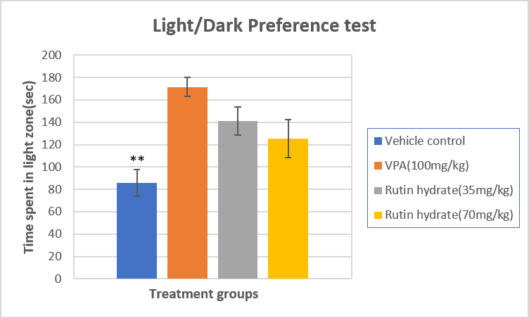
Fig. 4: Light/Dark preference test- Time spent in light zone
All data were expressed as the mean ± SEM (n=10). Data were analysed with One-way analysis of variance (ANOVA) followed by Dunnett’s t-test for multiple comparisons. A value of ** p<0.01 were considered statistically significant compared to positive control group.
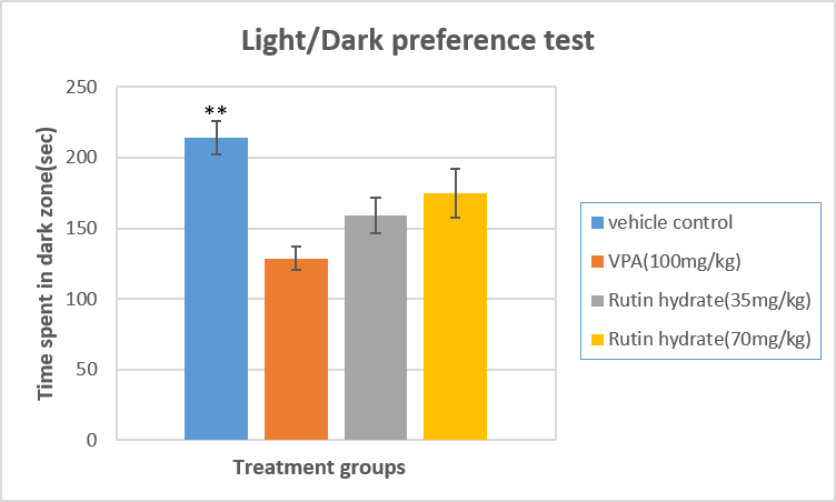
Fig. 5: Light/Dark preference test- Time spent in dark zone
All data were expressed as the mean ± SEM (n=10). Data were analysed with One-way analysis of variance (ANOVA) followed by Dunnett’s t-test for multiple comparisons. A value of ** p<0.01 were considered statistically significant compared to positive control group.
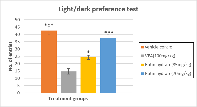
Fig. 6: Light/Dark preference test- No. of entries
All data were expressed as the mean ± SEM (n=10). Data were analysed with One-way analysis of variance (ANOVA) followed by Dunnett’s t-test for multiple comparisons. A value of *p<0.05, *** p <0.001 were considered statistically significant compared to positive control group.
Effect of Rutin Hydrate on Shoaling behaviour:
In the vehicle control group, zebrafish displayed strong shoaling behavior with a lower inter-individual distance (IID) of 1.833 ± 0.05774 cm and a nearest neighbour distance (NND) of 0.8 ± 0.1202 cm. Valproic acid (100 mg/kg) treatment significantly disrupted shoaling, as indicated by an increased IID of 4.667 ± 0.5774 cm and NND of 2.3 ± 0.2028 cm. Co-treatment with rutin hydrate (35 mg/kg) improved shoaling tendency, reducing the IID to 3 ± 0.5774 cm and NND to 1.6 ± 0.1155 cm compared to the VPA group. A dose of 70 mg/kg produced a more pronounced effect, bringing the IID closer to control values (2.233 ± 0.5774 cm) and restoring the NND to 1 ± 0.08819 cm, indicating significant reversal of VPA-induced shoaling deficits as mentioned in table 4 & fig 7,8.
Table 4: Shoaling behavior results of zebrafish across experimental groups.
|
Group |
Treatment protocol |
Inter-individual distance-IID (cm) |
Nearest neighbour distance-NND (cm) |
|
Group-1 |
Vehicle control |
1.833±0.05774*** |
0.8±0.1202*** |
|
Group-2 |
Valproic acid (100 mg/kg) |
4.667±0.5774 |
2.3±0.2028 |
|
Group-3 |
Valproic acid (100 mg/kg) + Rutin hydrate (35 mg/kg) |
3±0.5774*** |
1.6±0.1155*** |
|
Group-4 |
Valproic acid (100 mg/kg) +Rutin hydrate (70 mg/kg) |
2.233±0.5774*** |
1±0.08819*** |
All data were expressed as the mean ± SEM (n=10). Data were analysed with One-way analysis of variance (ANOVA) followed by Dunnett’s t-test for multiple comparisons. A value of *** p <0.001& ***p<0.001 were considered statistically significant compared to positive control group.

Fig. 7: Shoaling behaviour test- Inter individual distance
All data were expressed as the mean ± SEM (n=10). Data were analysed with One-way analysis of variance (ANOVA) followed by Dunnett’s t-test for multiple comparisons. A value of *** p <0.001 were considered statistically significant compared to positive control group.
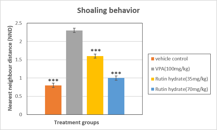
Fig. 8: Shoaling behaviour test- Nearest neighbour distance
All data were expressed as the mean ± SEM (n=10). Data were analysed with One-way analysis of variance (ANOVA) followed by Dunnett’s t-test for multiple comparisons. A value of *** p <0.001 were considered statistically significant compared to positive control group.
Effect of Rutin Hydrate on Mirror attack test:
In the vehicle control group, zebrafish showed normal aggressive behavior with a short latency to first attack (21 ± 2.082 sec), a higher number of attacks (12 ± 1.155), and longer time spent near the mirror (65 ± 2.887 sec). Valproic acid (100 mg/kg) markedly suppressed aggression, as evidenced by a prolonged attack latency (81.67 ± 4.410 sec), a drastic reduction in the number of attacks (2 ± 0.5774), and reduced mirror interaction time (20 ± 1.155 sec). Co-treatment with rutin hydrate (35 mg/kg) partially restored aggression, decreasing attack latency to 50 ± 2.887 sec, increasing the number of attacks to 7 ± 0.5774, and extending mirror interaction to 41 ± 2.082 sec. At a dose of 70 mg/kg rutin hydrate, zebrafish exhibited near-normal aggressive behavior, with a latency of 28.33 ± 0.8819 sec, 11 ± 0.5774 attacks, and 57.67 ± 1.453 sec spent near the mirror, suggesting a strong reversal of VPA-induced behavioral deficits as mentioned in table 5 & fig. 9,10,11.
Table 5: Mirror attack results of zebrafish across experimental groups.
|
Group |
Treatment protocol |
Latency to first attack (sec) |
No. of attacks |
Time spent near the mirror (sec) |
|
Group-1 |
Vehicle control |
21±2.082*** |
12±1.155*** |
65±2.887*** |
|
Group-2 |
Valproic acid (100 mg/kg) |
81.67±4.410 |
2±0.5774 |
20±1.155 |
|
Group-3 |
Valproic acid (100 mg/kg) + Rutin hydrate (35 mg/kg) |
50±2.887*** |
7±0.5774** |
41±2.082*** |
|
Group-4 |
Valproic acid (100 mg/kg) +Rutin hydrate (70 mg/kg) |
28.33±0.8819*** |
11±0.5774*** |
57.67±1.453*** |
All data were expressed as the mean ± SEM (n=10). Data were analysed with One-way analysis of variance (ANOVA) followed by Dunnett’s t-test for multiple comparisons. A value of *** p <0.001, ***p<0.001 & ***p<0.001 were considered statistically significant compared to positive control group.
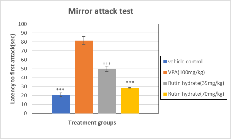
Fig. 9: Mirror attack test- Latency to first attack
All data were expressed as the mean ± SEM (n=10). Data were analysed with One-way analysis of variance (ANOVA) followed by Dunnett’s t-test for multiple comparisons. A value of *** p <0.001 were considered statistically significant compared to positive control group.

Fig. 10: Mirror attack test- No. of attacks
All data were expressed as the mean ± SEM (n=10). Data were analysed with One-way analysis of variance (ANOVA) followed by Dunnett’s t-test for multiple comparisons. A value of **p<0.01& *** p <0.001 were considered statistically significant compared to positive control group.
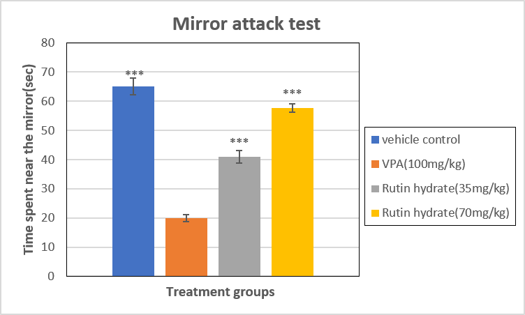
Fig. 11: Mirror attack test- Time spent near the mirror
All data were expressed as the mean ± SEM (n=10). Data were analysed with One-way analysis of variance (ANOVA) followed by Dunnett’s t-test for multiple comparisons. A value of *** p <0.001 were considered statistically significant compared to positive control group.
Effect of Rutin Hydrate on Social contact test:
In the vehicle control group, zebrafish spent significantly more time in the social zone (180 ± 2.887 sec) with a higher number of entries (12 ± 0.5774) and less time in the non-social zone (120 ± 2.887 sec). Valproic acid (100 mg/kg) treatment markedly impaired social interaction, reducing the time spent in the social zone to 69 ± 2.082 sec, with fewer entries (3.667 ± 0.3333) and increased time in the non-social zone (228.3 ± 4.410 sec). Co-treatment with rutin hydrate (35 mg/kg) improved sociability, as zebrafish spent 117.7 ± 1.453 sec in the social zone with 7.667 ± 0.3333 entries, while reducing non-social zone preference (182.3 ± 1.453 sec). At a dose of 70 mg/kg rutin hydrate further restored social preference, as indicated by increased social zone duration (160 ± 1.155 sec) and entries (11 ± 0.5774) along with a marked reduction in non-social zone time (140 ± 1.155 sec), suggesting reversal of VPA-induced social deficits as mentioned in table 6 & fig.12,13,14.
Table 6: Social contact test results of zebrafish across experimental groups.
|
Group |
Treatment protocol |
Time spent in social zone (sec) |
No. of entries into social zone |
Time spent in non-social zone (sec) |
|
Group-1 |
Vehicle control |
180±2.887*** |
12±0.5774*** |
120±2.887*** |
|
Group-2 |
Valproic acid (100 mg/kg) |
69±2.082 |
3.667±0.3333 |
228.3±4.410 |
|
Group-3 |
Valproic acid (100 mg/kg) + Rutin hydrate (35 mg/kg) |
117.7±1.453*** |
7.667±0.3333*** |
182.3±1.453*** |
|
Group-4 |
Valproic acid (100 mg/kg) +Rutin hydrate (70 mg/kg) |
160±1.155*** |
11±0.5774*** |
140±1.155*** |
All data were expressed as the mean ± SEM (n=10). Data were analysed with One-way analysis of variance (ANOVA) followed by Dunnett’s t-test for multiple comparisons. A value of *** p <0.001, ***p<0.001 & ***p<0.001 were considered statistically significant compared to positive control group.
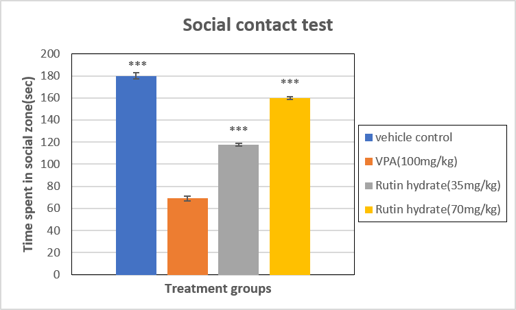
Fig. 12: Social attack test- Time spent in social zone
All data were expressed as the mean ± SEM (n=10). Data were analysed with One-way analysis of variance (ANOVA) followed by Dunnett’s t-test for multiple comparisons. A value of *** p <0.001 were considered statistically significant compared to positive control group.
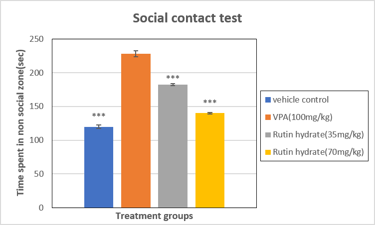
Fig. 13: Social attack test- Time spent in non- social zone
All data were expressed as the mean ± SEM (n=10). Data were analysed with One-way analysis of variance (ANOVA) followed by Dunnett’s t-test for multiple comparisons. A value of *** p <0.001 were considered statistically significant compared to positive control group.
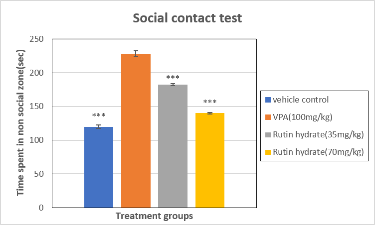
Fig. 14: Social attack test- No. of entries into social zone
All data were expressed as the mean ± SEM (n=10). Data were analysed with One-way analysis of variance (ANOVA) followed by Dunnett’s t-test for multiple comparisons. A value of *** p <0.001 were considered statistically significant compared to positive control group.
Whole body cortisol Estimation:
In the vehicle control group, zebrafish exhibited a baseline cortisol level of 0.5856 ± 0.1735 µg/dL. Valproic acid (100 mg/kg) treatment significantly elevated cortisol levels to 1.785 ± 0.04294 µg/dL, indicating heightened stress. Co-treatment with rutin hydrate at 35 mg/kg reduced cortisol to 1.057 ± 0.02973 µg/dL, while the dose of 70 mg/kg rutin hydrate further attenuated cortisol levels to 0.618 ± 0.03705 ng/g tissue, demonstrating a dose-dependent protective effect against VPA-induced stress as mentioned in table 7 & fig.15.
Table 7: Effect of treatments on whole-body cortisol levels in zebrafish
|
Group |
Treatment protocol |
Cortisol level (mg/dL) |
|
Group-1 |
Vehicle control |
0.5856±0.1735** |
|
Group-2 |
Valproic acid (100 mg/kg) |
1.785±0.04294 |
|
Group-3 |
Valproic acid (100 mg/kg) + Rutin hydrate (35 mg/kg) |
1.057±0.02973* |
|
Group-4 |
Valproic acid (100 mg/kg) +Rutin hydrate (70 mg/kg) |
0.618±0.03705*** |
All data were expressed as the mean ± SEM (n=10). Data were analysed with One-way analysis of variance (ANOVA) followed by Dunnett’s t-test for multiple comparisons. A value of * p <0.05, **p<0.01 & ***p<0.001 were considered statistically significant compared to positive control group.
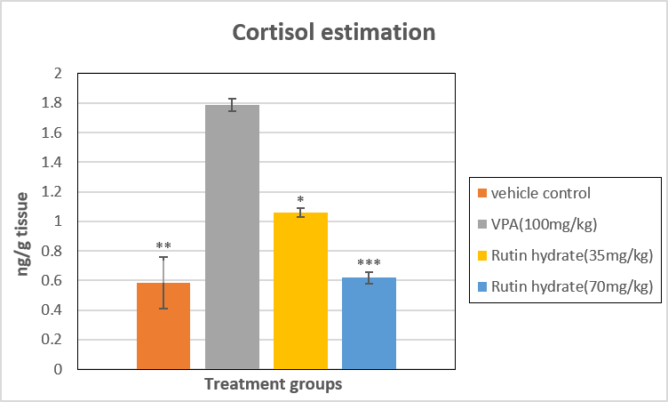
Fig. 15: Estimation of whole-body cortisol
All data were expressed as the mean ± SEM (n=10). Data were analysed with One-way analysis of variance (ANOVA) followed by Dunnett’s t-test for multiple comparisons. A value of * p <0.05, **p<0.01 & ***p<0.001 were considered statistically significant compared to positive control group.
Estimation of GSH:
In the vehicle control group, zebrafish exhibited normal glutathione levels of 1.624 ± 0.0059 nmol/mg protein. Valproic acid (100 mg/kg) administration significantly reduced glutathione to 0.8211 ± 0.0047 nmol/mg protein, indicating oxidative stress. Co-treatment with rutin hydrate at 35 mg/kg partially restored glutathione levels to 1.108 ± 0.0064 nmol/mg protein, while the dose 70 mg/kg of rutin hydrate further improved glutathione to 1.354 ± 0.0047 nmol/mg protein, approaching control values and demonstrating a dose-dependent protective effect as mentioned in table 8 & fig.16.
Table 8: Effect of treatments on whole-brain reduced glutathione (GSH) levels in zebrafish
|
Group |
Treatment protocol |
Glutathione levels (nmol/mg protein) |
|
Group-1 |
Vehicle control |
1.624±0.005894*** |
|
Group-2 |
Valproic acid (100 mg/kg) |
0.8211±0.004739 |
|
Group-3 |
Valproic acid (100 mg/kg) + Rutin hydrate (35 mg/kg) |
1.108±0.006376*** |
|
Group-4 |
Valproic acid (100 mg/kg) +Rutin hydrate (70 mg/kg) |
1.354±0.004724*** |
All data were expressed as the mean ± SEM (n=10). Data were analysed with One-way analysis of variance (ANOVA) followed by Dunnett’s t-test for multiple comparisons. A value of ***p<0.001 were considered statistically significant compared to positive control group.
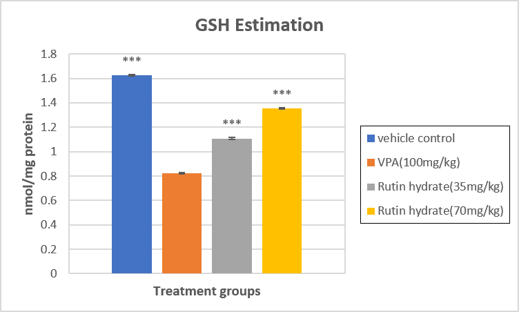
Fig. 16: Estimation of reduced GSH level
All data were expressed as the mean ± SEM (n=10). Data were analysed with One-way analysis of variance (ANOVA) followed by Dunnett’s t-test for multiple comparisons. A value of *** p <0.001 were considered statistically significant compared to positive control group.
Estimation of TBARS:
TBARS levels in the vehicle control group were 0.666 ± 0.0040 nmol/mg protein. Administration of valproic acid (100 mg/kg) significantly elevated TBARS levels to 1.215 ± 0.0156 nmol/mg protein, indicating enhanced lipid peroxidation and oxidative stress. Co-treatment with rutin hydrate (35 mg/kg) reduced the TBARS concentration to 0.936 ± 0.0113 nmol/mg protein, while the higher dose of rutin hydrate (70 mg/kg) further decreased the levels to 0.722 ± 0.0085 nmol/mg protein, approaching near-control values as mentioned in table 9 & fig.17.
Table 9: Effect of treatments on whole-brain TBARS levels in zebrafish
|
Group |
Treatment protocol |
TBARS levels (nmol/mg protein) |
|
Group-1 |
Vehicle control |
0.666±0.004041*** |
|
Group-2 |
Valproic acid (100 mg/kg) |
1.215±0.01562 |
|
Group-3 |
Valproic acid (100 mg/kg) + Rutin hydrate (35 mg/kg) |
0.9357±0.01129*** |
|
Group-4 |
Valproic acid (100 mg/kg) +Rutin hydrate (70 mg/kg) |
0.7217±0.008452*** |
All data were expressed as the mean ± SEM (n=10). Data were analysed with One-way analysis of variance (ANOVA) followed by Dunnett’s t-test for multiple comparisons. A value of ***p<0.001 were considered statistically significant compared to positive control group.
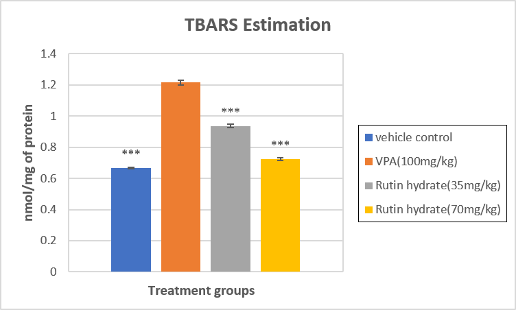
Fig. 17: Estimation of TBARS level
All data were expressed as the mean ± SEM (n=10). Data were analysed with One-way analysis of variance (ANOVA) followed by Dunnett’s t-test for multiple comparisons. A value of *** p <0.001 were considered statistically significant compared to positive control group.
Estimation of TNF-α
The TNF-α levels in the zebrafish brain were significantly elevated in the valproic acid (100 mg/kg) group (22.85 ± 1.075 pg/mg protein) compared to the vehicle control (10.36 ± 0.4068 pg/mg protein). Co-treatment with rutin hydrate at 35 mg/kg reduced TNF-α levels to 17.54 ± 0.6411 pg/mg protein, while rutin hydrate at 70 mg/kg further attenuated the increase, bringing TNF-α levels down to 13.79 ± 0.3605 pg/mg protein, approaching near-control values as mentioned in table 10 & fig.18.
Table 10: Effect of treatments on whole-brain TNF-α levels in zebrafish
|
Group |
Treatment protocol |
TNF-α levels (pg/mg protein) |
|
Group-1 |
Vehicle control |
10.36±0.4068*** |
|
Group-2 |
Valproic acid (100 mg/kg) |
22.85±1.075 |
|
Group-3 |
Valproic acid (100 mg/kg) + Rutin hydrate (35 mg/kg) |
17.54±0.6411** |
|
Group-4 |
Valproic acid (100 mg/kg) +Rutin hydrate (70 mg/kg) |
13.79±0.3605*** |
All data were expressed as the mean ± SEM (n=10). Data were analysed with One-way analysis of variance (ANOVA) followed by Dunnett’s t-test for multiple comparisons. A value of **p<0.01 & ***p<0.001 were considered statistically significant compared to positive control group.
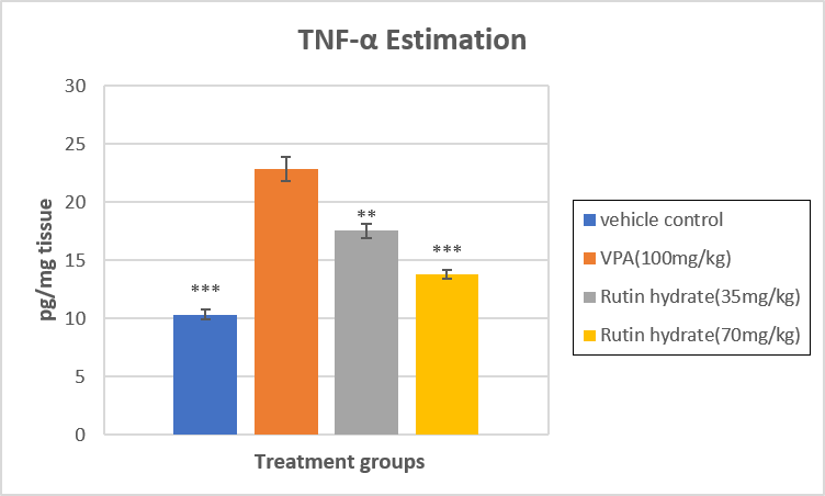
Fig. 18: Estimation of TNF-α level
All data were expressed as the mean ± SEM (n=10). Data were analysed with One-way analysis of variance (ANOVA) followed by Dunnett’s t-test for multiple comparisons. A value of **p<0.01 &*** p <0.001 were considered statistically significant compared to positive control group.
Estimation of IL-6
The ELISA analysis of brain homogenates revealed a significant elevation in IL-6 levels in the valproic acid (VPA)–treated group (12.15 ± 0.3525 pg/mg protein) compared with the vehicle control (3.768 ± 0.1328 pg/mg protein). Co-treatment with rutin hydrate at 35 mg/kg markedly reduced IL-6 levels to 8.481 ± 0.2113 pg/mg protein, while the dose of 70 mg/kg rutin hydrate further attenuated IL-6 to 5.084 ± 0.1753 pg/mg protein, approaching near-control values as mentioned in table 11 & fig.19.
Table 11: Effect of treatments on whole-brain IL-6 levels in zebrafish
|
Group |
Treatment protocol |
IL-6 levels (pg/mg protein) |
|
Group-1 |
Vehicle control |
3.768±0.1328*** |
|
Group-2 |
Valproic acid (100 mg/kg) |
12.15±0.3525 |
|
Group-3 |
Valproic acid (100 mg/kg) + Rutin hydrate (35 mg/kg) |
8.481±0.2113*** |
|
Group-4 |
Valproic acid (100 mg/kg) +Rutin hydrate (70 mg/kg) |
5.084±0.1753*** |
All data were expressed as the mean ± SEM (n=10). Data were analysed with One-way analysis of variance (ANOVA) followed by Dunnett’s t-test for multiple comparisons. A value of ***p<0.001 were considered statistically significant compared to positive control group.
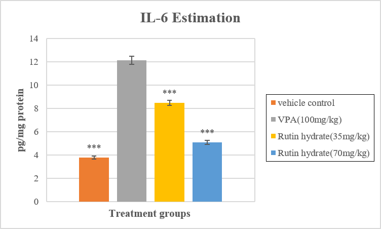
Fig. 19: Estimation of IL-6 level
All data were expressed as the mean ± SEM (n=10). Data were analysed with One-way analysis of variance (ANOVA) followed by Dunnett’s t-test for multiple comparisons. A value of *** p <0.001 were considered statistically significant compared to positive control group.
Estimation of Total brain protein
Total brain protein levels were measured across all groups. The vehicle control group showed a level of 1.279 ± 0.0145 mg/mL. Administration of valproic acid (100 mg/kg) significantly increased protein levels to 1.914 ± 0.02318 mg/mL. Co-treatment with Rutin hydrate at 35 mg/kg reduced the protein level to 1.455 ± 0.02714 mg/mL, whereas a dose of 70 mg/kg Rutin hydrate restored the protein level close to control values (1.282 ± 0.0382 mg/mL).
Table 12: Effect of treatments on total protein levels in zebrafish
|
Group |
Treatment protocol |
Total brain protein levels (mg/mL) |
|
Group-1 |
Vehicle control |
1.279±0.0145*** |
|
Group-2 |
Valproic acid (100 mg/kg) |
1.914±0.02318 |
|
Group-3 |
Valproic acid (100 mg/kg) + Rutin hydrate (35 mg/kg) |
1.455±0.02714*** |
|
Group-4 |
Valproic acid (100 mg/kg) +Rutin hydrate (70 mg/kg) |
1.282±0.0382*** |
All data were expressed as the mean ± SEM (n=10). Data were analysed with One-way analysis of variance (ANOVA) followed by Dunnett’s t-test for multiple comparisons. A value of ***p<0.001 were considered statistically significant compared to positive control group.
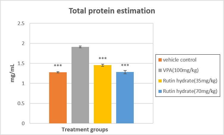
Fig. 20: Estimation of total protein
All data were expressed as the mean ± SEM (n=10). Data were analysed with One-way analysis of variance (ANOVA) followed by Dunnett’s t-test for multiple comparisons. A value of *** p <0.001 were considered statistically significant compared to positive control group.
DISCUSSION
Autism spectrum disorder (ASD) is a multifactorial neurodevelopmental disorder characterized by deficits in social interaction, communication impairments, repetitive behaviors, oxidative stress, neuroinflammation, and dysregulated stress responses. The present study demonstrated that valproic acid (VPA) administration successfully induced ASD-like behavioral and biochemical alterations in adult zebrafish, while treatment with rutin hydrate significantly attenuated these abnormalities. These findings support the therapeutic potential of rutin hydrate in ameliorating ASD-like pathology through modulation of oxidative stress, neuroinflammatory responses, and physiological stress.
Behavioral assessments demonstrated that VPA exposure significantly altered anxiety-related behavior, impaired shoaling and social interaction, and reduced aggressive responses in zebrafish. These behavioral impairments are characteristic of ASD-like phenotypes and are consistent with previous reports validating the VPA-induced zebrafish model. Wang et al. (2024) reported reduced locomotor activity,[27] impaired social interaction, and repetitive swimming behavior following VPA exposure, whereas Hodin et al. (2023) observed impaired locomotor responses and reduced survival following embryonic VPA exposure.[28] The restoration of behavioral performance following rutin hydrate administration, particularly at 70 mg/kg, indicates that rutin effectively ameliorates VPA-induced behavioral deficits. Similar improvements in locomotor activity and neurobehavioral function following rutin treatment have been reported in zebrafish by Hu et al. (2023) and Mahati et al. (2025), suggesting that the neuroprotective actions of rutin extend across different models of neurological dysfunction.[29,30]
Whole-body cortisol analysis further demonstrated significant activation of the hypothalamic–pituitary–interrenal (HPI) axis following VPA administration. Elevated cortisol levels have been associated with increased physiological stress and behavioral dysfunction in ASD models. The marked reduction in cortisol observed after rutin hydrate treatment indicates normalization of stress responses and supports its anxiolytic properties. Comparable findings have been reported by Dwivedi et al. (2019), who demonstrated enhanced stress responses following VPA exposure, while Wang et al. (2024) showed that pharmacological intervention reduced stress-associated biomarkers and improved behavioral outcomes.[27,31] The present findings therefore suggest that regulation of the stress axis may contribute to the behavioral improvements produced by rutin hydrate.
Oxidative stress is considered one of the principal mechanisms contributing to ASD pathogenesis. Consistent with previous studies, VPA administration significantly reduced glutathione (GSH) levels and increased lipid peroxidation, as indicated by elevated TBARS concentrations. These findings demonstrate impaired antioxidant defense and excessive generation of reactive oxygen species following VPA exposure. Rutin hydrate significantly restored GSH levels while reducing TBARS, indicating effective attenuation of oxidative stress. These observations are in agreement with Abdel Aleem et al. (2017), who demonstrated that rutin enhanced endogenous antioxidant defenses and protected against oxidative neuronal injury.[14] The antioxidant activity of rutin is likely attributable to its ability to scavenge free radicals, preserve cellular antioxidant capacity, and reduce oxidative damage within neural tissues.
Neuroinflammation represents another critical mechanism implicated in ASD. In the present study, VPA exposure significantly increased brain concentrations of the pro-inflammatory cytokines TNF-α and IL-6, indicating activation of inflammatory pathways. Treatment with rutin hydrate significantly reduced both cytokines, suggesting effective suppression of neuroinflammation. Similar anti-inflammatory effects of rutin have been reported by Hu et al. (2023), who demonstrated reduced inflammatory cytokine production following rutin administration in zebrafish exposed to inflammatory stimuli.[30] The reduction in TNF-α and IL-6 observed in the present study supports the hypothesis that inhibition of neuroinflammatory signaling contributes substantially to the neuroprotective effects of rutin hydrate.
Alterations in total brain protein observed following VPA exposure may reflect disturbed protein synthesis, neuronal remodeling, or impaired proteostasis associated with ASD pathology. Administration of rutin hydrate restored total brain protein levels toward those observed in the control group, suggesting preservation of neuronal homeostasis. Comparable findings reported by Abdel Aleem et al. (2017) further support the role of rutin in maintaining neuronal integrity through modulation of oxidative and inflammatory pathways.[14]
Collectively, the behavioral and biochemical findings indicate that rutin hydrate exerts a multifaceted neuroprotective effect against VPA-induced ASD-like alterations in zebrafish. Improvement in social behavior, anxiety-related responses, aggression, oxidative stress, inflammatory cytokines, cortisol levels, and protein homeostasis suggests that rutin acts through multiple interconnected mechanisms rather than targeting a single pathological pathway. Since oxidative stress, neuroinflammation, and dysregulated stress responses are closely linked in ASD pathogenesis, simultaneous modulation of these processes may explain the broad therapeutic efficacy observed in the present study.
Despite these promising findings, certain limitations should be considered. Although zebrafish provide a well-established and translationally relevant model for investigating ASD-associated behavioral and biochemical alterations, species-specific differences limit direct extrapolation to humans. Furthermore, the molecular signaling pathways underlying the protective effects of rutin hydrate were not investigated. Future studies should evaluate gene and protein expression associated with oxidative stress, neuroinflammation, synaptic plasticity, and neurotransmitter signaling. Validation of these findings in mammalian models and clinical studies will be necessary to establish the translational potential of rutin hydrate as a therapeutic intervention for ASD.
CONCLUSION
The present study demonstrated that rutin hydrate effectively attenuated valproic acid-induced autism spectrum disorder-like behavioral and biochemical abnormalities in adult zebrafish. Valproic acid administration produced characteristic ASD-associated alterations, including impaired social interaction, anxiety-related behavior, reduced aggression, elevated cortisol levels, oxidative stress, increased neuroinflammatory cytokines, and altered total brain protein. These findings further validate the VPA-induced zebrafish model as an effective experimental model for investigating ASD pathophysiology.
Treatment with rutin hydrate, particularly at a dose of 70 mg/kg, significantly improved behavioral performance and restored biochemical homeostasis. The observed neuroprotective effects were associated with normalization of cortisol levels, restoration of antioxidant defense through increased glutathione, reduction of lipid peroxidation, suppression of the pro-inflammatory cytokines TNF-α and IL-6, and maintenance of protein homeostasis. These findings indicate that rutin hydrate exerts therapeutic effects through complementary antioxidant, anti-inflammatory, and stress-regulating mechanisms.
Overall, the present findings identify rutin hydrate as a promising natural therapeutic candidate for the management of ASD. Further investigations involving mammalian models, mechanistic studies, pharmacokinetic evaluation, and well-designed clinical trials are warranted to confirm its efficacy, establish long-term safety, and determine its translational applicability in the treatment of autism spectrum disorder.
REFERENCES


P Aswathy, Surendra Vada, Harshitha G, K Keerthana, Manjunatha PM, Rutin Hydrate Attenuates Valproic Acid-Induced Autism Spectrum Disorder-Like Behavioral and Biochemical Alterations in Adult Zebrafish, Int. J. of Pharm. Sci., 2026, Vol 4, Issue 7, 5988-6012. https://doi.org/10.5281/zenodo.21713559
 10.5281/zenodo.21713559