

We use cookies to ensure our website works properly and to personalise your experience. Cookies policy
1 Pharm D Interns, Ezhuthachan College of Pharmaceutical Sciences, Marayamuttom, Thiruvananthapuram, Kerala, India.
2 Assistant Professor, Department of Pharmacy Practice, Ezhuthachan College of Pharmaceutical Sciences, Marayamuttom, Thiruvananthapuram, Kerala, India.
3 Consultant Neurologist, NIMS Medicity (Noorul Islam Multi-Speciality Hospital) Thiruvananthapuram, Kerala, India.
4 Principal/HOD, Department of Pharmacy Practice, Ezhuthachan College of Pharmaceutical Sciences, Marayamuttom, Thiruvananthapuram, Kerala, India.
Neuromyelitis optica spectrum disorder (NMOSD) is a rare autoimmune inflammatory disorder of the central nervous system that primarily affects the optic nerves and spinal cord. Although optic neuritis and longitudinally extensive transverse myelitis (LETM) are the classical manifestations, some patients initially present with isolated spinal cord involvement, making early diagnosis challenging. We report the case of a 46-year-old woman who presented with progressive bilateral lower limb pain and painful muscle spasms for six weeks. Initial investigations excluded vascular and peripheral neurological disorders. Further evaluation suggested LETM, and serological testing showed positive aquaporin-4 immunoglobulin G (AQP4-IgG) antibodies with negative myelin oligodendrocyte glycoprotein (MOG-IgG) antibodies, confirming AQP4-IgG-positive NMOSD. The patient was treated with high-dose intravenous methylprednisolone followed by supportive care and rehabilitation, resulting in gradual clinical improvement. This case highlights the importance of considering NMOSD in patients presenting with isolated LETM, even in the absence of optic neuritis. Early antibody testing enabled prompt diagnosis and timely initiation of appropriate immunotherapy, contributing to a favourable clinical outcome.
Definition and Epidemiology
Neuromyelitis optica spectrum disorder (NMOSD) is a rare autoimmune inflammatory disorder of the central nervous system that predominantly affects the optic nerves and spinal cord, although lesions may also involve the brainstem, area postrema, and other regions of the brain. Once regarded as a variant of multiple sclerosis, NMOSD is now recognized as a distinct disease entity because of its characteristic immunological, clinical, and radiological features. The disorder can occur at any age but is most frequently diagnosed in adults and shows a marked female predominance. Although NMOSD is relatively uncommon, its clinical impact is considerable because recurrent disease relapses may result in irreversible neurological deficits, emphasizing the importance of early diagnosis and appropriate treatment. (1)
Pathophysiology
The pathogenesis of NMOSD is primarily mediated by aquaporin-4 immunoglobulin G (AQP4-IgG) antibodies directed against aquaporin-4 water channels expressed on astrocytes within the central nervous system. Binding of these antibodies activates the complement cascade and triggers an inflammatory response that results in astrocyte injury, disruption of the blood–brain barrier, demyelination, and secondary neuronal damage. Although the majority of patients are positive for AQP4-IgG antibodies, a smaller proportion are seronegative and may instead have myelin oligodendrocyte glycoprotein (MOG-IgG)-associated disease, which differs in its clinical presentation, prognosis, and therapeutic approach. (3)
Clinical Manifestations
The clinical manifestations of NMOSD vary depending on the location and extent of central nervous system involvement. The disease most commonly presents with optic neuritis, characterized by painful visual loss, and longitudinally extensive transverse myelitis (LETM), which may manifest as limb weakness, sensory disturbances, neuropathic pain, painful muscle spasms, gait impairment, and bowel or bladder dysfunction. Some patients may also experience area postrema syndrome with persistent nausea, vomiting, or hiccups, while others develop brainstem or cerebral symptoms. Since these manifestations overlap with several inflammatory, vascular, infectious, and compressive disorders of the spinal cord, establishing an early diagnosis may be challenging, particularly in patients who present without optic neuritis. (2)
Diagnosis and Management
The diagnosis of NMOSD is established through a combination of detailed clinical evaluation, characteristic magnetic resonance imaging (MRI) findings, cerebrospinal fluid analysis when indicated, and detection of aquaporin-4 immunoglobulin G (AQP4-IgG) antibodies using highly sensitive cell-based assays. Early recognition of the disease is essential because timely treatment can limit inflammatory injury, preserve neurological function, and reduce the risk of permanent disability. Acute attacks are primarily treated with high-dose intravenous methylprednisolone, while plasma exchange is recommended for patients who respond inadequately to corticosteroid therapy. Intravenous immunoglobulin may be considered in selected situations when standard therapies are contraindicated or ineffective. Long-term management aims to prevent disease relapse through maintenance immunotherapy with agents such as rituximab, azathioprine, mycophenolate mofetil, eculizumab, satralizumab, or inebilizumab. In addition, comprehensive supportive care, including pain management, treatment of spasticity, physiotherapy, rehabilitation, psychological support, and regular neurological follow-up, plays an important role in improving functional recovery, preserving quality of life, and minimizing long-term neurological disability. (14)
CASE PRESENTATION
A 46-year-old woman presented to the neurology department with a six-week history of progressively worsening pain in both lower limbs accompanied by recurrent painful muscle spasms. The symptoms gradually increased in severity, resulting in difficulty walking and limiting her ability to perform routine daily activities. She denied any history of visual loss, painful eye movements, diplopia, bowel or bladder dysfunction, fever, recent infection, trauma, or previous episodes of neurological illness.
Her past medical history was significant for hypertrophic cardiomyopathy, for which she had been receiving regular treatment with propranolol. She had no known history of autoimmune disease, demyelinating disorders, or any significant family history of neurological illness.
On admission, the patient was conscious, alert, and oriented, with stable vital signs. Her blood pressure was 130/90 mmHg, pulse rate was 82 beats/min, respiratory rate was 23 breaths/min, and oxygen saturation was 98% on room air. General physical examination was unremarkable. Neurological examination revealed marked bilateral lower limb pain associated with painful muscle spasms that significantly impaired ambulation. Cranial nerve examination, including assessment of visual function, was normal, with no clinical evidence of optic neuritis.
Considering the nature of her symptoms, vascular and peripheral neurological causes were initially investigated. Arterial and venous Doppler ultrasonography of both lower limbs demonstrated no evidence of arterial stenosis, arterial occlusion, or deep vein thrombosis. Nerve conduction studies were also within normal limits, making peripheral neuropathy an unlikely explanation for her clinical presentation. As the patient's symptoms continued to progress despite these negative investigations, further evaluation of the central nervous system was undertaken.
Magnetic resonance imaging (MRI) of the thoracic spine was subsequently performed to investigate a possible spinal cord pathology. The imaging findings were suggestive of longitudinally extensive transverse myelitis (LETM), raising suspicion of an underlying inflammatory demyelinating disorder of the spinal cord.
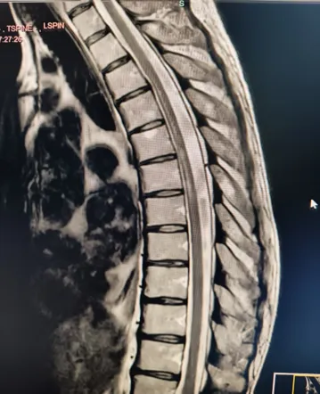
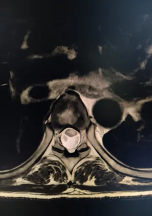
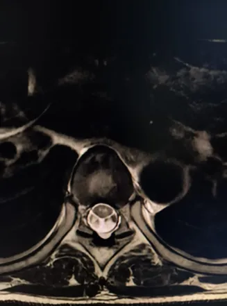
Figure 1. Magnetic resonance imaging (MRI) of the thoracic spine showing a long-segment intramedullary T2-weighted hyperintense lesion extending from the D3 to D7 vertebral levels with associated thoracic spinal cord atrophy, consistent with longitudinally extensive transverse myelitis (LETM) in aquaporin-4 antibody-positive neuromyelitis optica spectrum disorder (NMOSD).
Based on the clinical presentation and MRI findings, an autoimmune etiology was considered. Serum testing using a cell-based immunofluorescence assay demonstrated positivity for aquaporin-4 immunoglobulin G (AQP4-IgG) antibodies, whereas myelin oligodendrocyte glycoprotein (MOG-IgG) antibodies were negative. The combination of compatible clinical features, characteristic MRI findings, and disease-specific serology established the diagnosis of aquaporin-4 antibody-positive neuromyelitis optica spectrum disorder (NMOSD)
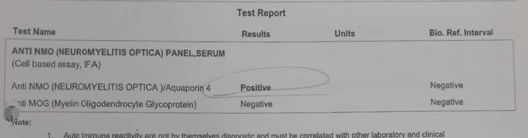
Figure 2. Cell-based immunofluorescence assay demonstrating positive aquaporin-4 (AQP4-IgG) antibody and negative myelin oligodendrocyte glycoprotein (MOG-IgG) antibody, confirming the diagnosis of AQP4-IgG-positive neuromyelitis optica spectrum disorder.
THERAPEUTIC INTERVENTION
Following confirmation of the diagnosis, the patient was started on high-dose intravenous methylprednisolone (1 g once daily) for five consecutive days as first-line therapy for the acute inflammatory episode. The primary objective of treatment was to suppress the ongoing autoimmune inflammatory process and minimize further neurological injury.
Along with corticosteroid therapy, comprehensive supportive care was provided to address the patient's symptoms. Pregabalin was prescribed for neuropathic pain, while baclofen and tizanidine were administered to relieve painful muscle spasms and spasticity. Tramadol was used for additional pain control, pantoprazole was given for gastrointestinal protection during corticosteroid therapy, and methylcobalamin supplementation was continued as supportive management. Bowel care measures and a structured physiotherapy program were also initiated to maintain functional mobility and support rehabilitation throughout the hospital stay.
During hospitalization, the patient's neurological status and vital parameters were monitored regularly to assess the response to treatment and detect any evidence of disease progression or treatment-related complications. Adequate hydration, nutritional support, and nursing care were maintained throughout the course of therapy to optimize recovery. A multidisciplinary approach involving neurologists, physiotherapists, and nursing staff was adopted to ensure comprehensive patient management and facilitate functional rehabilitation.
CLINICAL OUTCOME AND FOLLOW-UP
Following completion of the treatment regimen, the patient showed a favorable clinical response. The severity of bilateral lower limb pain gradually decreased, and the painful muscle spasms became less frequent, resulting in improved mobility and functional status. She was able to ambulate more comfortably with continued physiotherapy and supportive care.
The patient remained clinically stable throughout her hospital stay, with no new neurological deficits or treatment-related complications. At the time of discharge, her symptoms had improved sufficiently to allow continuation of rehabilitation on an outpatient basis. She was advised to attend regular neurology follow-up visits for ongoing clinical assessment, monitoring for disease recurrence, and evaluation of the need for long-term immunosuppressive therapy based on her future disease course. She was also counselled regarding the importance of adherence to follow-up and early reporting of any new neurological symptoms suggestive of relapse.
DISCUSSION
Neuromyelitis optica spectrum disorder (NMOSD) is a severe autoimmune inflammatory disorder that can present with a broad range of neurological manifestations. Although optic neuritis and longitudinally extensive transverse myelitis (LETM) are regarded as the hallmark features of the disease, some patients initially present with isolated spinal cord involvement, making diagnosis challenging. The present case represents one such atypical presentation, where progressive bilateral lower limb pain and painful muscle spasms occurred in the absence of optic neuritis. Careful clinical evaluation combined with disease-specific antibody testing enabled early diagnosis and timely treatment, resulting in a favourable clinical outcome.
Weinshenker and Wingerchuk described NMOSD as a relapsing autoimmune disorder in which optic neuritis and LETM are the predominant clinical manifestations. They also noted that a proportion of patients may initially present with isolated myelitis before developing visual symptoms. The present case closely resembles these observations, as the patient presented with progressive lower limb pain and painful muscle spasms without any evidence of optic neuritis. This atypical presentation made the diagnosis more challenging because several neurological conditions had to be excluded before NMOSD was considered. (2)
Papadopoulos and Verkman demonstrated that aquaporin-4 immunoglobulin G (AQP4-IgG) antibodies play a pivotal role in the pathogenesis of NMOSD and are highly specific for establishing the diagnosis. Similarly, the present patient tested positive for AQP4-IgG antibodies while MOG-IgG antibodies were negative. These findings confirmed AQP4-IgG-positive NMOSD and helped differentiate the disease from MOG antibody-associated disorders, allowing appropriate treatment to be initiated at an early stage. (3)
Kim et al. reported that isolated longitudinally extensive transverse myelitis can be the initial manifestation of NMOSD, even in the absence of optic neuritis. The clinical presentation of the present patient was consistent with these findings, as spinal cord involvement was the only neurological manifestation during the initial evaluation. This similarity highlights the importance of considering NMOSD in patients presenting with unexplained LETM, regardless of whether visual symptoms are present. (7)
Lennon et al. demonstrated that the identification of AQP4-IgG antibodies significantly improved the ability to distinguish NMOSD from multiple sclerosis and other inflammatory demyelinating disorders. In the present case, antibody testing played a decisive role after vascular disorders and peripheral neuropathy had been excluded. Early serological confirmation enabled prompt diagnosis and ensured that appropriate immunotherapy was started without unnecessary delay. (5)
Trebst et al. recommended high-dose intravenous methylprednisolone as the first-line treatment for acute attacks of NMOSD, with plasma exchange reserved for patients who do not respond adequately to corticosteroid therapy. The management of the present patient was consistent with these recommendations. She received high-dose intravenous methylprednisolone along with supportive treatment and rehabilitation, leading to gradual improvement in lower limb pain, muscle spasms, and mobility without requiring escalation of therapy. (14)
Kitley et al. observed that early diagnosis and timely initiation of immunotherapy are associated with better neurological recovery and may reduce the risk of long-term disability in patients with AQP4-IgG-positive NMOSD. The present case supports these findings, as early antibody testing facilitated rapid diagnosis and prompt treatment, resulting in clinical improvement during hospitalization. This case further emphasizes that maintaining a high index of suspicion for NMOSD in patients presenting with isolated LETM can facilitate earlier diagnosis and improve patient outcomes. (10)
CONCLUSION
Neuromyelitis optica spectrum disorder remains a rare but important cause of inflammatory myelopathy that should be considered in patients presenting with unexplained longitudinally extensive transverse myelitis, even in the absence of optic neuritis. As demonstrated in this case, the initial clinical presentation may be nonspecific and overlap with several neurological conditions, making the diagnosis difficult during the early stages of the disease. Such atypical presentations can delay appropriate treatment if NMOSD is not included in the differential diagnosis.
The confirmation of aquaporin-4 immunoglobulin G antibody positivity was crucial in establishing the diagnosis and enabled the prompt initiation of appropriate therapy. Early recognition and timely treatment are essential because they help limit ongoing inflammatory damage, preserve neurological function, reduce the risk of future relapses, and improve long-term clinical outcomes. This case also highlights the importance of integrating careful clinical assessment with disease-specific antibody testing to achieve an accurate diagnosis in patients presenting with longitudinally extensive transverse myelitis.
Furthermore, the favorable clinical response observed following corticosteroid therapy emphasizes the benefits of initiating immunotherapy as early as possible after diagnosis. A multidisciplinary approach involving neurologists, rehabilitation specialists, physiotherapists, and other healthcare professionals is equally important in optimizing recovery, improving functional independence, and enhancing the patient's quality of life.
Greater awareness of the diverse clinical presentations of NMOSD among healthcare professionals may facilitate earlier diagnosis and timely referral for specialist care. Continued long-term follow-up remains essential because of the relapsing nature of the disease and the potential for cumulative neurological disability. This case adds to the growing clinical evidence that isolated longitudinally extensive transverse myelitis may be the initial manifestation of AQP4-IgG-positive NMOSD and reinforces the value of a systematic diagnostic approach, early antibody testing, and prompt evidence-based management in improving patient outcomes.
REFERENCES


Ancy U, Nandana R S, Alnon L J, Vipin Venugopalan, Shaiju S Dharan, Aquaporin-4 Antibody-Positive Neuromyelitis Optica Spectrum Disorder Presenting as Isolated Longitudinally Extensive Transverse Myelitis: A Case Report, Int. J. of Pharm. Sci., 2026, Vol 4, Issue 7, 5981-5987. https://doi.org/10.5281/zenodo.21713479
 10.5281/zenodo.21713479