
We use cookies to ensure our website works properly and to personalise your experience. Cookies policy
1,2 Department of Pharmaceutical Sciences, School of Pharmacy, Massachusetts College of Pharmacy and Health Sciences, Boston, Massachusetts, USA 02115
1 Bioanalytical Science/Predictive Science Group, Genentech, San Francisco, California, USA 94080
Aqueous vaccine formulations are highly susceptible to degradation at elevated temperatures, necessitating cold storage throughout their lifecycle, which increases overall costs. Additionally, most vaccines are administered via the parenteral route, requiring trained professionals and often leading to pain and distress, thereby reducing patient compliance. A major challenge in vaccine development is the in vivo evaluation of candidates during the screening stage, a process that is labor-intensive, costly, and time-consuming, with variable reproducibility. This study aims to develop a nanoparticulate vaccine formulation while concurrently establishing a dendritic cell-based model for efficient vaccine screening in vitro. Fluorescence imaging and staining assays were employed to assess the uptake of vaccine-loaded nanoparticles in HeLa and dendritic cells. Cell Mask Orange assay confirmed intracellular localization of the nanoparticles. Enzyme-linked immunosorbent assay (ELISA) was performed to quantify cytokine (IL-6, TNF-?, and IL-1?) secretion by dendritic cells following stimulation with the nanoparticulate vaccine. Statistical analysis revealed significant cytokine release, indicating successful immune activation. These findings suggest that vaccines can be successfully encapsulated into nanoparticulate formulations and that dendritic cells provide a promising in vitro model for high-throughput vaccine screening. Additionally, this study paves the way for further investigation into optimizing nanoparticle formulations and in vitro screening methodologies to enhance vaccine development efficiency. This study highlights the importance of in vitro dendritic cell models as predictive tools for vaccine screening, reducing reliance on animal testing while accelerating vaccine development timelines.
The rapid rise in morbidity and mortality caused by the coronavirus disease 2019 (COVID-19) created an urgent demand to accelerate vaccine development. This led to the integration of different clinical trial phases to assess safety and efficacy more quickly (1). In light of the COVID vaccine development experiences, finding faster and more accessible vaccine testing methods along with discovering and optimizing their delivery systems has become an important goal in vaccine drug development (2).
A major challenge in vaccine development is screening the vaccine candidates for safety and efficacy before clinical trials. Traditional evaluation methods using preclinical animal models are expensive, require institutional approval, and demand a significant number of animals for toxicity, immune response, potency, safety, and efficacy assessments (3). These factors not only increase costs but also extend the time needed for vaccines to reach the market. In pandemic situations, such as COVID-19, rapid vaccine development and distribution are critical to saving lives. Given these challenges, the development of alternative in vitro models that can reliably predict immune responses and accelerate the screening of vaccine candidates is imperative.
Vaccine efficacy is highly dependent on the interaction between antigen-presenting cells (APCs) and immune system components. Various antigen-presenting cell (APC) based models – such as peripheral blood mononuclear cells (PBMC) (4), dendritic cells (DC) (5), MUTZ3 cell line (6), tissue constructs and/or 3D tissue constructs – have been employed to gain insight into how the immune system responds to invading antigens (7, 8). In these models, antigens were presented to the APCs resulting in the activation of immune response cascade (5, 9) and data obtained from these models have shown consistency. Thus, in-vitro models offer a promising approach for vaccine screening, reducing both time and cost.
To address vaccine screening challenges, our research focuses on developing a dendritic cell-based APC model. This model is advantageous due to its simplicity, ability to test large numbers of molecules/formulations rapidly, and reproducibility. Moreover, it eliminates the need for animal testing in early research stages, avoiding ethical concerns and institutional approvals, thereby expediting vaccine development. Among APCs, DCs play a crucial role in immunity, bridging innate and adaptive responses. Table 1 describes the key differences in the characteristics of mature and immature dendritic cells (10). These cells continuously patrol the body for foreign antigens, recognizing them through pathogen-associated molecular patterns via toll-like receptors (TLRs). Upon detection, TLRs signal the nucleus to activate an immune response, leading to secretion of cytokines such as interleukin-6 (IL-6), tumor necrosis factor-alpha (TNF-α), and interleukin-1 beta (IL-1β). These cytokines play crucial roles in recruiting immune cells and enhancing adaptive immune responses, making DC-based models an excellent platform for vaccine evaluation in vitro.
Table 1 Key differences in the characteristics of mature and immature dendritic cells
|
Characteristics |
Immature DCs |
Mature DCs |
|
Co-stimulatory molecules |
¯ |
|
|
MHC II expression |
¯ |
|
|
Secretion of pro-inflammatory cytokines |
¯ |
|
|
Phagocytic capacity |
|
¯ |
|
CCR7 expression |
¯ |
|
|
Glycolysis |
¯ |
|
Vaccine formulations must also address challenges related to stability, storage, and administration. Most vaccines are formulated as aqueous solutions administered via injection, presenting distribution and administration challenges. Liquid formulations are highly susceptible to physical and chemical degradation (11), requiring cold storage, which increases financial burdens (12, 13). Additionally, injectable vaccines require trained professionals, pose risks of needle injuries, and may cause pain or distress, emphasizing the need for alternative formulations that enhance stability while maintaining safety and efficacy at lower manufacturing costs (14).
One promising strategy to overcome these issues is the development of nanoparticle-based vaccine delivery systems. Nanoparticles exhibit unique physicochemical properties—such as particle size, surface charge, and hydrophobicity—that influence their interaction with biological systems. Studies have demonstrated that nanoparticle-based vaccines can improve antigen stability, facilitate targeted delivery to APCs, and enhance antigen uptake through receptor-mediated endocytosis. By tuning nanoparticle characteristics, researchers can design vaccine formulations that optimize immune responses while maintaining safety and biocompatibility.
Spray-dried solid nanoparticle (SNP) formulations offer several advantages, including enhanced antigen stability, the ability to circumvent cold-chain storage requirements, and the potential for mucosal administration via nasal, pulmonary, or oral routes. Encapsulating vaccines in nanoparticles allows for controlled antigen release, thereby optimizing immune system stimulation and potentially reducing the need for multiple booster doses. Furthermore, nanoparticles can be engineered to improve antigen uptake by APCs, enhancing immune activation and facilitating a more robust immune response compared to conventional vaccines. SNPs have been explored as a large-scale alternative for vaccine distribution and administration. (15) These SNPs can either be reconstituted with sterile water or can be administered through the mucosal route via inhalation (nasal/pulmonary) (16) or orally thus providing flexibility of dosage forms (17). SNP based vaccines can help eliminate the risk of instability/inactivity of components along with problems associated with cold-chain distribution and maintenance. Administering them through the mucosal route can overcome poor patient compliance, and dependency on an expert healthcare professional thus reducing the overall cost of the final product (18). Encapsulating vaccines in the SNPs has the potential to be a cost-effective method of vaccination that may enhance patient compliance and stability, and also offer a faster immune response when given through the mucosal route (19, 20). Furthermore, nanoparticle-based vaccines prevent antigen degradation, enhance immunological responses, and induce both systemic and mucosal immunity (17-19, 21, 22).
Effective vaccine administration and delivery are crucial to achieving the desired immune response. In pursuit of improved vaccine delivery methods, our research focused on encapsulating vaccines in spray-dried albumin nanoparticles. We investigated and quantified the ability of traditional and encapsulated spray-dried vaccine formulations to stimulate dendritic cells to release cytokines using ELISA. Spray drying was chosen for its ability to produce vaccine-loaded nanoparticles efficiently in a single-step continuous process, unlike freeze-drying. Albumin, known for its non-toxic, non-immunogenic, biocompatible, and biodegradable properties, serves as an ideal carrier for drug delivery, making it a promising candidate for vaccine encapsulation. Simultaneously, we sought to establish a dendritic cell-based in vitro model to screen the immunogenicity of vaccine candidates, providing a cost-effective and efficient alternative to traditional in vivo animal testing. By combining nanotechnology with immunological screening approaches, this research aims to contribute to the development of next-generation vaccine platforms that are both effective and accessible.
MATERIALS AND METHODS
Materials
Influenza vaccine - Flucelvax quadrivalent was purchased from Nationwide Medical Surgical. Bovine serum albumin powder (Fraction V. Nuclease and protease-free), and 4',6'-Diamidino-2-phenylindole dihydrochloride (DAPI) were purchased from VWR USA. Polysorbate 80 NF (Tween 80) was purchased from Professional Company of America (PCCA), TX, USA. Dulbecco's Modified Eagle's Medium (DMEM), fetal bovine serum (FBS), JAWS II cell line, and L-Glutamine Solution, 200 mM was purchased from ATCC, USA. Trypsin EDTA 1X, and Minimum Essential Medium (MEM) was purchased from Corning. Gibco PBS (pH 7.4), GibcoTM Penicillin/Streptomycin (5000U/ml), ProLong Gold antifade reagent, CellMask™ Orange Plasma Membrane Stain Invitrogen™, and paraformaldehyde 4% w/v was purchased from Thermo Fischer Scientific, USA. Recombinant mouse GM-CSF protein was purchased from R&D systems, USA. LPS was purchased from Sigma Aldrich, USA. ELISA kits for IL-6, TNF-α, and IL-1β were purchased from ABClonal, USA. Flow cytometry sub-micron particle size reference kit (green-fluorescent beads ranging in diameter from 0.02 μm to 2.0 μm) packaged in individual size vials was purchased from Thermo Fischer Scientific, USA.
Methods
Preparation of albumin nanoparticles
Preparation of albumin nanoparticles with a conventional spray dryer has been known to be extremely challenging due to the low collection efficiency of fine particles smaller than 2 µm in size. Thus, various parameters of the spray drying process such as albumin concentration, spray mesh size, inlet temperature, drying gas flow rate, spray rate, pump rate and frequency were optimized. Albumin NPs were prepared by dissolving 1% w/v of BSA solution in 18 ? deionized water. The solution was then crosslinked with 0.5% w/v of glutaraldehyde and stirred continuously at low speed for 12 hours. The crosslinked solution was then sprayed using a BUCHI B90 HP nano spray dryer with a 4 µm spray mesh. To prevent agglomeration, the gas flow rate was maintained between 140–150 L/min. The inlet temperature was set at 110 °C, the pump rate at 22%, the spray rate at 80%, and the frequency at 110 kHz to achieve the desired particle characteristics. The collected particles were scraped from the electrode collector region, weighed, and stored in glass vials.
Preparation of influenza vaccine – loaded nanoparticles
Crosslinked BSA solution was prepared as described above. To this, 8.33 mL of the marketed vaccine formulation was added to 1% w/v of crosslinked BSA solution (0.5% w/v drug loading). The mixture was stirred gently and subsequently sprayed using BUCHI B90 HP spray dryer under the same optimized conditions. The resulting particles were collected from the spray dryer walls by gentle scraping, weighed, and stored at 4°C in glass vials.
Characterization of nanoparticles
For characterization, 5 mg of nanoparticles were suspended in 15 mL of an aqueous 0.01% Tween 80 solution (stock solution) and sonicated for 1 min to ensure uniform dispersion and prevent aggregation. The particle size, zeta potential, and polydispersity index (PDI) were determined using a Nanoparticle Size Analyzer (90 Plus, Brookhaven Instruments). An average of three measurements were taken for particle size and zeta potential.
In Vitro cell culture
Complete media (CM) for HeLa cells was prepared by supplementing Dulbecco’s Modified Eagle Medium (DMEM) with 1% penicillin-streptomycin and 10% fetal bovine serum (FBS). CM for dendritic cells was prepared using Alpha Minimum Essential Medium supplemented with 1% penicillin-streptomycin, 20% FBS, 5 mL of 200 mM L-glutamine stock solution (final concentration: 4 mM), and 25 µL of 100 µg/mL murine granulocyte-macrophage colony-stimulating factor (GM-CSF) stock solution (final concentration: 5 ng/mL).
Preparation of albumin nanoparticle suspension
Desired quantity of serum – free media was taken in a beaker covered with aluminum foil. Serum-free media was used to prepare BSA nanoparticle suspensions at concentrations of 50 μg/mL, 100 μg/mL, and 200 μg/mL. Tween 80 stock solution was added to facilitate uniform dispersion before introducing the nanoparticles. The suspension was sonicated in 30-second intervals for up to 3 minutes to ensure even distribution.
Preparation of commercial nanoparticle suspension
Commercial nanoparticles (green-fluorescent beads) with a particle size of 1000 nm and a density of ∼1× 10⁶ beads/mL were dispersed in serum-free media. One drop of the commercial nanoparticle suspension was sonicated for 1 min to ensure uniform distribution before use in experiments.
Evaluation of cellular uptake
After 24 h of seeding, well plates were treated with BSA nanoparticle suspension and commercial nanoparticle suspension followed by incubation at 37°C, 5% CO2 for 4 hr, 8 hr, 24 hr, and 48 hr. Serum-free media was used as a negative control. After incubating the nanoparticles with cells for the desired time, wells were rinsed with PBS 3X times to remove the nanoparticles that were not taken up by the cells. The plate was then visualized for fluorescence using a Keyence fluorescence microscope. Various concentrations of BSA-NP suspensions (50 μg/mL, 100 μg/mL, and 200 μg/mL) and Tween 80 (0.25% v/v, 1% v/v, 4.5% v/v) were evaluated for optimizing the cellular uptake.
Cell Mask Orange assay for confirming nanoparticle uptake
To further validate the cellular uptake of nanoparticles, cell mask orange assay was conducted. Dendritic cells were seeded and incubated with the desired concentration of the BSA nanoparticles and commercial nanoparticles for 4 hr and 8 hr followed by PBS wash to remove any non-internalized nanoparticles. For fixation and permeabilization, 0.5 mL of 4% (w/v) paraformaldehyde solution in PBS and 0.1% Triton X-100 in PBS were added to the wells, followed by a 2-minute incubation at room temperature. The wells were then washed three times with PBS to remove the fixative solution. Subsequently, 0.5 mL of 4% (w/v) paraformaldehyde solution in PBS was added, and the plate was incubated for 15 minutes at room temperature, followed by three additional PBS washes.
Cells were then stained with CellMask Orange dye and incubated for 30 minutes at room temperature, followed by PBS washes to remove excess stain. To counterstain the nuclei, wells were treated with 4′,6-diamidino-2-phenylindole (DAPI) for 5 minutes at room temperature, followed by a final PBS wash. ProLong Gold antifade reagent was applied in a sufficient quantity to cover the wells, and cellular uptake was visualized using a Keyence fluorescence microscope.
Activation of dendritic cells
Blank nanoparticles and vaccine-loaded nanoparticles were dispersed in serum-free media containing Tween 80 stock solution to achieve a final concentration of 100 µg/mL. The vaccine loading in the nanoparticles was targeted at 0.5% (w/w). To prepare an equivalent concentration of the marketed formulation (commercial flu vaccine) containing 0.5 µg of vaccine antigen, 100 µL of the marketed formulation was diluted in 24 mL of serum-free media, yielding a final vaccine antigen concentration of 0.5 µg/mL. Additionally, 5 µg/mL of lipopolysaccharide (LPS) was diluted to obtain a final concentration of 100 ng/mL in serum-free media.
Following preparation, dendritic cells were incubated with each treatment group for 24 and 48 hours (Table 2). Cytokine secretion, including interleukin-6 (IL-6), tumor necrosis factor-alpha (TNF-α), and interleukin-1 beta (IL-1β), was quantified at defined time points using an ELISA kit obtained from ABClonal.
Table 2 Treatment groups for stimulating the dendritic cells to release cytokines
|
Treatment group |
Concentration |
Role |
|
Serum free media |
1 mL/well |
(-)ve control |
|
Blank nanoparticles |
100 µg/ mL dispersed in 1% Tween 80 |
Test |
|
Vaccine-loaded nanoparticles |
100 µg/mL dispersed in 1% Tween 80 |
Test |
|
Marketed flu vaccine formulation |
0.5 µg/mL of vaccine antigen |
Test |
|
LPS |
100 ng/mL |
(+)ve control |
Statistical analysis
The data was represented as mean values along with the standard deviation (SD). The one-way ANOVA test determined statistical significance, with the probability value (p) set at < 0.05 indicating significance. The GraphPad Prism 8 software was used to perform the analysis of variance (ANOVA).
RESULTS
Characterization of nanoparticles
The physicochemical properties of the formulated nanoparticles were assessed in terms of particle size and zeta potential. Blank nanoparticles exhibited a mean particle size of 706 nm, with a mean polydispersity index (PDI) of 0.15, and a mean zeta potential of -29.4 mV (Fig. 1A). In contrast, vaccine-loaded nanoparticles demonstrated an increased mean particle size of 1090 nm, a PDI of 0.19, and a zeta potential of -36.85 mV (Fig. 1B). The PDI values for both formulations remained below 0.2, indicating a narrow particle size distribution.
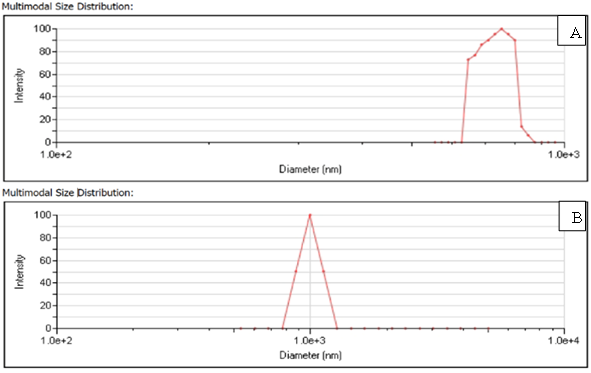
Figure 1 Multimodal particle size distribution of (A) blank BSA nanoparticles and (B) vaccine loaded BSA nanoparticles suspended in 0.01% Tween 80 aqueous solution
Optimization of BSA-NP concentration and Tween 80 for preparing NP suspension
HeLa cells treated with BSA-NP suspension at the concentration of 100 μg/mL for 4 hours are shown in Figure 2. BSA-NP suspensions were prepared with Tween 80 at 0.25% (v/v), 1% (v/v) and 4.5% (v/v) solution. All images were acquired at a magnification of 20X using a Keyence fluorescence microscope. It is observed that at high Tween 80 concentration (4.5% v/v), BSA-NP appeared to clump together resulting in higher localized density around the cell surface while at low Tween 80 concentration (0.25% v/v), BSA-NP appeared to aggregate due to insufficient dispersion leading to lower localized density around the cell surface. An intermediate Tween 80 concentration (1.0 % v/v) was found to be optimal for both optimal dispersion as well as localization on the cell surface. Tween 80 (1% v/v) solution was thus used to prepare all BSA-NP suspension henceforth. Figure 3 shows fluorescent images of HeLa cells treated with BSA-NP at 200 μg/mL for 4, 8, 24 and 48 hours respectively. It was observed that exposure to a nanoparticle suspension at a concentration of 200 μg/mL led to excessive nanoparticle accumulation, overcrowding the well plate, whereas a concentration of 50 μg/mL (Fig. 4) was found to be suboptimal for effective cellular treatment. Based on these observations, an intermediate concentration of 100 μg/mL (Fig. 5) was identified as optimal for subsequent experimental analyses.
Cellular uptake of albumin nanoparticles and effect on cellular morphology
Following incubation with varying concentrations (50–200 μg/mL) of the nanoparticle suspension for 4, 8, 24, and 48 hours (Fig. 3–5), no significant alterations in cellular morphology were observed. The cells retained their structural integrity across all tested conditions. Furthermore, BSA-NP exhibited a tendency to aggregate on and around the cell surface, suggesting their interaction with the cells. A progressive increase in fluorescence intensity from 4 hr to 48 hr (Fig. 3-5) further confirmed nanoparticle association with HeLa cells in a time-dependent manner.

Figure 2 Fluorescent images of HeLa cells treated with BSA-NP (100 μg/mL)
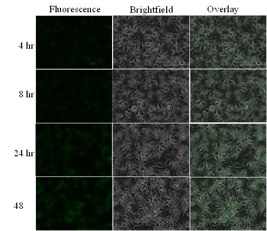
Figure 3 Fluorescent images of HeLa cells treated with BSA-NP (200 μg/mL)

Figure 4 Fluorescent images of HeLa cells treated with BSA-NP (50 μg/mL)

Figure 5 Fluorescent images of HeLa cells treated with BSA-NP (100 μg/mL)
Comparison of prepared albumin nanoparticles with commercial nanoparticles
The uptake of commercial silica nanoparticles by cells was minimal at 4 hours but increased noticeably by 8 hours (Fig. 6). When wells were treated with BSA nanoparticle suspension and commercial nanoparticle suspension in the absence of cells, BSA nanoparticles adhered to the well surface, whereas commercial nanoparticles were easily removed by PBS washing (data not shown), indicating weaker adherence. Additionally, prolonged incubation of HeLa cells with either BSA or commercial nanoparticles resulted in a comparable increase in fluorescence intensity, suggesting similar uptake potential.

Figure 6 Fluorescent images of HeLa cells treated with BSA-NP and commercial nanoparticles
Uptake of nanoparticles - Cell Mask Orange assay
The internalization of nanoparticles by dendritic cells is a crucial process for their activation in response to external stimuli. Initial experiments demonstrated a progressive increase in fluorescence intensity over time, indicating enhanced nanoparticle uptake. However, nanoparticle aggregation around the cells complicated precise localization. To distinguish between surface association and intracellular uptake, a CellMask Orange assay was performed. CellMask™ Orange, a lipophilic amphipathic membrane probe, selectively associates with the outer layer of the plasma membrane, resulting in a uniform delineation of the cell boundary. In contrast, DAPI preferentially binds to nuclear DNA, producing blue fluorescence of the cell nucleus. The observed purple fluorescence within the cytoplasmic region arises from the spatial colocalization of the intrinsic autofluorescence of albumin nanoparticles in proximity to the DAPI-stained nucleus. Staining nanoparticle-treated cells with CellMask Orange dye and DAPI revealed nanoparticle accumulation around the nucleus, confirming their internalization (Fig. 7).

Figure 7 Fluorescent images of dendritic cells treated with BSA-NP and commercial nanoparticles.
Cytokines Release by the Activated Dendritic Cells
Dendritic cell activation in response to external stimuli plays a critical role in the adaptive immune system. To utilize dendritic cells as an in vitro model for early-stage vaccine screening, it is essential to confirm their activation upon exposure to antigenic stimuli. In this study, JAWS II cells, an immature dendritic cell line, were used as a model. These cells remain in an immature state at rest but become activated upon antigen invasion, leading to cytokine secretion and the expression of specific surface molecules. Since cytokines act as key immune messengers bridging innate and adaptive immunity, the activation of JAWS II cells was assessed by quantifying cytokine levels using ELISA.
JAWS II cells were stimulated with various treatment groups, including serum-free media, blank nanoparticles, vaccine-loaded albumin nanoparticles, a marketed formulation, and lipopolysaccharide (LPS) as a positive control. Supernatants were collected at 24 hr and 48 hr for cytokine analysis using ELISA. The data was represented as mean values along with the standard error of the mean (SEM). The one-way ANOVA test followed by multiple comparisons with the control group was used for analysis. The probability value (p) of less than 0.05 indicated significance; ns represents no significance, ****p < 0.0001. The secretion of interleukin-6 (IL-6), tumor necrosis factor-alpha (TNF-α), and interleukin-1 beta (IL-1β) was evaluated. LPS strongly stimulated dendritic cells, leading to significant secretion of all tested cytokines (Fig. 8).

Figure 9 Activation of dendritic cells stimulated with different groups at 24 hr and 48 hr
For IL-6, blank nanoparticles and the marketed formulation induced cytokine levels comparable to the serum free media control, whereas vaccine-loaded nanoparticles significantly increased IL-6 secretion (p < 0.0001) at both 24 h and 48 h, except in comparison to LPS. These results indicate that vaccine-loaded nanoparticles effectively stimulate dendritic cells to release IL-6.
For TNF-α, both vaccine-loaded nanoparticles and the marketed formulation induced secretion levels comparable to the serum free media control. However, blank nanoparticles triggered significantly higher TNF-α secretion than all groups except LPS at both time points.
For IL-1β, blank nanoparticles, vaccine-loaded nanoparticles, and the marketed formulation induced comparable secretion levels to the serum free media control at 24 hours. However, the marketed formulation significantly increased IL-1β secretion compared to blank and vaccine-loaded nanoparticles. At 48 hours, vaccine-loaded nanoparticles and the marketed formulation-maintained IL-1β levels comparable to the serum free media control, with no statistically significant difference between them. A time-dependent decrease in IL-1β secretion was observed across all groups at 48 hours compared to 24 hours. Specifically, IL-1β secretion following vaccine-loaded nanoparticle stimulation was 68.0 ± 42.70 pg/mL at 24 hours, which declined to 38.4 ± 8.6 pg/mL at 48 hours, indicating peak IL-1β levels at 24 hours.
DISCUSSION
Nanoparticles are increasingly being explored for diverse biomedical applications, including drug delivery, imaging, and diagnostics. Depending on their intended use, various fabrication techniques such as desolvation, self-assembly, gelation, spray-drying, and emulsification are under investigation to produce different types of nanoparticles, including carbon-based, metal-based, lipid-based, and polymeric nanoparticles, with a focus on ensuring safety and efficacy. Critical physicochemical parameters, such as particle size and zeta potential, play a crucial role in determining nanoparticle interactions with cellular components. Nanoparticles exceeding 250 nm in size are primarily internalized by cells via phagocytosis, while zeta potential is a key determinant of nanoparticle stability, aggregation behavior, and overall biocompatibility. Aggregation of nanoparticles may lead to potential cytotoxicity and adverse cellular effects.
Although preparation of nanoparticles via a single step spray drying process appears to be simple and cost-effective, the process is very challenging as evident by the scarcity of literature reports. To this date, Lee et. al. were the first and only ones to report preparation of albumin nanoparticles using a Nano Spray Dryer B-90 which was equipped with a vibrating mesh spray technology and an electrostatic particle collector (23). It was found that particle size was predominantly influenced by spray mesh size and the albumin concentration. In this study, solid albumin nanoparticles were synthesized via spray-drying and loaded with a commercially available influenza vaccine formulation. Albumin was selected as the nanoparticle matrix due to its high binding capacity, biocompatibility, biodegradability, and cost-effectiveness. Various factors such as particle size, surface charge, surface coating, and surface area-to-volume ratio influence the uptakes of nanoparticles by cells (24). HeLa cells were initially used in cellular uptake studies because of their robust growth, reproducibility, and well-characterized endocytic pathways. Their consistent morphology and high transfection efficiency make them a reliable in vitro model for evaluating intracellular internalization and trafficking of drugs, nanoparticles, and other delivery systems. Characterization of the prepared nanoparticles revealed an increase in particle size following the incorporation of the vaccine into crosslinked bovine serum albumin (BSA), while polydispersity index (PDI) values remained below 0.2, indicating a monodisperse formulation (25). A zeta potential of approximately ±30 mV is considered optimal for stable colloidal dispersions; the formulations in this study exhibited zeta potential values near –30 mV, suggesting good physical stability and suitability for long-term storage at controlled room temperature.
The surface charge of nanoparticles also plays a critical role in cellular uptake. Positively charged nanoparticles exhibit strong electrostatic interactions with the negatively charged cell membrane, which may lead to cytotoxic effects and apoptosis. In contrast, the nanoparticles in this study were negatively charged, potentially influencing their uptake by dendritic cells in a manner conducive to immune response activation. Initial optimization studies involving cellular uptake were done using HeLa cells as quick screening tools. While CellMask Orange studies were done using DC cells to further validate the cellular uptake of the nanoparticles. Cellular viability assays indicated that HeLa cells retained their morphology upon incubation with nanoparticles for 48 hours, whereas morphological changes observed in dendritic cells suggested cellular maturation. These findings highlight the potential of the formulated nanoparticles for influenza vaccine delivery in a spray-dried solid dosage form, which could be reconstituted for injection or administered via mucosal routes. However, further investigations are required to elucidate the impact of nanoparticle encapsulation on morphology, encapsulation efficiency, surface area-to-volume ratio, and immune activation.
Fluorescence imaging and staining techniques were employed to evaluate nanoparticle-cell interactions in both HeLa and dendritic cells. Intrinsic green fluorescence from albumin nanoparticles was used to analyze the location and evaluate the uptake of the nanoparticles. No external fluorescent labels were attached to albumin nanoparticles since albumin itself has autofluorescence (26, 27), Fluorescence intensity increased with incubation time, with nanoparticles predominantly localizing around or on the cell surface. The CellMask™ Orange assay confirmed nanoparticle uptake, demonstrating their presence throughout the cytoplasm and near the nucleus.
The immune-stimulatory potential of the vaccine-loaded nanoparticles was assessed using a dendritic cell-based model to evaluate cytokine secretion, including interleukin-6 (IL-6), tumor necrosis factor-alpha (TNF-α), and interleukin-1 beta (IL-1β) (28). This study demonstrated that the vaccine-loaded nanoparticles induced the dendritic cells to release cytokines at a significantly higher level for IL-6 and at comparable levels for TNF-α, and IL1-β to the marketed formulation (commercially available flu vaccine) indicating the potential of the spray-dried nanoparticle-based vaccine delivery system to activate the immune responses. The spike in IL-6 levels as compared to the TNF-α, and IL-1β levels could be due to the role of different cytokines on the immune system or the influence of the particulate system on activating the immune cells through different pathways. IL-6 is a multifunctional cytokine and is known to have pro and anti-inflammatory properties whereas TNF-α and IL-1β possess only pro-inflammatory properties. The IL-6 cytokines released by the cells competes with binding site to the same receptors as TNF-α and IL1-β thus preventing its signaling which could be the possible reason for the decreased levels of these cytokines (29). Additionally, immune activation may be influenced by various pathways, such as JAK-STAT signaling for IL-6 release, TNF-α activation by binding to TNFR1 and TNFR2 on the cell surface, and activation of IL-1β through pro-inflammatory receptor complexes (30-32). Furthermore, variations in dendritic cell receptor populations and their differential activation at specific time points may contribute to the observed cytokine expression patterns (33-35).
As explained by Fernandez-Botran R. (36), cytokine release is tightly regulated through transcriptional and post-transcriptional mechanisms, including proteolytic cleavage of precursor molecules. Proteolytic cleavage by different stimulations can result in the production of soluble endogenous receptors such as IL-1 receptors which can attenuate the maturation of pro-IL-1β precursor, thereby reducing its activity. In contrast, the soluble IL-6 receptors (sIL-6 α) amplify the release of IL-6 by forming complexes with released IL-6 thus increasing its half-life and further enhancing the signaling cascades (37). These regulatory mechanisms may explain the observed cytokine secretion profiles upon nanoparticle-mediated dendritic cell stimulation.
A comprehensive understanding of cytokine signaling pathways in response to vaccine formulations is critical for rational vaccine development. Future research should focus on elucidating the effects of different vaccine components on dendritic cell maturation, receptor expression, antigen-receptor interactions, and immune activation pathways. Investigating the influence of nanoparticle physicochemical properties on dendritic cell function will provide valuable insights for optimizing nanoparticle-based vaccine formulations and advancing novel immunization strategies.
CONCLUSION
The commercially available influenza (Flu) vaccine was successfully encapsulated within spray-dried albumin nanoparticles, and an in vitro dendritic cell-based model was developed for screening vaccine candidates. Characterization of the vaccine-loaded nanoparticles revealed an increase in mean particle size, attributed to vaccine incorporation within the albumin matrix. Initial uptake studies using HeLa cells demonstrated a progressive increase in fluorescence intensity from 4 to 48 hours, indicating nanoparticle association with the cells. A comparable uptake pattern was observed for commercial nanoparticles, further confirming the cellular internalization of the developed formulation. To further investigate nanoparticle uptake, dendritic cells were incubated with the formulations and subjected to differential staining techniques to assess nanoparticle localization. Results indicated that the nanoparticles clustered around the nucleus, confirming their intracellular presence. The encapsulated Flu vaccine was subsequently evaluated for its ability to stimulate dendritic cells and induce cytokine release, a critical factor in immune system activation. The vaccine-loaded nanoparticles elicited TNF-α and IL-1β secretion at levels comparable to the marketed formulation, while significantly enhancing IL-6 secretion. These findings demonstrate the feasibility of utilizing albumin-based nanoparticle formulations for vaccine delivery and highlight the potential of the developed in vitro dendritic cell model as a rapid and efficient screening platform for vaccine candidates.
DECLARATION OF COMPETING INTEREST
The authors declare that they have no known competing financial interests or personal relationships that could have appeared to influence the work reported in this paper.
ACKNOWLEDGEMENTS
The authors would like to thank the department of pharmaceutical sciences, school of pharmacy at Massachusetts College of Pharmacy and Health Sciences for providing the funding for this project.
REFERENCES


Sayali Rane, Sanjaykumar Gayakwad, Evaluation of a Spray-dried Nanoparticulate Vaccine Formulation in a Dendritic Cell-based Model as an In Vitro Screening Tool, Int. J. of Pharm. Sci., 2026, Vol 4, Issue 2, 1974-1990. https://doi.org/10.5281/zenodo.18623211
 10.5281/zenodo.18623211