
We use cookies to ensure our website works properly and to personalise your experience. Cookies policy
Dr. J.J. Magdum Pharmacy Collage, Jaysingpur
Urolithiasis is a common urinary tract disorder characterized by the formation of kidney stones due to the crystallization of minerals in the urinary system. The present study was carried out to evaluate the anti-urolithiatic activity of Vitis vinifera leaves extract using an in-vitro nucleation assay method. The leaves of Vitis vinifera were collected, dried, powdered, and extracted using ethanol by Soxhlet extraction method. Preliminary phytochemical screening of the extract revealed the presence of flavonoids, tannins, phenolic compounds, saponins, and alkaloids, which are known for their medicinal properties.The anti-urolithiatic activity was assessed by measuring the inhibition of calcium oxalate crystal nucleation at different concentrations of the extract and comparing it with the standard drug Cystone. Absorbance values were recorded using a UV-Visible spectrophotometer, and percentage inhibition was calculated. The extract showed concentration-dependent inhibition of calcium oxalate crystal formation, indicating significant anti-urolithiatic potential. Higher concentrations of the extract exhibited greater inhibitory activity, comparable to the standard drug.The study suggests that Vitis vinifera leaves possess promising anti-urolithiatic activity and may be useful in the prevention and management of kidney stone formation. Further in-vivo and clinical studies are required to confirm its therapeutic efficacy and safety.
The kidneys are two bean-shaped organs, each approximately the size of a fist, located on either side of the spine just below the rib cage. They play a vital role in maintaining overall body health by filtering waste products, toxins, and excess fluids from the blood to produce urine. In addition to waste removal, the kidneys help regulate electrolyte balance, maintain fluid levels, control blood pressure, and support the production of red blood cells through hormone secretion. Their proper functioning is essential for maintaining the body's internal balance and overall well-being. [1]
Urolithiasis is a common disorder of the urinary system characterized by the formation of stones (calculi) within the kidneys, ureters, bladder, or urethra. The term is derived from the Greek words ouron (urine) and lithos (stone), referring to the formation of urinary stones. Among the various types of urinary calculi, calcium oxalate stones are the most prevalent. These stones develop when calcium ions combine with oxalate in the urine, leading to the formation and aggregation of crystals. Over time, these crystals grow into larger stone masses, which can cause pain, urinary obstruction, and other complications if left untreated.[2]
Nephrolithiasis, commonly known as kidney stone disease, is a significant public health concern worldwide. In India, approximately two million people are affected by kidney stones each year. Certain regions, including Gujarat, Maharashtra, Punjab, Rajasthan, Delhi, Haryana, and parts of northern India, are commonly referred to as the "stone belt" due to the high prevalence of the disease. Kidney stones are also frequently reported in southern India, where dietary habits, including the regular consumption of oxalate-rich foods such as tamarind, may contribute to stone formation.
Globally, the incidence and prevalence of kidney stones vary across different regions. Studies indicate that approximately 0.1–0.4% of the population in the United States and Europe develops kidney stones annually. The lifetime prevalence is estimated to be 2–5% in Asia, 8–15% in Europe and North America, and up to 20% in Saudi Arabia. Kidney stone disease is also known for its high recurrence rate, with nearly 75% of affected individuals experiencing a recurrence within 20 years of the initial episode. These statistics highlight the growing burden of urolithiasis and the importance of effective preventive and therapeutic strategies.[3]
The treatment of kidney stones depends on the size, type, and location of the stone as well as the severity of symptoms. Small stones are usually treated conservatively by increasing fluid intake to 2–3 liters per day, which helps dilute urine and promotes stone passage. Large or obstructive stones require surgical or minimally invasive procedures such as Extracorporeal Shock Wave Lithotripsy, which uses shock waves to break stones into smaller fragments, Ureteroscopy, where a scope is inserted through the urinary tract to remove or fragment the stone, and Percutaneous Nephrolithotomy, which is used for large kidney stones through a small incision in the back.[4]
Synthetic drug used in treatment of Kidney Stones involves the use of chemically synthesized drugs and modern medical procedures to relieve pain, dissolve stones, prevent crystal formation, and promote stone expulsion. the first step in treatment is pain management because kidney stones cause severe flank pain due to obstruction of urine flow. Non-steroidal anti-inflammatory drugs such as Ibuprofen and Diclofenac are commonly used to reduce pain and inflammation, while stronger analgesics may be given in severe cases.
Drugs like Tamsulosin and Nifedipine relax the smooth muscles of the ureter and facilitate the passage of stones through urine. In patients with uric acid stones, alkalinizing agents such as potassium citrate and sodium bicarbonate are administered to increase urinary pH and dissolve the stones. Allopurinol is prescribed to reduce uric acid production and prevent recurrence of uric acid stones. For calcium stone prevention, thiazide diuretics are used to decrease urinary calcium excretion,[5] while antibiotics are administered.
Conventional medications used in the management of kidney stones, including citrate supplements and other pharmacological agents, can be associated with several adverse effects such as nausea, vomiting, diarrhea, and abdominal discomfort. Prolonged use of these medications may also lead to complications, including hyperkalemia (elevated potassium levels) in patients receiving citrate therapy and metabolic acidosis associated with certain carbonic anhydrase inhibitors. These limitations have encouraged the exploration of alternative therapeutic approaches for the prevention and treatment of urolithiasis.
Herbal medicines have gained considerable attention as potential alternatives to synthetic drugs due to their relatively lower cost, wider availability, and perceived safety profile. Many medicinal plants contain a variety of bioactive compounds that can act on multiple stages of kidney stone formation. These phytochemicals may inhibit crystal nucleation, growth, and aggregation while also providing antioxidant and anti-inflammatory effects that help protect renal tissues.
Among the important phytochemicals reported to possess anti-urolithiatic activity are flavonoids, polyphenols, tannins, and resveratrol. These compounds have been shown to reduce calcium oxalate crystal formation, minimize oxidative stress, and support normal kidney function. In addition, they exhibit diuretic, antioxidant, and anti-inflammatory properties, which contribute to the prevention and management of urinary stone disease.
Vitis vinifera (grapevine) leaves are known to contain many of these beneficial phytoconstituents. Therefore, the present study was undertaken to evaluate the anti-urolithiatic activity of Vitis vinifera leaf extract and to investigate its potential role in the prevention and management of kidney stone formation.
METHODS:
In-Vivo Assay of Anti Urolithiatic Activity of Vitis vinifera:
Table No.1: In- Vivo Methods
|
Sr.No |
Test Name/Model |
Animal |
Principle |
|
1 |
Ethylene Glycol (EG)– Induced Urolithiasis Model |
Wistar rats / Sprague– Dawley rats |
EG (0.75–1% in drinking Water) induces hyperoxaluria, causing calcium oxalate stone formation. Test drug prevents or reduces crystal deposition, improves urine/serum markers.[24] |
|
2 |
Ethylene Glycol + Ammonium Chloride (EG + AC) Model |
Wistar rats |
AC causes metabolic acidosis and enhances stone formation with EG. Test drug reduces biochemical abnormalities and kidney CaOx deposits.[25]
|
|
3 |
Sodium Oxalate–Induced Urolithiasis |
Wistar rats / Mice |
Sodium oxalate injection rapidly increases urinary oxalate, causing acute CaOx crystal deposition. Test drug decreases crystalluria and Renal damage.[26] |
|
4 |
Zinc Disc Implantation Method |
Rats |
A zinc disc is surgically implanted into the bladder, acting as a nidus for stone growth. Test drug reduces stone size, weight, and deposition rate.[27] |
|
5 |
Ammonium Oxalate and Vitamin D3–Induced Model |
Rats |
Vitamin D3 increases calcium absorption; ammonium oxalate increases oxalate load→ CaOx crystals form. Test drug prevents hypercalciuria, stone.[28] |
In vitro Assay of Anti-Urolithiatic Activity of Vitis vinifera:
Table No.2: In-Vitro Methods
|
Sr. No. |
METHODS
|
PRINCIPLE
|
|
1 |
Aggregation Assay
|
Induced Model Ethylene glycol causes hyperoxaluria, leading to calcium oxalate stone formation. Test drug prevents or reduces crystal deposition.[29] |
|
2 |
Dissolution/ Litholytic Assay
|
Ammonium chloride causes metabolic acidosis, which enhances stone formation with EG. Test drug reduces biochemical abnormalities and CaOx deposits. [30] |
|
3 |
Microscopic Evaluation
|
Sodium oxalate injection increases urinary oxalate, rapidly forming CaOx crystals. Test drug decreases crystalluria and kidney damage. [31] |
|
4 |
Crystal Growth Assay |
Assesses whether a test sample inhibits the enlargement of calcium oxalate crystals. [32]
|
|
5 |
Nucleation Assay
|
Determines how effectively a sample prevents the initial formation of calcium oxalate crystal nuclei. [33] |
|
6 |
Egg membrane assay |
The egg membrane acts as a natural semi-permeable membrane in which calcium oxalate crystals are enclosed. The test drug diffuses through the membrane and dissolves the calcium oxalate crystals. The extent of dissolution indicates anti-urolithiatic activity. |
PLANT PROFILE:
GRAPES LEAVES:
Plant description:
Vitis vinifera is a woody climbing plant belonging to the family Vitaceae, commonly known as the grapevine the leaves contain various phytoconstituents such as flavonoids, tannins, polyphenols, saponins, and resveratrol, which are responsible for antioxidant, anti-inflammatory
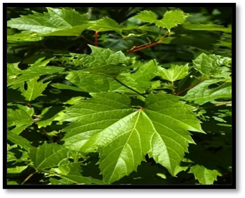
Fig No. 1: Vitis vinifera Leaves
Synonym: European grape vine, Grapevine, wine grape
Scientific name: Vitis vinifera
Biological source: Vitis vinifera consists of the dried leaves, fruits, and seeds of Vitis vinifera Linn, belonging to the family Vitaceae.
Geographical classification: Vitis vinifera is native to the Mediterranean region and Western Asia. It is widely cultivated in many countries including India, Italy, France, Spain, the United States, and China. In India, grape cultivation is mainly carried out in Maharashtra, Karnataka, Andhra Pradesh, Tamil Nadu, and Punjab due to favourable climatic conditions.
Scientific classification:
|
Kingdom |
Plantae |
|
Division |
Magnoliophyte |
|
Class |
Magnoliopsida |
|
Order |
Vitales |
|
Family |
Vitaceae |
|
Genus |
Vitis |
|
Species |
Vitis vinifera |
Chemical constituents: Vitis vinifera contains various important phytochemical constituents responsible for its medicinal properties. The major chemical constituents present in the Leaves, Fruits, and Seeds include Flavonoids, Polyphenols, Tannins, Saponins, Anthocyanins, Terpenoids, Organic Acids, Vitamins, Minerals, and Resveratrol.
Pharmacological Action: Acts as an antioxidant by scavenging free radicals, exhibits anti- inflammatory activity by reducing inflammatory mediators, shows cardio protective effect by improving blood circulation and reducing oxidative damage and possesses antimicrobial activity against various microorganisms.
PLANT MATERIAL PROCESSING:
Collection of Plant and Authentication:
The leaves samples of the Vitis vinifera collected from the nursery and authenticated by Department of Botany, Jaysingpur College, Jaysingpur having authentication Outward No. AES/JCJ/ By hand. [ Ref. Annexure No. 1]
Preparation of the plant extract:
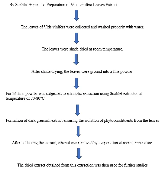
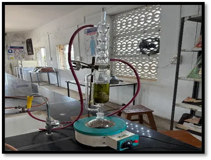
Fig No.2: Soxhlet Apparatus
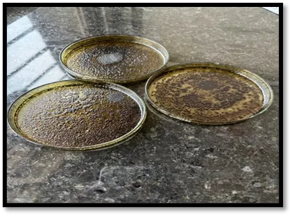
Fig No.3: Extract of Vitis vinifera
Yield Calculation:
Preliminary Phytochemical Investigation:
Chemical Test:
Various chemical tests are performed for the determination of phytoconstituents present in extract [35]
1) TEST FOR ALKALOIDS:
Take a small quantity of the plant extract in a test tube and dissolve it in a few millilitres of dilute hydrochloric acid. Filter the solution if necessary to obtain a clear extract. Add 2–3 drops of Dragendorff’s reagent to the solution and mix gently. Observe the formation of an orange, reddish-brown, or brick-red precipitate. The appearance of this precipitate indicates the presence of alkaloids in the plant extract.
Take a small amount of the plant extract in a test tube and add a few ml of dilute hydrochloric acid. Filter the solution if required to obtain a clear extract. Then add 2–3 drops of Mayer’s reagent to the solution and shake gently. The formation of a cream or white coloured precipitate indicates the presence of alkaloids in the plant extract.
Take a small quantity of the plant extract in a test tube and dissolve it in a few ml of dilute hydrochloric acid. Filter the solution if necessary to obtain a clear extract. Add 2–3 drops of Wagner’s reagent to the extract and shake gently. The formation of a brown or reddish-brown precipitate indicates the presence of alkaloids in the plant extract.
2) TEST FOR FLAVONOIDS:
Add a few drops of lead acetate solution to the plant extract. Formation of a yellow precipitate indicates the presence of flavonoids.
Take a small quantity of the plant extract in a test tube and add a small amount of zinc dust. Then add a few drops of concentrated hydrochloric acid carefully and mix gently. The formation of a red or orange-red colour indicates the presence of flavonoids in the plant extract.
Take a small quantity of the plant extract in a test tube and add a few ml of dilute ammonia solution. Then add concentrated sulfuric acid carefully along the sides of the test tube. The appearance of a yellow colour, which disappears on standing, indicates the presence of flavonoids in the plant extract.
3) TANNINS TEST:
Take a small quantity of the plant extract in a test tube and add 1% gelatin solution containing sodium chloride. Mix the solution gently and observe the formation of a white precipitate. The appearance of the precipitate indicates the presence of tannins in the plant extract.
Take a small quantity of the plant extract in a test tube and add a few drops of 10% alcoholic ferric chloride solution. Mix the contents gently and observe the colour change. The formation of a blue, green, or black coloration indicates the presence of tannins in the plant extract.
Take a small quantity of the plant extract in a test tube and add a few drops of bromine water solution slowly. Mix the solution gently and observe the reaction. The formation of a buff-coloured precipitate indicates the presence of tannins in the plant Extract.
Take a small quantity of the plant extract in a test tube and add sodium acid phosphate solution followed by a few drops of phenazone solution. Mix the contents gently and observe the formation of a bulky precipitate. The appearance of the precipitate indicates the presence of tannins in the plant extract.
4) CARDIAC GLYCOSIDE:
Pyridine and sodium nitroprusside or sodium NaOH added to extract to observe red or faded to brownish colour.
Add sodium picrate solution to the plant extract and mix gently. Development of an orange or yellow colour confirms the presence of cardiac glycosides.
Take a small quantity of the plant extract in a test tube and add glacial acetic acid containing a trace of ferric chloride solution. Carefully add concentrated sulfuric acid along the sides of the test tube. The formation of a brown ring at the junction of the two layers and a bluish-green colour in the upper layer indicates the presence of cardiac glycosides.
5) CARBOHYDRATES:
Add Barfoed’s reagent to the plant extract and heat in a water bath. Formation of a red precipitate within a few minutes indicates the presence of monosaccharides.
Take a small quantity of the plant extract in a test tube and add Seliwanoff’s reagent. Heat the mixture gently in a water bath for a few minutes. The appearance of a cherry-red colour indicates the presence of ketose sugars such as fructose in the plant extract.
Take a small quantity of the plant extract in a test tube and add a few drops of resorcinol reagent along with concentrated hydrochloric acid. Heat the mixture gently in a water bath for a few minutes. The formation of a red or cherry-red colour indicates the presence of ketose sugars in the plant extract.
IN- VITRO METHODS
Egg Membrane Assay [34]:
Preparation of calcium oxalate stones:
The following procedure was used to make the calcium oxalate (CaOx) experimental kidney stones by homogenous precipitation. In 100 mL of distilled water, 1.47 g of calcium chloride dihydrate was dissolved, and in 100 mL of 1 M H2SO4, 1.34 g of sodium oxalate was dissolved. Both were combined in a beaker and stirred to precipitate calcium oxalate. Ammonia solution was used to free the crystals from H2SO4. Finally, the crystal was washed in distilled water and dried for four hours at 60?. Further research was carried out using the artificial urine and stone that had been prepared.
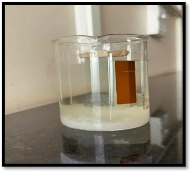
Fig No.4: Calcium Oxalate Crystal
Preparation of the semipermeable membrane from eggs:
The semipermeable membrane was prepared from egg. A glass rod was used to puncture the apex of the eggs and the entire contents were squeezed out. Empty egg shells were washed thoroughly in distilled water, after that the shells were placed in a beaker containing 4.0 mL concentrated HCl in 200 mL distilled water. The semi-permeable membrane was completely decalcified after being kept for an overnight period. The next day the semi permeable membranes were carefully removed from the egg shell and washed thoroughly with distilled water. The trace of acid present in the membrane is neutralised by placing the shell membranes in an ammonia solution, and rinsed with distilled water. It was kept in a moistened state in the refrigerator at a pH of 7–7.4.
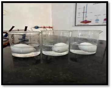
Fig No.5: Decalcification of Egg Membrane
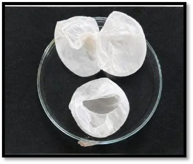
Fig No.6: Semipermeable Membrane of Eggs
Methods:
The studies were carried out in three groups as per experimental design
All the above groups were packed in semi permeable membrane by suturing. They were suspended in a conical flask containing 100 ml of 0.1 M TRIS buffer. The conical flasks of all groups were placed in an incubator and preheated to 37°C for 2 hours. The contents of semi permeable membrane from each group were taken into test tubes. 2 ml of sulphuric acid was added to each test tube and titrated with 0.2 M KMnO? till a light pink colour end point was obtained. 1 ml of 0.2 M KMnO? is equivalent to 0.1898 mg of calcium. Percentage dissolution of calcium oxalate in various groups was calculated.
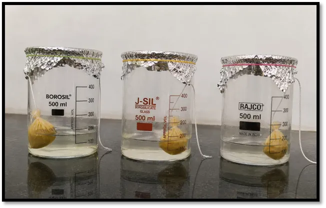
Fig No. 7: In –Vitro model used to evaluate anti-urolithiatic activity
Crystal Nucleation Assay:
A buffer containing 0.05 M Tris–HCl and 0.15 M NaCl at pH 6.5, a solution of calcium chloride (5 mM) and sodium oxalate (7.5 M) was prepared. A 9 mL calcium chloride solution was combined with 1 mL extracts at various concentrations (50, 100,250, 500, and 1000 mg/mL). Crystallization was stimulated by the insertion of 1 ml of sodium oxalate solution and the absorbance shift was recorded at 620 nm in a UV spectrophotometer (UV- 1800, Shimadzu Pvt. Ltd.) for 30 minutes at 37oC.
The procedure was followed for the control, substituting distilled water instead of the extract. All samples were inspected in triplicate. Standard drug Cystone was used as a positive control for comparison at distinct concentrations including 50, 100,250, 500, and 1000 mg/mL. The percentage inhibition of nucleation rate was calculated by given formul.
RESULTS:
Collection and Authentication of plant material:
The whole plant was identified and authenticated by Prof. Manisha V. Kale Head Dept. of Botany Jaysingpur College, Jaysingpur.
Preparation of Plant Extract:
The total yield of Ethanolic Extract was found to be
Table No.3: % yield of Ethnolic Extract
|
Sr. No |
Extract |
% Yield (w/w) |
|
1. |
Leaves |
2.7 |
PRELIMINARY PHYTOCHEMICAL INVESTIGATION:
Table No.4: Phytochemical evaluation test:
|
Sr. No. |
Test |
EEVV |
|
1 |
Test for Alkaloids |
|
|
Mayer’s test |
+ |
|
|
Wagner’s test |
+ |
|
|
Dragendroff’s test |
+ |
|
|
Hager’s test |
- |
|
|
|
|
|
|
2 |
Test for flavonoids |
|
|
Lead acetate test |
+ |
|
|
Pew’s test |
+ |
|
|
Ammonia test |
+ |
|
|
|
Ferric chloride test |
- |
|
|
|
|
|
3 |
Test for Glycosides |
|
|
|
Cardenoloides test |
+ |
|
Baljet test |
+ |
|
|
Keller killani test |
+ |
|
|
Kedee’s test |
- |
|
|
|
|
|
|
4 |
Test for Carbohydrates |
|
|
|
Barfoed test |
+ |
|
Seliwanoff’s test |
+ |
|
|
Resorcinol test |
+ |
|
|
Test for pentose |
- |
|
|
|
|
|
|
5 |
Test for saponins |
|
|
|
Foam test |
- |
|
|
|
|
|
6 |
Test for tannins |
|
|
|
Gelatine test |
+ |
|
Braymer’s test |
+ |
|
|
Bromine water test |
+ |
|
|
Phenadzone test |
+ |
|
Various chemical tests are performed for the phytochemical investigation. From the results as we found that all chemical tests are positive for the flavonoids, alkaloids, tannins, carbohydrate test in Vitis vinifera leaves Which indicates that the major phyto-constituents like flavonoids, alkaloids, carbohydrate, glycoside and tannins are present in the leaves extract of Vitis vinifera.
In-Vitro Evaluation of Anti-Urolithiatic Activity
Egg Membrane Assay
Groups as per experimental design
Table No.5: Percent of Calcium Oxalate dissolution by various groups under study
|
Groups |
Vol. of Standard KMnO4(ml) |
Wt. Of Calcium Oxalate Taken |
Wt. Of Calcium Estimated (Mg) |
Wt. Of Calcium Reduced (Mg) |
% Dissolution |
|
1 |
0.1 |
1 |
- |
- |
- |
|
2 |
0.6 |
1 |
0.11388 |
0.88612 |
88.61 |
|
3 |
0.9 |
1 |
0.17082 |
0.82918 |
82.91 |
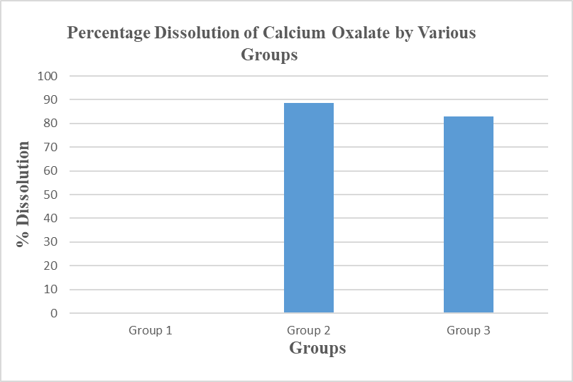
Fig. No. 13: Percentage Dissolution of Calcium Oxalate by Various Groups under Study
The egg membrane assay showed considerable dissolution of calcium oxalate crystals in the treated groups. The standard drug (Cystone) exhibited 88.61% dissolution, while the Ethanolic extract of Vitis viniferra leaves showed 82.91% dissolution of calcium oxalate crystals. The findings indicate that the leaves extract possesses significant Anti-urolithiatic Activity.
Nucleation Assay:
Table N0.6: Nucleation Assay of Standard (Cystone) drug
|
GROUP |
CONCENTRATION µg/mL |
ABSORBANCE |
%INHIBITION |
IC 50 |
|
Standard (Cystone) |
50 |
0.486±0.0004 |
48.44 |
56.5µg/mL |
|
100 |
0.373±0.0008 |
60.48 |
||
|
250 |
0.297±0.0008 |
68.58 |
||
|
500 |
0.177±0.0004 |
81.17 |
||
|
1000 |
0.107±0.0012 |
88.59 |
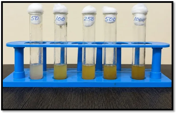
Fig. No. 14: Cystone (Standard Drug) at Various Concentration
Table No.7: Nucleation Assay of Ethanolic extract of Vitis vinifera leaves
|
GROUP |
CONCENTRATION µg/mL |
ABSORBANCE |
%INHIBITION |
IC50 |
|
TEST (EEVV) |
50 |
0.566±0.0001 |
39.97 |
78.6µg/mL |
|
100 |
0.401±0.0004 |
57.48 |
||
|
250 |
0.375±0.0008 |
60.27 |
||
|
500 |
0.302±0.0012 |
67.93 |
||
|
1000 |
0.217±0.001 |
76.97 |
The results of the nucleation assay demonstrated that the standard drug Cystone exhibited significant anti-urolithiatic activity by inhibiting calcium oxalate crystal nucleation in a concentration-dependent manner. The percentage inhibition is 48.44, 60.48, 68.58, 81.17,88.59 at concentration 50, 100, 250, 500, 1000 µg/mL respectively the IC?? value of 56.5 µg/mL suggests good inhibitory potential against kidney stone formation.
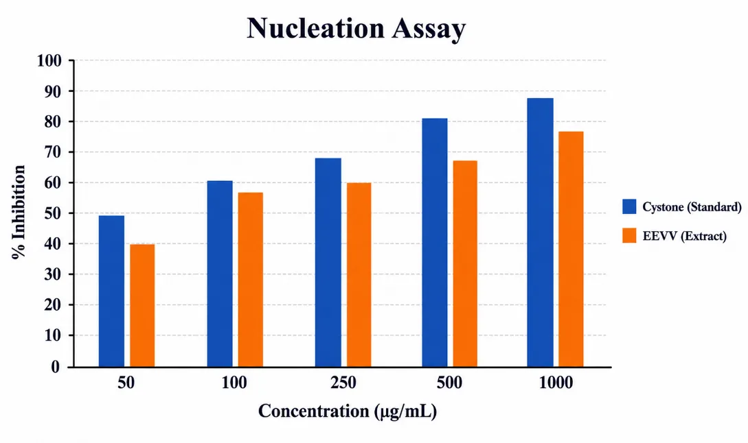
Fig No. 15: Ethanolic extract of Vitis vinifera leaves at various concentration
DISCUSSION:
Kidney stone formation mainly occurs due to supersaturation and crystallization of calcium oxalate in the urinary tract. Therefore, inhibition of calcium oxalate crystal formation is considered an important approach in the prevention and treatment of urolithiasis. The present study was carried out to evaluate the anti-urolithiatic activity of the ethanolic extract of Vitis vinifera leaves using different in-vitro models such as phytochemical screening, egg membrane assay, and crystal nucleation assay
The preliminary phytochemical investigation of the ethanolic extract revealed the presence of important phytoconstituents such as flavonoids, alkaloids, tannins, glycosides, and carbohydrates. These phytochemicals are known to possess antioxidant, anti-inflammatory, and diuretic properties which may contribute to anti-urolithiatic activity. Flavonoids and polyphenolic compounds such as catechin and Gallic acid help reduce oxidative stress and prevent crystal aggregation, while tannins such as Hydrolysable tannins may inhibit calcium oxalate crystal formation.
In the egg membrane assay, the ethanolic extract showed significant dissolution of calcium oxalate crystals. The extract produced 82.91% dissolution, whereas the standard drug Cystone showed 88.61% dissolution. The results indicate that the extract possesses considerable litholytic activity and can dissolve preformed calcium oxalate crystals effectively. Although the activity was slightly lower than the standard drug, the extract demonstrated promising anti-urolithiatic potential.
The nucleation assay further confirmed the anti-urolithiatic activity of the extract. The ethanolic extract inhibited calcium oxalate crystal nucleation in a concentration-dependent manner. The percentage inhibition increased from 39.97% at 50 µg/mL to 76.97% at 1000 µg/mL. Similarly, the standard drug Cystone showed inhibition ranging from 48.44% to 88.59% at corresponding concentrations.
The IC?? value of the ethanolic extract was found to be 78.6 µg/mL, while the standard drug showed an IC?? value of 56.5 µg/mL. A lower IC?? value of the standard indicates stronger activity; however, the extract still exhibited good inhibitory potential against calcium oxalate crystallization. The activity of the extract may be attributed to the synergistic action of flavonoids, tannins, saponins, and other phenolic compounds present in the leaves.
Overall, the findings of the study suggest that the ethanolic extract of Vitis vinifera leaves possesses significant anti-urolithiatic activity by inhibiting nucleation and promoting dissolution of calcium oxalate crystals. The study supports the traditional use of herbal medicines in the management of kidney stones and indicates that the plant may serve as a potential natural therapeutic agent for urolithiasis. However, further in-vivo studies and clinical investigations are required to confirm its safety, efficacy, and mechanism of action.
CONCLUSION:
The present study demonstrated that the ethanolic extract of Vitis vinifera leaves possesses significant anti-urolithiatic activity against calcium oxalate crystal formation. Phytochemical screening confirmed the presence of important bioactive constituents such as flavonoids, tannins, alkaloids, glycosides, and carbohydrates, which may contribute to its therapeutic effect. In the in-vitro evaluation, the extract showed considerable calcium oxalate crystal dissolution in the egg membrane assay and effectively inhibited crystal nucleation in a concentration-dependent manner, although slightly lower than the standard drug Cystone.
The findings suggest that Vitis vinifera leaves have promising potential as a natural remedy for the prevention and management of kidney stones due to their crystal inhibitory and antioxidant properties. However, further in-vivo and clinical studies are required to establish their safety, efficacy, and mechanism of action for therapeutic use.
REFERENCES


Sumit Shinge, Sumayya Shaikh, Sujit Chavan, Sujit Ukande, Sushant Patil, Dhanshree Rajput,Evaluation of Anti Urolethiatic Potential of Vitis Vinifera Leaves, Int. J. of Pharm. Sci., 2026, Vol 4, Issue 6, 6353-6371. https://doi.org/ 10.5281/zenodo.20844672
 10.5281/zenodo.20844672