

We use cookies to ensure our website works properly and to personalise your experience. Cookies policy
1Student, Bachelor of Pharmacy, S. C. S. M. S. S. Institute of Pharmacy, Maregaon, Yavatmal, Maharashtra, India.
2Student, Bachelor of Pharmacy, S. C. S. M. S. S. Institute of Pharmacy, Maregaon, Yavatmal, Maharashtra, India.
3Assistant Professor, Bachelor of Pharmacy, S. C. S. M. S. S. Institute of Pharmacy, Maregaon, Yavatmal, Maharashtra, India.
4Principal, Bachelor of Pharmacy, S. C. S. M. S. S. Institute of Pharmacy, Maregaon, Yavatmal, Maharashtra, India
This investigation aimed to develop and assess a mucoadhesive buccal patch loaded with Psidium guajava (guava) leaf extract for the site-specific management of oral ulcers. Oral ulcers are distressing mucosal wounds marked by pain, burning discomfort, and impairment of routine activities such as speaking, eating, and swallowing. Current therapeutic options including mouthwashes, topical ointments, and gels exhibit limited efficacy due to insufficient retention at the application site caused by saliva-induced washout. To address these drawbacks, mucoadhesive buccal patches were fabricated using HPMC K15M and Carbopol 934 polymer matrices via the solvent casting technique. Fresh guava leaves were harvested, botanically authenticated, shade dried, milled into powder, and subjected to maceration-based extraction using a hydro-alcoholic solvent. Preliminary characterization through organoleptic assessment, solubility profiling, phytochemical screening, TLC, and UV spectroscopy confirmed the presence of therapeutically active phytoconstituents including quercetin, flavonoids, tannins, and phenolic compounds. The formulated patches were assessed for thickness, weight uniformity, folding endurance, surface pH, swelling index, drug content, mucoadhesion time, and in vitro drug permeation. Formulation F3 exhibited optimal physicochemical attributes, adequate mechanical integrity, storage stability, and controlled drug release. Permeation studies via the Franz diffusion cell revealed 87.5% cumulative drug release over 8 hours. These findings establish the guava leaf mucoadhesive buccal patch as a viable, safe, and efficacious plant-based drug delivery system for oral ulcer treatment.
1.1. Mucoadhesive Patch:
Mucoadhesive buccal patches are sophisticated drug delivery devices engineered to bond firmly to the oral mucosa and maintain continuous, site-specific therapeutic release [1]. These systems effectively bypass the drawbacks of conventional oral therapy, namely hepatic first-pass metabolism, gastrointestinal enzymatic degradation, and premature salivary clearance [2,3]. Polymer systems such as HPMC allow these flexible carriers to adhere at the ulcer site, establishing a physical barrier while continuously delivering bioactive components from guava leaf extract, particularly its flavonoid and tannin constituents [5,6]. This approach promotes patient acceptability, accelerates wound healing, diminishes pain and inflammatory responses, and represents a reliable treatment modality for oral ulcers [7,8].
1.2. Oral Ulcer:
Oral ulcers are described as painful erosive lesions of the oral mucosal lining that significantly impair basic functions including eating, swallowing, and speech [9,10]. A broad range of etiological factors has been implicated, encompassing mechanical trauma, psychological stress, microbial infections, pharmacological agents, nutritional deficiencies, autoimmune disorders, and idiopathic causes [11]. Based on their duration, oral ulcers may be categorized as acute (resolving within three weeks) or chronic (persisting and recurring over extended periods). Frequent non-neoplastic types include traumatic ulcers from sharp dental restorations or ill-fitting prostheses, minor and major aphthous ulcers, herpetic lesions, and immune-mediated disorders such as oral lichen planus. Minor aphthous ulcers typically resolve without intervention, whereas major variants tend to persist, result in scarring, and may closely resemble malignant lesions [12]. Chronic presentations may also arise from persistent mucosal irritation or underlying systemic conditions, underscoring the necessity of accurate diagnosis and timely management [13].
1.3. Guava Plant:
Guava (Psidium guajava) is a tropical medicinal shrub classified under the family Myrtaceae, recognized globally for its nutritional and pharmacological significance [14]. Due to its high adaptability and rapid growth habit, this plant, which originated in Central and South America, has since naturalized throughout tropical and subtropical regions, including China, India, Brazil, and Thailand [15]. Traditional medicine has traditionally used the plant's various anatomical components, including as its leaves, fruits, bark, and roots, to treat problems like diarrhea, diabetes, cough, hypertension, cutaneous wounds, mouth infections, and gastrointestinal disorders. Contemporary scientific research has corroborated many of these ethnomedicinal applications, attributing their efficacy to bioactive phytoconstituents including quercetin, flavonoids, tannins, and polyphenols [16,17].
The guava plant grows as a perennial shrub or modest-sized tree, distinguished by smooth reddish-brown exfoliating bark, glossy dark green foliage, aromatic white blossoms, and pulpy fruits enclosing numerous seeds [18]. Guava leaves hold special medicinal significance and are traditionally prepared as aqueous decoctions or alcoholic infusions. Their conventional applications include oral hygiene maintenance, wound management, gastrointestinal relief, and glycemic regulation [19]. The fruit serves as an excellent source of ascorbic acid (Vitamin C), antioxidant carotenoids such as lutein and lycopene, and dietary fibre, collectively contributing to improved immune function and digestive wellness, while the bark and roots are predominantly valued for their pronounced astringent and antidiarrheal activities [20,21].
Guava leaves exhibit outstanding pharmacological efficacy in the treatment of oral ulcers owing to their well-documented anti-inflammatory, antimicrobial, antioxidant, and tissue-regenerative properties. The flavonoid quercetin reduces inflammation by inhibiting inflammatory mediators, thereby decreasing redness, swelling, and pain at the ulcer site. The high tannin content forms a protective barrier over the ulcer, shielding exposed nerve endings from irritation caused by food, speech, and chewing [22].
Furthermore, guava extract demonstrates potent antimicrobial efficacy against oral pathogens such as Streptococcus mutans, Streptococcus oralis, and Candida albicans, thereby preventing opportunistic secondary infections that otherwise impede the healing process. The combined influence of ascorbic acid, polyphenols, and triterpenoids stimulates collagen biosynthesis and promotes cellular proliferation, expediting tissue restoration and reducing ulcer dimensions more effectively [25]. These synergistic pharmacological attributes collectively establish guava leaf extract as a promising phytomedicinal candidate for the targeted local treatment of oral ulcers [26].
1.4. Chemical constituents:
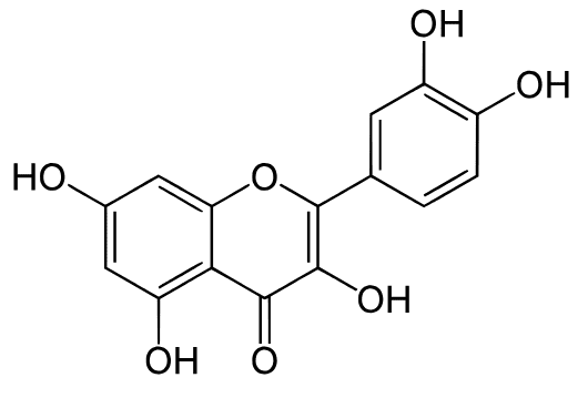
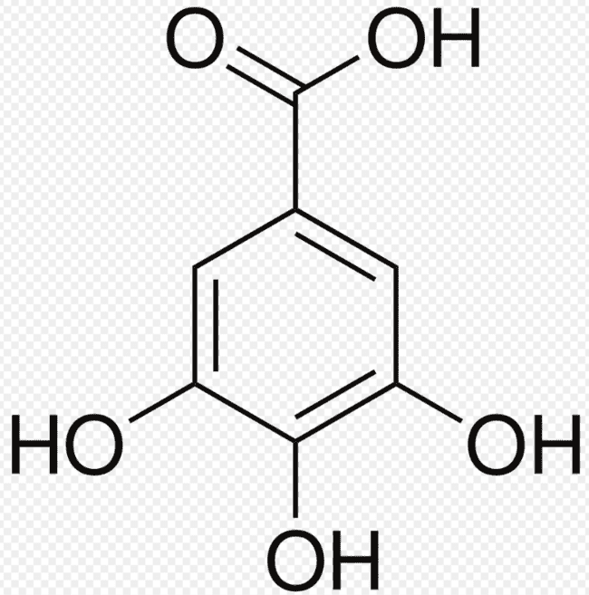
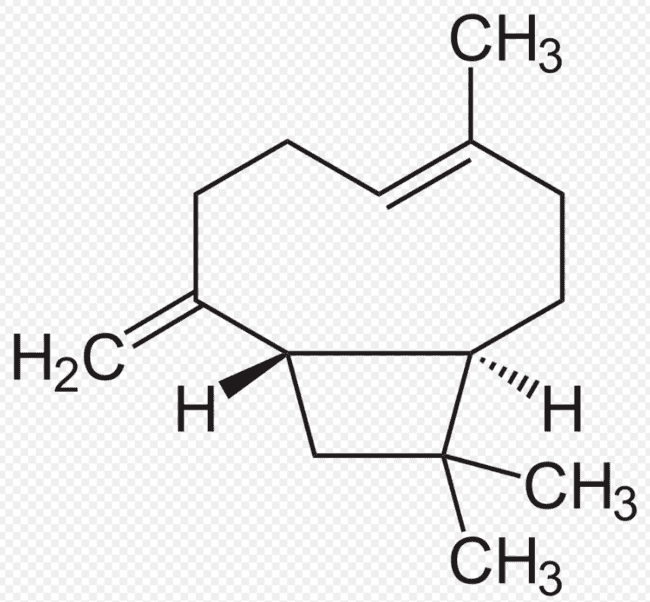
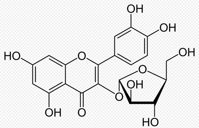
Table No. 1: Structure of chemical constituents
|
Sr. no |
Compounds |
Molecular formula |
Molecular structure
|
activity |
|
1 |
Quercetin |
C15H10O7 |
|
Antioxidant, anti-inflammatory, and antimicrobial |
|
2 |
Gallic Acid |
C7H6O5 |
|
Antimicrobial and Antifungal |
|
3 |
B-Caryophyllene |
C15H24
|
|
Analgesic, Potent Anti-Inflammatory |
|
4 |
Avicularin |
C20H18O11 |
|
Anti-Inflammatory Action |
|
5 |
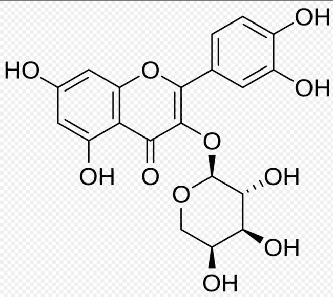
Guaijaverin |
C20H18O11 |
|
Broad-Spectrum Antibacterial |
|
6 |
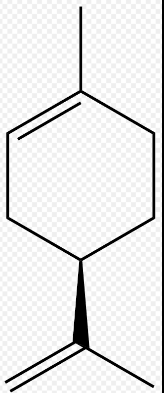
Limonene |
C10H16 |
|
Skin Penetration Enhancement, Antioxidant and Anti-Inflammatory |
2. Plan of Work:
2.1. Plant Material: Psidium Guavaja L.
Table No. 2: Ingredients along with their roles used in formulation on Mucoadhesive patches.
|
Ingredient |
Category |
|
Guava Leaf Extract |
Active Pharmaceutical Ingredient (API) |
|
HPMC K15M |
Hydrophilic Polymer |
|
Carbopol 934 |
Bio adhesive Polymer |
|
Ethanol & Water |
Solvent System |
|
Propylene Glycol |
Plasticizer |
2.2. Procedure:
2.2.1 Phase I: Botanical Processing and Extraction
2.2.2 Phase II: Preformulation Studies
1. Organoleptic Characterization:
Physical characteristics of the extract including colour, odour, taste, and textural consistency were systematically assessed using standard sensory evaluation techniques.
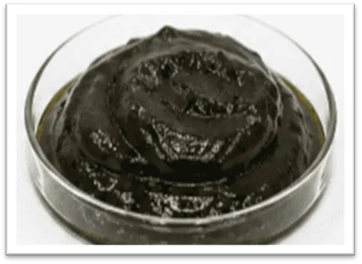
Fig.No. 1: Guava Leaves Extract.
2. Solubility:
The dissolution behaviour of the extract was investigated in distilled water, ethanol, and phosphate buffer at pH 6.8 to determine its solvent compatibility profile.
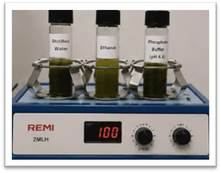
Fig.No. 2: Solubility testing
3. Physicochemical Screening.
Qualitative chemical tests were conducted to detect and confirm the presence of key phytoconstituents, particularly flavonoids and tannins, within the guava leaf extract.
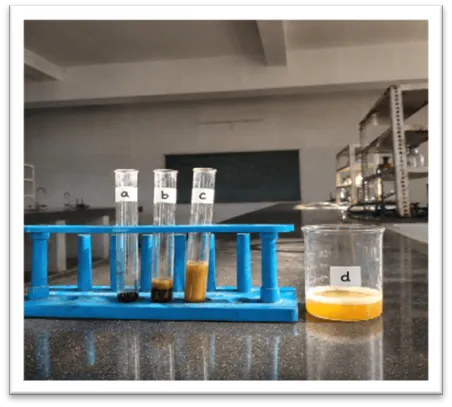
Fig.No. 3: Chemical identification test
4. Identification of Quercetin using TLC:
Using toluene: ethyl acetate: formic acid (5:4:1) as the mobile phase, thin-layer chromatographic analysis was performed on silica gel 60 F254 stationary phase. The plate was inspected under UV light at 254 nm and 366 nm after chromatographic development. The presence of quercetin, flavonoids, and polyphenolic chemicals in the extract was confirmed by the formation of distinctive fluorescent spots with matching Rf values.
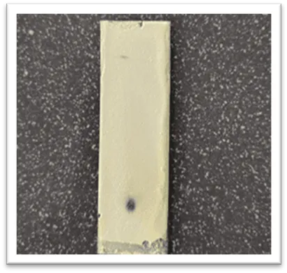
Fig.No. 4: TLC of quercetin
5. Polymer Hydration and Viscosity.
Hydration and viscometric studies of the selected polymers were performed to characterize their swelling tendency and gel-network forming capacity upon contact with aqueous media.
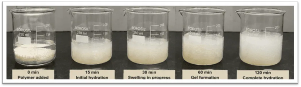
Fig.No. 5: Polymer Hydration and Viscosity.
2.2.3 Phase III: Formulation and Development (Solvent Casting Method)
2.2.3 Phase IV: Evaluation of Mucoadhesive Patch
The fabricated mucoadhesive patches were visually inspected for uniformity of colour, surface smoothness, optical clarity, mechanical pliability, and absence of defects such as air entrapment, cracks, or surface irregularities to confirm overall quality compliance.
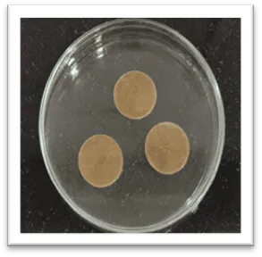
Fig.No. 6: Visual Inspection
Patch thickness was quantified at multiple locations using a calibrated screw gauge, and the arithmetic mean was reported to verify consistent film formation and homogeneous drug distribution throughout the matrix.
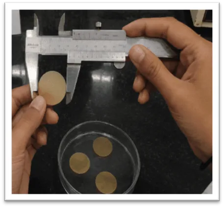
Fig.No. 7: Thickness Uniformity
Mechanical flexibility was evaluated by repeatedly folding each patch at an identical position until fracture occurred. The total number of folding cycles endured before breakage served as a quantitative indicator of the patch's mechanical integrity and elasticity.
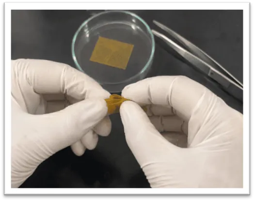
Fig,No. 8: Folding Endurance
The surface pH was measured after slight hydration of the patch using a calibrated digital pH meter to confirm physiological compatibility with the salivary pH range and to minimize the risk of mucosal irritation upon application.
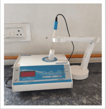
Fig.No. 9: Surface PH
To ensure equal drug loading across the patch matrix, the patch was extracted in phosphate buffer pH 6.8, and the guava leaf extract concentration was spectrophotometrically quantified at the predefined λmax using UV-visible spectroscopy.
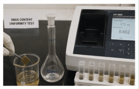
Fig.No. 10: Drug Content Uniformity
The swelling index was quantified by gravimetrically comparing pre-immersed and post-immersed patch weights at predetermined time intervals, providing a measure of fluid absorption kinetics and mucoadhesive potential under simulated physiological conditions.
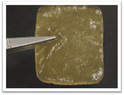
Fig.No. 11: Swelling Index
7. In Vitro Residence Time:
In vitro residence time is assessed by recording the duration for which the patch adheres to a simulated mucosal substrate, such as agar gel or excised sheep buccal mucosa, under standardized experimental conditions. This evaluation provides critical data on the mucoadhesive performance and site-retention capability of the patch, both essential prerequisites for sustained localized drug delivery.
8. In Vitro Drug Release:
Drug permeation behaviour is routinely characterized using the Franz diffusion cell apparatus, which is widely accepted as the standard methodology for evaluating topical and mucoadhesive formulations. In this setup, the test patch is positioned in the donor compartment while a synthetic or biological membrane separates it from the receptor chamber containing an appropriate physiological buffer. Aliquots withdrawn at scheduled intervals are spectrophotometrically analyzed to quantify the proportion of drug diffused. This methodology enables comprehensive assessment of release kinetics, release rate consistency, and formulation reproducibility prior to any clinical evaluation.
3. RESULTS:
[A] Formula:
Table No.3: Composition of Mucoadhesive Patch.
|
Sr No |
Materials |
F1(mg) * |
|
1 |
Guava Leaves Extract |
100mg |
|
2 |
HPMC K15M |
1.5g |
|
3 |
Carbopol 934 |
0.5g |
|
4 |
Propylene Glycol |
0.5 ml |
|
5 |
Water: Ethanol (7:1) |
q.s. to 20ml |
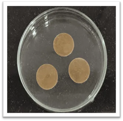
Fig.No. 12: Batch of Mucoadhesive Patches
[B] Phytochemical Test:
Table No. 4: Determination of phytochemical tests
|
SR. NO. |
Chemical constituents |
test |
Results |
|
1 |
Flavonids |
Shinoda test |
Present (Pink or Crimson Red colour) |
|
2 |
Tannins |
Ferric chloride test |
Present (Blue-Black colour) |
|
3 |
Saponins |
Froth test |
Present (Stable Honeycomb forth apper) |
|
4 |
Phenolic compound |
Lead acetate test |
Present (White Precipitate occurs) |
A] Ferric Chloride Test.
B] Shinoda Test.
C] Lead Acetate Test.
D] Saponions Test.
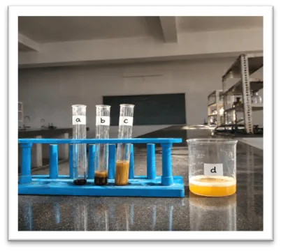
Fig. No.13; Observation of Chemical Test.
Table No. 5: TLC of Quercetin
|
SR. NO |
Test Material |
Procedure |
Observed RF Value |
Inference |
|
1. |
Guava Leaf Extract (quercetin) |
Thin Layer Chromatography (TLC) utilizing silica gel plates and a mobile phase of toluene, ethyl acetate, and formic acid (5:4:1) was used to identify quercetin in guava leaf extract. Following development, the chromatogram was examined under UV illumination at 254 and 366 nm. The presence of quercetin in the extract was verified by the distinctive fluorescent spot and corresponding Rf value. |
RF Value= 0.42 |
The appearance of an extract spot matching the exact R_f value and yellow orange UV fluorescence of the standard confirms the successful presence of Quercetin. |
[D] Preformulation Studies:
Table No.6: Preformulation Studies of Guava Leaves Extract
|
Parameters |
Method |
Trail 1 |
Trial 2 |
Trial 3# |
|
Physical appearance |
Visual |
Dark Green |
Dark Green |
Dark Green |
|
Loss on drying (%) |
Oven method |
4.2% |
4.5% |
4.3% |
|
Solubility(mg/ml) |
Buffer PH 6.8 |
28.5 |
29.0 |
28.5 |
|
Lambda max(nm) |
UV Spectroscopy |
256 |
257 |
256 |
|
Surface PH |
Digital PH Meter |
6.8 |
6.9 |
6.8 |
[E] Evaluation Test:
Table NO. 7: Evaluation Parameters and Results for Buccal Patches.
|
Evaluation Parameters |
Method/ Instruments |
Results |
Acceptance Criteria |
|
Weight Variation (mg) |
Digital Balance |
150.2 ± 2.1 |
+- 10% of mean weight |
|
Thickness (mm)
|
Vernier caliper |
0.28 ± 0.02 |
Uniform thickness (+-5%) |
|
Surface pH |
Digital pH Meter |
6.75 ± 0.12
|
6.2 – 7.2 (Neutral) |
|
Folding Endurance |
Repeated manual folding |
245 ± 12 |
> 200 folds |
|
Drug Content (%) |
UV-Vis Spectroscopy |
98.45 ± 1.15
|
90% – 110% |
|
Swelling Index (%) |
Gravimetric method |
45.2 ± 3.4
|
Consistent hydration |
|
Residence Time (h) |
Modified USP Apparatus |
3.5 ± 0.4 h |
2 – 4 hours |
[F] In Vitro Drug Release :
Franz diffusion cell results
Table. No. 8: In Vitro Drug Release Results
|
Time (hr) |
Absorbance |
Drug Released (%) |
|
0 |
0.000 |
0 |
|
0.5 |
0.118 |
12.4 |
|
1 |
0.201 |
21.3 |
|
2 |
0.336 |
35.8 |
|
4 |
0.512 |
54.6 |
|
6 |
0.671 |
71.2 |
|
8 |
0.824 |
87.5 |
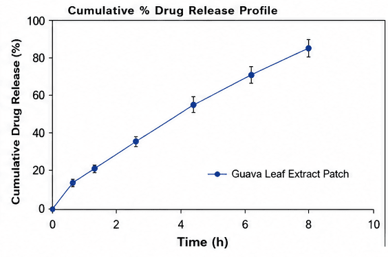
Fig No. 14: Cumulative Drug Release of Mucoadhesive Patch
4. Discussion:
The current research was directed toward the systematic formulation and comprehensive evaluation of a mucoadhesive buccal patch incorporating guava leaf extract for the localized treatment of oral ulcers. Oral ulcers represent a frequently encountered category of inflammatory lesions associated with significant pain, mucosal irritation, and functional interference during speaking and eating. Existing therapeutic platforms such as mouthwashes and topical ointments typically offer only transient symptomatic relief owing to rapid salivary dilution and inadequate tissue retention at the ulcer site. Accordingly, the development of a mucoadhesive buccal patch was identified as a strategically appropriate approach to prolong drug dwell time at the affected mucosa and sustain pharmacological activity. The patch system, prepared via solvent casting using HPMC K15M and Carbopol 934, yielded smooth, flexible, and dimensionally consistent films exhibiting acceptable physicochemical characteristics.
5. CONCLUSION:
The present investigation successfully accomplished the formulation and systematic characterization of a mucoadhesive buccal patch incorporating Psidium guajava (guava) leaf extract for the targeted local management of oral ulcers. Oral ulcers represent a commonly encountered category of painful mucosal lesions manifesting with inflammation, persistent irritation, burning discomfort, and functional impairment including difficulties in speaking, swallowing, and eating. Conventional pharmaceutical formats such as mouthwashes, gels, and ointments are inherently limited in therapeutic durability due to their rapid displacement by salivary secretions and tongue movement. Accordingly, the development of a mucoadhesive buccal drug delivery system was conceived as an innovative and clinically advantageous approach to prolong drug-mucosa contact and ensure sustained local availability of the therapeutic agent.
REFERENCES




Pavan Nikhade*, Kalyani Bhendare, Aakanksha Panajwar , Nilesh Chachda, Formulation And Evaluation Of Mucoadhesive Patch Using Guava Leaves Extract, Int. J. of Pharm. Sci., 2026, Vol 4, Issue 8, 890-907. https://doi.org/10.5281/zenodo.21812881
 10.5281/zenodo.21812881