

We use cookies to ensure our website works properly and to personalise your experience. Cookies policy
Department of Pharmacology, Geetanjali Institute of Pharmacy, Geetanjali University, Udaipur, Rajasthan, India.
Background: The rapid emergence of antimicrobial resistance (AMR) has become a major global health concern, necessitating the search for alternative antimicrobial agents from natural sources. Leucas aspera (Willd.) Link is a medicinal plant widely used in traditional medicine and is known to contain several bioactive phytochemicals with potential antimicrobial properties. Objective: The present study aimed to evaluate the antibacterial activity of the ethanolic leaf extract of Leucas aspera against selected multidrug-resistant (MDR) bacterial strains using the agar well diffusion method and minimum inhibitory concentration (MIC) assay. Methods: Fresh leaves of Leucas aspera were collected, shade-dried, powdered, and extracted using ethanol by the maceration method. The antibacterial activity of the extract was assessed against selected MDR bacterial strains by measuring the zone of inhibition, while MIC values were determined to evaluate the minimum concentration required to inhibit bacterial growth. Results: The ethanolic leaf extract exhibited appreciable antibacterial activity against several bacterial strains, with the highest activity observed against Haemophilus influenzae (13.0 ± 1.0 mm) and Staphylococcus aureus (12.5 ± 0.5 mm). Lower antibacterial activity was observed against Streptococcus pneumoniae, while no detectable inhibition was found against Bacillus subtilis, Proteus vulgaris, and Neisseria gonorrhoeae. Conclusion: The findings demonstrate that the ethanolic leaf extract of Leucas aspera possesses promising antibacterial activity against selected multidrug-resistant bacteria and supports its traditional medicinal use. Further studies focusing on phytochemical characterization, toxicity evaluation, and in vivo investigations are warranted to establish its therapeutic potential and facilitate the development of plant-based antimicrobial agents.
Antimicrobial resistance (AMR) has emerged as one of the most serious public health challenges of the twenty-first century. The widespread and often inappropriate use of antibiotics in human medicine, veterinary practice, and agriculture has accelerated the evolution of resistant microorganisms, reducing the effectiveness of many conventional antimicrobial agents. As a consequence, infections that were once readily treatable have become increasingly difficult to manage, leading to prolonged hospital stays, higher healthcare costs, increased morbidity, and mortality. The World Health Organization (WHO) has identified AMR as one of the top global health threats and has emphasized the urgent need for the discovery of novel antimicrobial agents capable of overcoming resistant pathogens (1).
Multidrug-resistant (MDR) bacteria are defined as microorganisms that exhibit resistance to at least one antimicrobial agent in three or more different classes of antibiotics. The increasing prevalence of MDR pathogens has significantly limited therapeutic options available to clinicians and has complicated the management of both community-acquired and hospital-acquired infections (2). Common MDR organisms, including Staphylococcus aureus, Escherichia coli, Klebsiella pneumoniae, Pseudomonas aeruginosa, and Proteus species, are responsible for a wide range of infections such as pneumonia, urinary tract infections, bloodstream infections, wound infections, and sepsis. Their growing resistance to β-lactams, fluoroquinolones, aminoglycosides, and other commonly prescribed antibiotics poses a major challenge to modern healthcare systems (2,3).
The rapid dissemination of antimicrobial resistance is driven by multiple factors, including irrational antibiotic prescribing, self-medication, poor infection control practices, inadequate surveillance, and horizontal transfer of resistance genes among bacteria. The ability of microorganisms to develop resistance through enzymatic drug degradation, alteration of target sites, reduced membrane permeability, and active efflux mechanisms has further contributed to the global AMR crisis. Consequently, there is an increasing demand for alternative therapeutic strategies that can either replace or complement existing antibiotics (1,3).
Natural products have historically played a pivotal role in drug discovery and continue to represent one of the most valuable sources of biologically active compounds. Numerous clinically important antimicrobial agents have originated from natural sources, particularly plants, microorganisms, and marine organisms. Medicinal plants produce a wide variety of secondary metabolites, including alkaloids, flavonoids, phenolic compounds, tannins, terpenoids, glycosides, and saponins, many of which possess potent antimicrobial, antioxidant, anti-inflammatory, and immunomodulatory activities. These phytochemicals often act through multiple mechanisms, reducing the likelihood of rapid resistance development compared with conventional antibiotics (4–6).
Traditional systems of medicine such as Ayurveda, Siddha, and Unani have relied extensively on medicinal plants for the prevention and treatment of infectious diseases for centuries. According to the World Health Organization, a substantial proportion of the global population continues to depend on traditional herbal medicine as a primary source of healthcare, particularly in developing countries. Scientific validation of these medicinal plants has gained increasing attention in recent years because of their therapeutic potential, favorable safety profile, and accessibility. Consequently, medicinal plants are increasingly being investigated as potential sources of novel antimicrobial agents for combating MDR bacterial infections (1,5,6).
Evaluation of antimicrobial activity is a critical step in identifying promising plant-derived compounds with therapeutic potential. Among the available laboratory techniques, the agar well diffusion method remains one of the most widely accepted screening methods because of its simplicity, reproducibility, and ability to provide preliminary information regarding the antibacterial efficacy of plant extracts. The method measures the diameter of the inhibition zone produced around wells containing the test extract and provides an initial assessment of antimicrobial susceptibility. Confirmation of antibacterial potency is often supplemented by determination of the minimum inhibitory concentration (MIC), which identifies the lowest concentration capable of inhibiting visible microbial growth (4). Standardized laboratory protocols, including those recommended by the Clinical and Laboratory Standards Institute (CLSI), ensure the reliability and reproducibility of antimicrobial susceptibility testing across different laboratories (7).
In response to the alarming increase in antimicrobial resistance, researchers worldwide are actively exploring medicinal plants with established ethnopharmacological uses as potential alternatives to conventional antibiotics. The discovery of plant-derived antimicrobial compounds may contribute not only to the development of novel therapeutic agents but also to the identification of lead molecules for future pharmaceutical research. These investigations have strengthened interest in medicinal herbs possessing broad-spectrum antimicrobial activity and rich phytochemical composition, including Leucas aspera (Willd.) Link, which has attracted considerable scientific attention because of its diverse biological activities and long history of traditional medicinal use.
MATERIALS AND METHODS
Fresh leaves of Leucas aspera (Willd.) Link were collected from the Botanical Garden of Meera Girls College, Udaipur, Rajasthan, India, and authenticated by the Department of Botany, Meera Girls College. The leaves were washed, shade-dried for 7–10 days, powdered using a mechanical grinder, and stored in airtight containers until extraction.
The powdered leaf material (50 g) was extracted with 250 mL ethanol by the maceration method for 48 h with intermittent shaking. The extract was filtered through Whatman No. 1 filter paper and concentrated using a rotary evaporator. The concentrated extract was stored at 4°C until further analysis.
The antibacterial activity of the ethanolic extract was evaluated against multidrug-resistant bacterial strains, including Staphylococcus aureus and Escherichia coli, obtained from the National Collection of Industrial Microorganisms (NCIM). Bacterial cultures were maintained on nutrient agar and subcultured in nutrient broth at 37°C for 24 h. The inoculum was adjusted to the 0.5 McFarland standard before testing.
Antimicrobial activity was assessed using the agar well diffusion method. Sterile nutrient agar plates were inoculated with standardized bacterial suspensions, and 6-mm wells were prepared using a sterile cork borer. Different concentrations of the ethanolic extract were added to the wells, while ciprofloxacin and the extraction solvent served as the positive and negative controls, respectively. After incubation at 37°C for 24 h, the diameter of the inhibition zones was measured in millimetres. All experiments were performed in triplicate, and the results were expressed as mean ± standard deviation.
Statistical analysis was performed using one-way analysis of variance (ANOVA), and a p-value of <0.05 was considered statistically significant.
RESULTS
The ethanolic leaf extract of Leucas aspera exhibited antibacterial activity against several multidrug-resistant (MDR) bacterial strains, although the degree of inhibition varied among the tested organisms. Antibacterial activity was evaluated using the agar well diffusion assay, and the results are presented as mean ± standard deviation (SD) of three independent experiments.
The extract showed the highest antibacterial activity against Haemophilus influenzae (13.0 ± 1.0 mm), followed by Staphylococcus aureus (12.5 ± 0.5 mm), Escherichia coli (12.0 ± 1.0 mm), Micrococcus luteus (12.0 ± 0.5 mm), Vibrio cholerae (11.5 ± 0.5 mm), Klebsiella pneumoniae (11.0 ± 1.0 mm), and Salmonella typhi (10.0 ± 1.0 mm). A comparatively smaller inhibition zone was observed against Streptococcus pneumoniae (6.0 ± 1.0 mm), whereas Bacillus subtilis, Proteus vulgaris, and Neisseria gonorrhoeae showed no detectable inhibition.
Table 1: Antibacterial Activity of Leucas aspera Ethanolic Leaf Extract Against Multidrug-Resistant Bacterial Strains
|
Sr. No. |
Bacterial Strain |
Zone of Inhibition (mm) Mean ± SD |
Standard Antibiotic (mm) |
|
1 |
Staphylococcus aureus |
12.5 ± 0.5 |
25 |
|
2 |
Streptococcus pneumoniae |
6.0 ± 1.0 |
15 |
|
3 |
Bacillus subtilis |
0.0 |
13 |
|
4 |
Micrococcus luteus |
12.0 ± 0.5 |
20 |
|
5 |
Escherichia coli |
12.0 ± 1.0 |
17 |
|
6 |
Vibrio cholerae |
11.5 ± 0.5 |
15 |
|
7 |
Salmonella typhi |
10.0 ± 1.0 |
15 |
|
8 |
Proteus vulgaris |
0.0 |
12 |
|
9 |
Haemophilus influenzae |
13.0 ± 1.0 |
21 |
|
10 |
Neisseria gonorrhoeae |
0.0 |
22 |
|
11 |
Klebsiella pneumoniae |
11.0 ± 1.0 |
16 |
Values are expressed as Mean ± SD (n = 3)
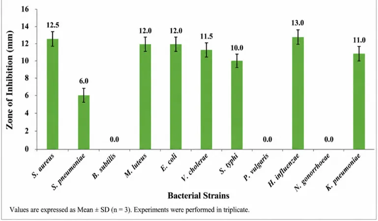
Figure 1: Zone of Inhibition Produced by Leucas aspera Ethanolic Leaf Extract Against Multidrug-Resistant Bacterial Strains
The minimum inhibitory concentration (MIC) assay further confirmed the antibacterial efficacy of the extract. The lowest MIC values were recorded for Haemophilus influenzae and Salmonella typhi (15.6 µL/mL), indicating greater susceptibility. Moderate MIC values were observed for Staphylococcus aureus, Escherichia coli, and Vibrio cholerae (31.2 µL/mL), while Micrococcus luteus and Klebsiella pneumoniae exhibited MIC values of 62.4 µL/mL. Higher MIC values were recorded for Proteus vulgaris (249.6 µL/mL), Bacillus subtilis and Neisseria gonorrhoeae (499.2 µL/mL), whereas Streptococcus pneumoniae demonstrated the highest MIC value (998.4 µL/mL), indicating the lowest susceptibility to the extract.
Table 2: Minimum Inhibitory Concentration (MIC) of Leucas aspera Ethanolic Leaf Extract
|
Sr. No. |
Test Organism |
MIC (µL/mL) |
Interpretation |
|
1 |
Staphylococcus aureus |
31.2 |
Moderate susceptibility |
|
2 |
Streptococcus pneumoniae |
998.4 |
Least susceptible |
|
3 |
Bacillus subtilis |
499.2 |
Low susceptibility |
|
4 |
Micrococcus luteus |
62.4 |
Moderate susceptibility |
|
5 |
Escherichia coli |
31.2 |
Moderate susceptibility |
|
6 |
Vibrio cholerae |
31.2 |
Moderate susceptibility |
|
7 |
Salmonella typhi |
15.6 |
High susceptibility |
|
8 |
Proteus vulgaris |
249.6 |
Low susceptibility |
|
9 |
Haemophilus influenzae |
15.6 |
High susceptibility |
|
10 |
Neisseria gonorrhoeae |
499.2 |
Low susceptibility |
|
11 |
Klebsiella pneumoniae |
62.4 |
Moderate susceptibility |
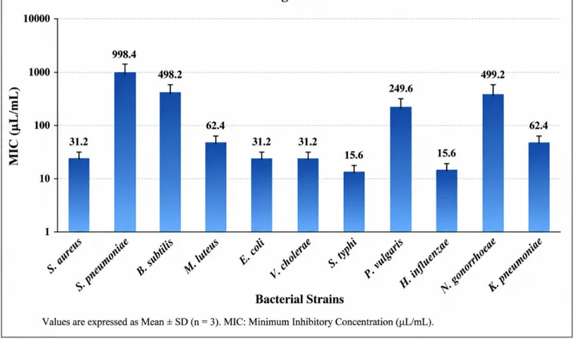
Figure 2: Minimum Inhibitory Concentration (MIC) of Leucas aspera Ethanolic Leaf Extract Against MDR Bacterial Strains
One-way analysis of variance (ANOVA) demonstrated significant differences in antibacterial activity among the tested bacterial strains (p < 0.05). Bacterial isolates exhibiting larger zones of inhibition generally showed lower MIC values, indicating a significant inverse relationship between inhibition zone diameter and MIC. These findings suggest that the antibacterial activity of Leucas aspera is concentration-dependent and varies according to bacterial species.
Comparison with the standard antibiotic showed that the antibiotic produced larger inhibition zones against all bacterial strains tested. Nevertheless, the ethanolic leaf extract demonstrated appreciable antibacterial activity against several clinically important MDR pathogens, particularly Haemophilus influenzae, Staphylococcus aureus, Escherichia coli, Micrococcus luteus, and Klebsiella pneumoniae.
Table 3: Comparative Antibacterial Activity of Leucas aspera Leaf Extract and Standard Antibiotic
|
Sr. No. |
Bacterial Strain |
Plant Extract (mm) |
Standard Antibiotic (mm) |
|
1 |
Staphylococcus aureus |
12.5 |
25 |
|
2 |
Streptococcus pneumoniae |
6.0 |
15 |
|
3 |
Bacillus subtilis |
0.0 |
13 |
|
4 |
Micrococcus luteus |
12.0 |
20 |
|
5 |
Escherichia coli |
12.0 |
17 |
|
6 |
Vibrio cholerae |
11.5 |
15 |
|
7 |
Salmonella typhi |
10.0 |
15 |
|
8 |
Proteus vulgaris |
0.0 |
12 |
|
9 |
Haemophilus influenzae |
13.0 |
21 |
|
10 |
Neisseria gonorrhoeae |
0.0 |
22 |
|
11 |
Klebsiella pneumoniae |
11.0 |
16 |
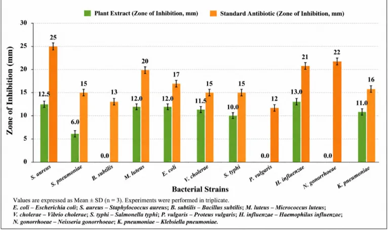
Figure 3: Comparison of Antibacterial Activity of Leucas aspera Ethanolic Leaf Extract and Standard Antibiotic
When the bacterial isolates were grouped according to Gram reaction, the extract exhibited antibacterial activity against both Gram-positive and Gram-negative bacteria. Among the Gram-positive organisms, Staphylococcus aureus was the most susceptible, whereas Bacillus subtilis and Streptococcus pneumoniae showed comparatively lower susceptibility. Among the Gram-negative bacteria, Haemophilus influenzae exhibited the highest susceptibility, followed by Escherichia coli, Vibrio cholerae, Klebsiella pneumoniae, and Salmonella typhi.
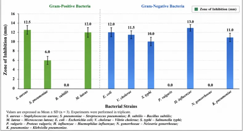
Figure 4: Comparative Antibacterial Activity of Leucas aspera Ethanolic Leaf Extract Against Gram-Positive and Gram-Negative MDR Bacteria
Representative agar well diffusion plates demonstrated distinct inhibition zones surrounding the wells containing the ethanolic leaf extract, confirming its antibacterial activity against susceptible bacterial strains and supporting the quantitative findings obtained from the zone of inhibition assay.
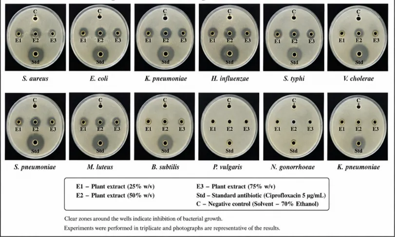
Figure 5: Representative agar well diffusion assay showing the zones of inhibition produced by the ethanolic leaf extract of Leucas aspera against multidrug-resistant bacterial strains
DISCUSSION
The present study demonstrated that the ethanolic leaf extract of Leucas aspera possesses appreciable antibacterial activity against several multidrug-resistant bacterial pathogens, although its efficacy varied among the tested microorganisms. The extract showed the highest activity against Haemophilus influenzae and Staphylococcus aureus, while little or no activity was observed against Bacillus subtilis, Proteus vulgaris, and Neisseria gonorrhoeae. The observed variation in susceptibility may be attributed to differences in bacterial cell wall structure, membrane permeability, and intrinsic resistance mechanisms.
The antibacterial activity observed in the present study is consistent with the findings of Mangathayaru et al. (2005), who reported significant antimicrobial activity of Leucas aspera flower extracts against both Gram-positive and Gram-negative bacteria. Similarly, Rahman et al. (2013) demonstrated that the ethanolic extract of Leucas aspera exhibited notable antibacterial activity against several pathogenic bacteria, indicating that ethanol is an effective solvent for extracting bioactive phytochemicals responsible for antimicrobial activity. Furthermore, Cowan (1999) reported that medicinal plants rich in flavonoids, alkaloids, tannins, terpenoids, and phenolic compounds exhibit broad-spectrum antimicrobial activity through disruption of bacterial cell membranes, inhibition of essential enzymes, and interference with nucleic acid synthesis. Gibbons (2008) also highlighted the therapeutic potential of plant-derived phytochemicals against multidrug-resistant microorganisms, supporting the concentration-dependent antibacterial activity and inverse relationship between the zone of inhibition and MIC observed in the present study.
Overall, the present findings support the traditional medicinal use of Leucas aspera and indicate that its ethanolic leaf extract may serve as a promising natural source of antibacterial compounds. However, further phytochemical characterization, toxicity evaluation, isolation of active constituents, and in vivo studies are required before its potential clinical application can be established.
CONCLUSION
The present study demonstrated that the ethanolic leaf extract of Leucas aspera possesses appreciable antibacterial activity against selected multidrug-resistant bacterial strains, supporting its traditional use as a medicinal plant. The extract exhibited the highest antibacterial activity against Haemophilus influenzae and Staphylococcus aureus, whereas comparatively lower or no activity was observed against Bacillus subtilis, Proteus vulgaris, and Neisseria gonorrhoeae. The agar well diffusion assay and minimum inhibitory concentration (MIC) analysis confirmed that the antibacterial activity of the extract varied among the tested organisms and was dependent on their susceptibility. Although the antibacterial efficacy of the extract was lower than that of the standard antibiotic, the findings indicate that Leucas aspera contains bioactive phytochemicals with promising antimicrobial properties. These results provide scientific evidence supporting the potential use of this medicinal plant as a natural source of antibacterial agents for the management of multidrug-resistant bacterial infections. Further studies involving phytochemical isolation, characterization of active constituents, toxicity evaluation, and in vivo investigations are recommended to validate its safety, mechanism of action, and therapeutic efficacy before considering its pharmaceutical or clinical application.
REFERENCES


Pooja Rao, Ghanshyam Sevak, In Vitro Evaluation of Antimicrobial Activity of Leucas aspera Leaf Extract against Multidrug Resistance Bacterial Strains, Int. J. of Pharm. Sci., 2026, Vol 4, Issue 8, 838-846. https://doi.org/10.5281/zenodo.21809192
 10.5281/zenodo.21809192