
We use cookies to ensure our website works properly and to personalise your experience. Cookies policy
Sagar Institute of Research and Technology – Pharmacy, Sanjeev Agrawal Global Educational University, Bhopal, Madhya Pradesh, India..
The present study evaluated the hepatoprotective activity of the ethanolic extract of Silybum marianum against carbon tetrachloride (CCl?)-induced liver damage in Wistar rats. The seeds were extracted using 70% ethanol and subjected to preliminary phytochemical screening, which confirmed the presence of flavonoids, phenols, tannins, saponins, glycosides, and steroids. Hepatotoxicity was induced by CCl? administration, and the extract was evaluated at doses of 100 and 200 mg/kg. Treatment with Silybum marianum significantly reduced elevated serum levels of ALT, AST, ALP, and bilirubin while restoring total protein levels. The extract also improved antioxidant status by increasing superoxide dismutase (SOD) and catalase (CAT) activities and reducing malondialdehyde (MDA) levels. Histopathological examination revealed marked restoration of normal hepatic architecture with reduced necrosis, steatosis, and inflammatory infiltration, particularly at the dose of 200 mg/kg. The hepatoprotective effects were comparable to those of the standard drug silymarin. The findings indicate that Silybum marianum possesses significant dose-dependent hepatoprotective and antioxidant activities, supporting its potential use in the management of liver disorders.
The liver is a vital organ responsible for numerous physiological functions, including metabolism of carbohydrates, proteins, and lipids, detoxification of xenobiotics, synthesis of plasma proteins, and regulation of biochemical homeostasis. Owing to its central role in the metabolism of endogenous and exogenous compounds, the liver is highly susceptible to injury caused by drugs, chemicals, alcohol, and environmental toxins [1, 2]. Liver diseases constitute a major global health problem, and the search for effective hepatoprotective agents remains an important area of biomedical research [3, 4].Carbon tetrachloride (CCl₄) is one of the most commonly used hepatotoxic agents for inducing experimental liver injury in laboratory animals. Following metabolic activation by the hepatic cytochrome P450 enzyme system, CCl₄ generates reactive free radicals, particularly trichloromethyl (CCl₃) and trichloromethyl peroxy (OOCCl₃) radicals. These radicals initiate lipid peroxidation, leading to oxidative stress, cellular membrane damage, inflammation, and hepatocellular necrosis [5,6]. Therefore, CCl₄-induced hepatotoxicity serves as a reliable experimental model for evaluating the efficacy of hepatoprotective compounds.
Oxidative stress is a key factor in the pathogenesis of liver injury. Excessive production of reactive oxygen species (ROS) overwhelms the body's antioxidant defense mechanisms, resulting in cellular damage and impaired liver function [7]. Consequently, natural antioxidants derived from medicinal plants have gained significant attention for their potential role in protecting the liver against oxidative damage [8].
Silybum marianum (Milk Thistle), a medicinal plant belonging to the family Asteraceae, has been traditionally used for the treatment of liver and biliary disorders for centuries[9, 10]. Its major bioactive constituent, silymarin, is a mixture of flavonolignans including silybin, silydianin, and silychristin. Silymarin possesses potent antioxidant, anti-inflammatory, membrane-stabilizing, and free radical scavenging properties, which contribute to its hepatoprotective effects [11,12,13]. Previous studies have demonstrated that silymarin can inhibit lipid peroxidation, enhance cellular antioxidant defenses, promote hepatocyte regeneration, and protect the liver against various toxic insults [14].
Therefore, the present study was undertaken to evaluate the hepatoprotective activity of Silybum marianum against carbon tetrachloride-induced liver damage in rats. The protective effect was assessed through biochemical and histopathological parameters to validate its potential as a natural hepatoprotective agent[15, 16].
MATERIALS AND METHODS:
Plant Material and Extraction
Dried seeds of Silybum marianum were procured from a reputed herbal supplier and authenticated by a qualified botanist. The seeds were cleaned, shade-dried, powdered, and subjected to Soxhlet extraction using 70% ethanol for 8 h. The extract was concentrated under reduced pressure using a rotary evaporator and stored at 4°C until use. Percentage yield was calculated based on the dry weight of the extract [17,18].
Phytochemical Screening
The ethanolic extract was screened qualitatively for major phytoconstituents such as flavonoids, alkaloids, tannins, phenolics, saponins, glycosides, and steroids using standard phytochemical tests described in the literature [19].
Experimental Animals and Ethical Approval
Healthy male Wistar rats (150–200 g) were obtained from a CPCSEA-approved animal facility and acclimatized for one week under standard laboratory conditions. The study protocol was approved by the Institutional Animal Ethics Committee (IAEC) and conducted according to CPCSEA guidelines [20,21].
Acute Oral Toxicity Study
Acute oral toxicity of the extract was evaluated according to OECD Guideline 423. Animals were observed for signs of toxicity and mortality for 14 days after administration. Based on the findings, doses of 100 and 200 mg/kg were selected for further studies [22].
Experimental Design
Table 1: Grouping of Animals for Antioxidant activity
|
Group |
Treatment |
|
I |
Normal control |
|
II |
CCl₄ control (1 mL/kg, i.p.) |
|
III |
CCl₄ + Silymarin (100 mg/kg) |
|
IV |
CCl₄ + S. marianum extract (100 mg/kg) |
|
V |
CCl₄ + S. marianum extract (200 mg/kg) |
Carbon tetrachloride (1:1 in olive oil) was administered intraperitoneally to induce hepatotoxicity, while treatments were given orally for 14 days [23].
Biochemical Analysis
Blood samples were collected at the end of the study, and serum was separated by centrifugation. Liver function markers including ALT, AST, ALP, total bilirubin, and total protein were estimated using standard diagnostic kits [24].
Estimation of Antioxidant Parameters
Liver tissues were homogenized in phosphate buffer (pH 7.4), centrifuged, and the supernatant was used for antioxidant assays [29].
Malondialdehyde (MDA)
MDA levels were determined using the TBARS method described by Ohkawa . Absorbance was measured at 532 nm, and results were expressed as nmol MDA/mg protein [30].
Superoxide Dismutase (SOD)
SOD activity was estimated by the Misra and Fridovich method based on inhibition of epinephrine auto-oxidation. Results were expressed as U/min/mg protein [31].
Catalase (CAT)
Catalase activity was measured according to Aebi’s method by monitoring the decomposition of hydrogen peroxide at 240 nm and expressed as μmol H₂O₂ decomposed/min/mg protein [32].
Histopathological Evaluation
Liver tissues were fixed in 10% neutral buffered formalin, processed routinely, and stained with hematoxylin and eosin (H&E). Histological examination was performed to assess necrosis, inflammation, fatty changes, and hepatocellular degeneration [33].
Statistical Analysis
Results were expressed as mean ± SEM. Statistical significance was analyzed using one-way ANOVA followed by Tukey’s post hoc test. Values of p < 0.05 were considered statistically significant [34].
RESULT AND DISCUSSION
Plant Material and Extraction
The seeds of Silybum marianum were procured from a reputed herbal supplier and authenticated by a qualified taxonomist. A voucher specimen was deposited in the departmental herbarium for future reference. The seeds were cleaned, shade-dried, powdered, and extracted using 70% ethanol in a Soxhlet apparatus for 8 hours. The extract was concentrated under reduced pressure using a rotary evaporator (40–50°C) and stored in an amber-colored container at 4°C until further use. The extraction process yielded a thick brownish semi-solid ethanolic extract with a percentage yield of 13.5% w/w. The obtained yield indicates efficient extraction of bioactive constituents and is consistent with previously reported yields for Silybum marianum seeds extracted using hydroalcoholic solvents.
Table 2: Comparative Yield of Silybum marianum Extracts Using Various Solvents and Methods
|
S. No. |
Extraction Method |
Solvent Used |
Plant Material (g) |
Yield (% w/w) |
|
1 |
Soxhlet Extraction |
70% Ethanol |
200 |
13.5 |
|
2 |
Cold Maceration |
70% Ethanol |
100 |
11.8 |
|
3 |
Soxhlet Extraction |
Absolute Ethanol |
200 |
10.2 |
|
4 |
Reflux Extraction |
Methanol |
150 |
14.3 |
|
5 |
Cold Maceration |
Distilled Water |
100 |
7.6 |
|
6 |
Soxhlet Extraction |
80% Methanol |
200 |
15.5 |
The use of 70% ethanol as the extraction solvent was selected because of its ability to efficiently extract both polar and moderately non-polar phytoconstituents. The obtained extractive yield of **13.5% w/w** was within the reported range for *Silybum marianum* seeds, indicating effective recovery of bioactive constituents, particularly flavonolignans such as silybin and silydianin.
The relatively high yield suggests the presence of important phytochemical groups including flavonoids, phenolic compounds, tannins, and sterols, which are associated with hepatoprotective and antioxidant activities. In addition, concentration of the extract under reduced pressure and storage at 4°C helped preserve thermolabile constituents, ensuring the stability and biological activity of the extract for subsequent pharmacological evaluation. Overall, the results demonstrate that Soxhlet extraction with 70% ethanol is a suitable method for obtaining a phytochemically rich extract of Silybum marianum.
Phytochemical Screening
Preliminary phytochemical analysis of the ethanolic extract of Silybum marianum confirmed the presence of flavonoids, phenols, saponins, tannins, glycosides, and steroids, while alkaloids were absent. The presence of these bioactive constituents may contribute to the antioxidant and hepatoprotective activity of the extract.
Table 3: Phytochemical Constituents in Ethanolic Extract of Silybum marianum
|
S. No. |
Phytochemical Class |
Test Method |
Result |
|
1 |
Flavonoids |
Alkaline reagent test |
+ |
|
2 |
Phenols |
Ferric chloride test |
+ |
|
3 |
Saponins |
Frothing test |
+ |
|
4 |
Tannins |
Lead acetate / Ferric chloride test |
+ |
|
5 |
Glycosides |
Keller–Killiani test |
+ |
|
6 |
Steroids |
Salkowski’s test |
+ |
|
7 |
Alkaloids |
Wagner’s and Mayer’s tests |
− |
Biochemical Analysis
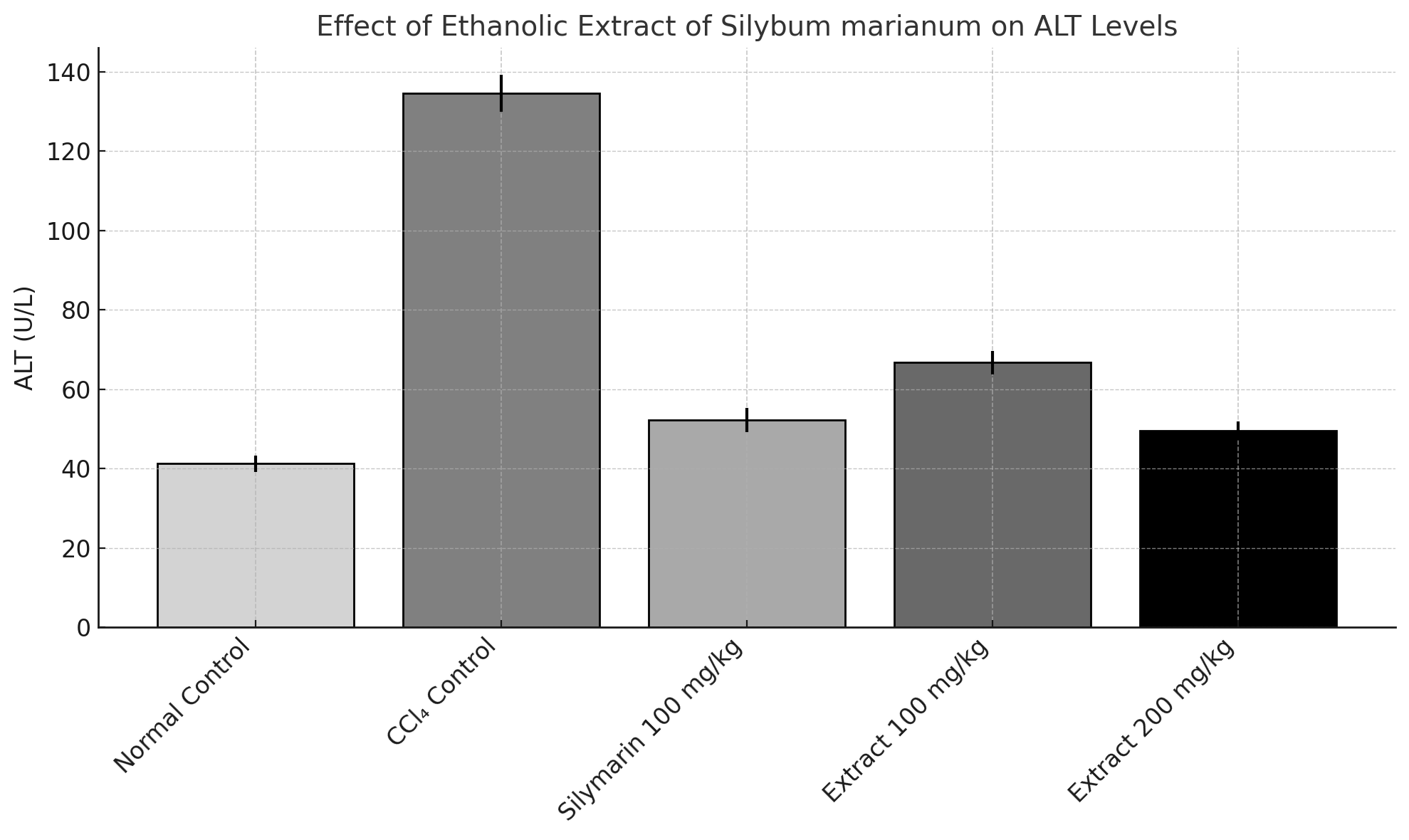
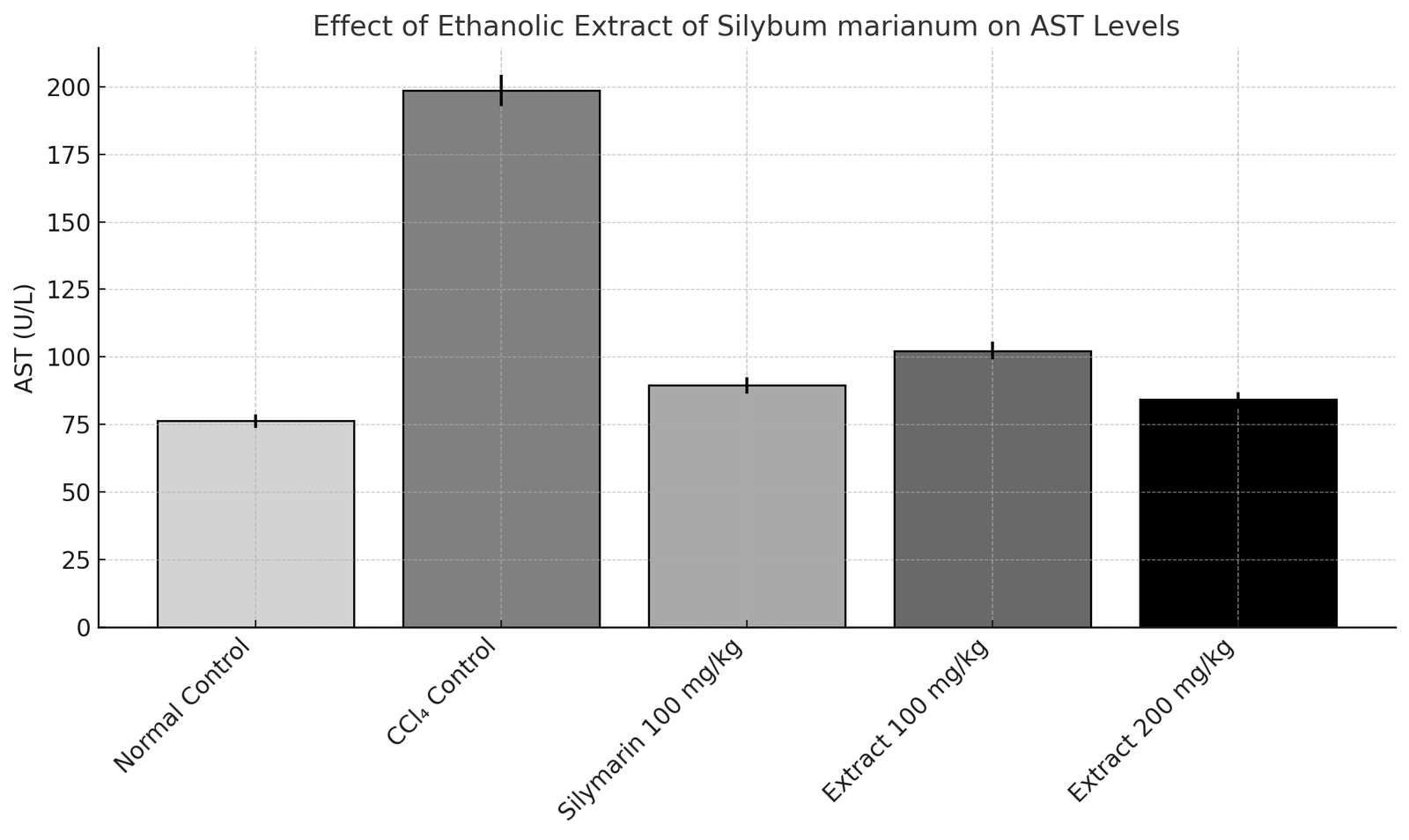
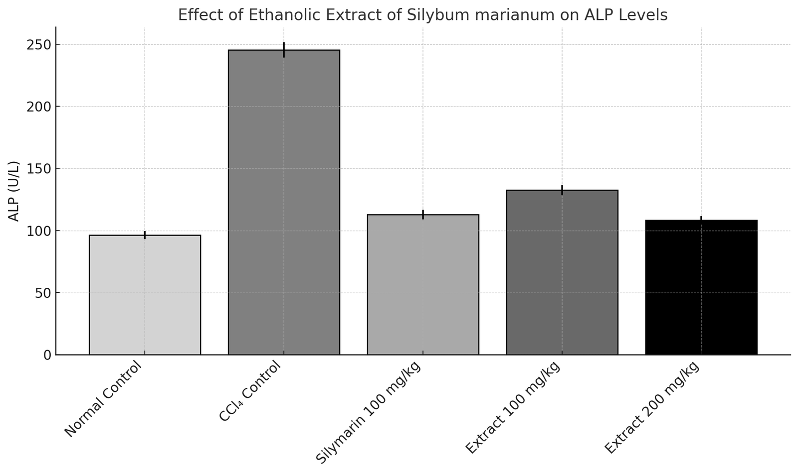
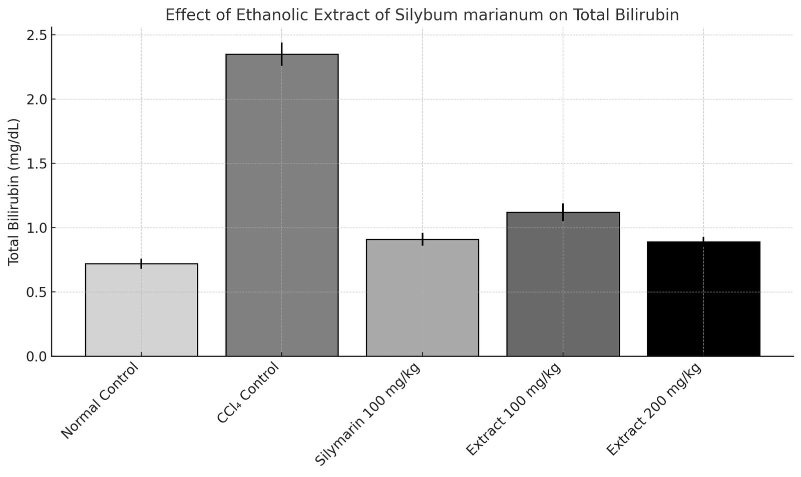
Table 4: Effect of Ethanolic Extract of Silybum marianum for Biochemical estimation
|
Group |
ALT (U/L) |
AST (U/L) |
ALP (U/L) |
Total Bilirubin (mg/dL) |
Total Protein (g/dL) |
|
I |
41.25 ± 2.11 |
76.32 ± 2.45 |
96.42 ± 3.18 |
0.72 ± 0.04 |
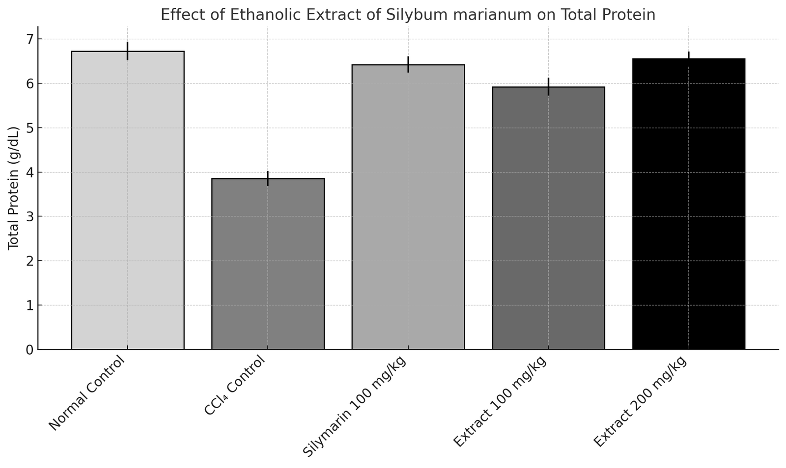
6.72 ± 0.21 |
|
II |
134.62 ± 4.58† |
198.56 ± 5.67† |
245.36 ± 6.01† |
2.35 ± 0.09† |
3.85 ± 0.17† |
|
III |
52.33 ± 3.04*** |
89.47 ± 3.01*** |
112.78 ± 3.92*** |
0.91 ± 0.05*** |
6.42 ± 0.18*** |
|
IV |
66.72 ± 2.95** |
102.36 ± 3.29** |
132.64 ± 4.11** |
1.12 ± 0.07** |
5.92 ± 0.20** |
|
V |
49.56 ± 2.38*** |
84.25 ± 2.74*** |
108.25 ± 3.44*** |
0.89 ± 0.04*** |
6.55 ± 0.16*** |
|
|
|
|
Effect of Ethanolic Extract of Silybum marianum on ALT Levels in CCl₄-Induced Hepatotoxic Rats |
Effect of Ethanolic Extract of Silybum marianum on AST Levels in CCl₄-Induced Hepatotoxic Rats |
|
|
|
|
Effect of Ethanolic Extract of Silybum marianum on ALP Levels in CCl₄-Induced Hepatotoxic Rats |
Effect of Ethanolic Extract of Silybum marianum on Total Bilirubin Levels in CCl₄-Induced Hepatotoxic Rats |
|
|
|
|
Effect of Ethanolic Extract of Silybum marianum on Total Protein Levels in CCl₄-Induced Hepatotoxic Rats |
|
Figure 1: Biochemical estimation of Silybum marianum for Antioxidant activity
Estimation of Antioxidant Parameters
Table 5: Effect of Ethanolic Extract of Silybum marianum for Antioxidant activity
|
Group |
MDA (nmol/mg protein) |
SOD (units/min/mg protein) |
CAT (μmol H₂O₂ decomposed/min/mg protein) |
|
I |
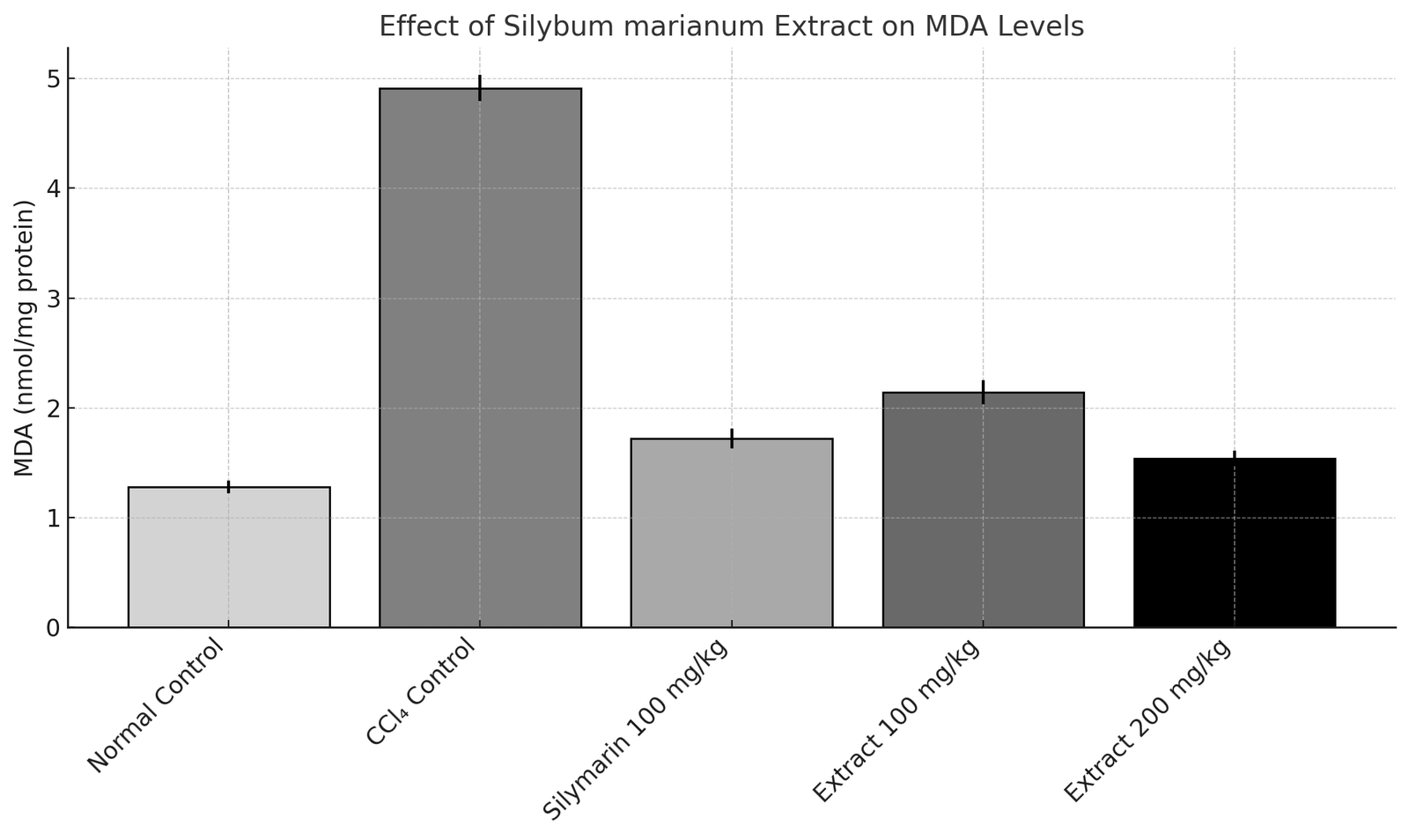
1.28 ± 0.06 |
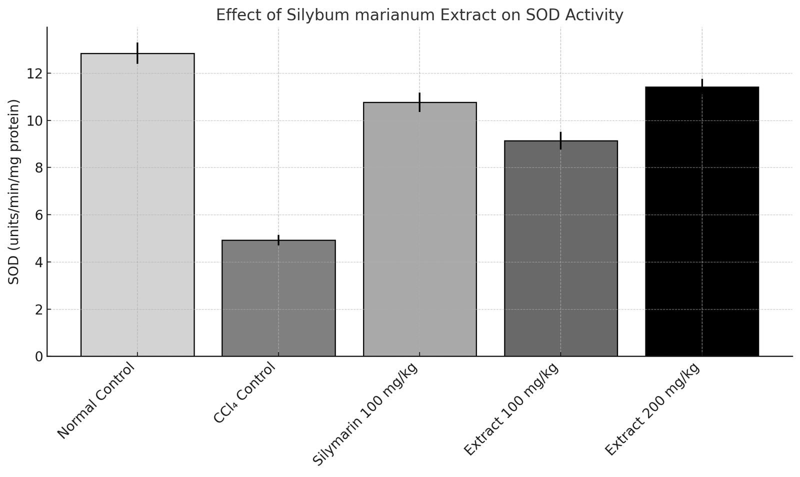
12.84 ± 0.45 |
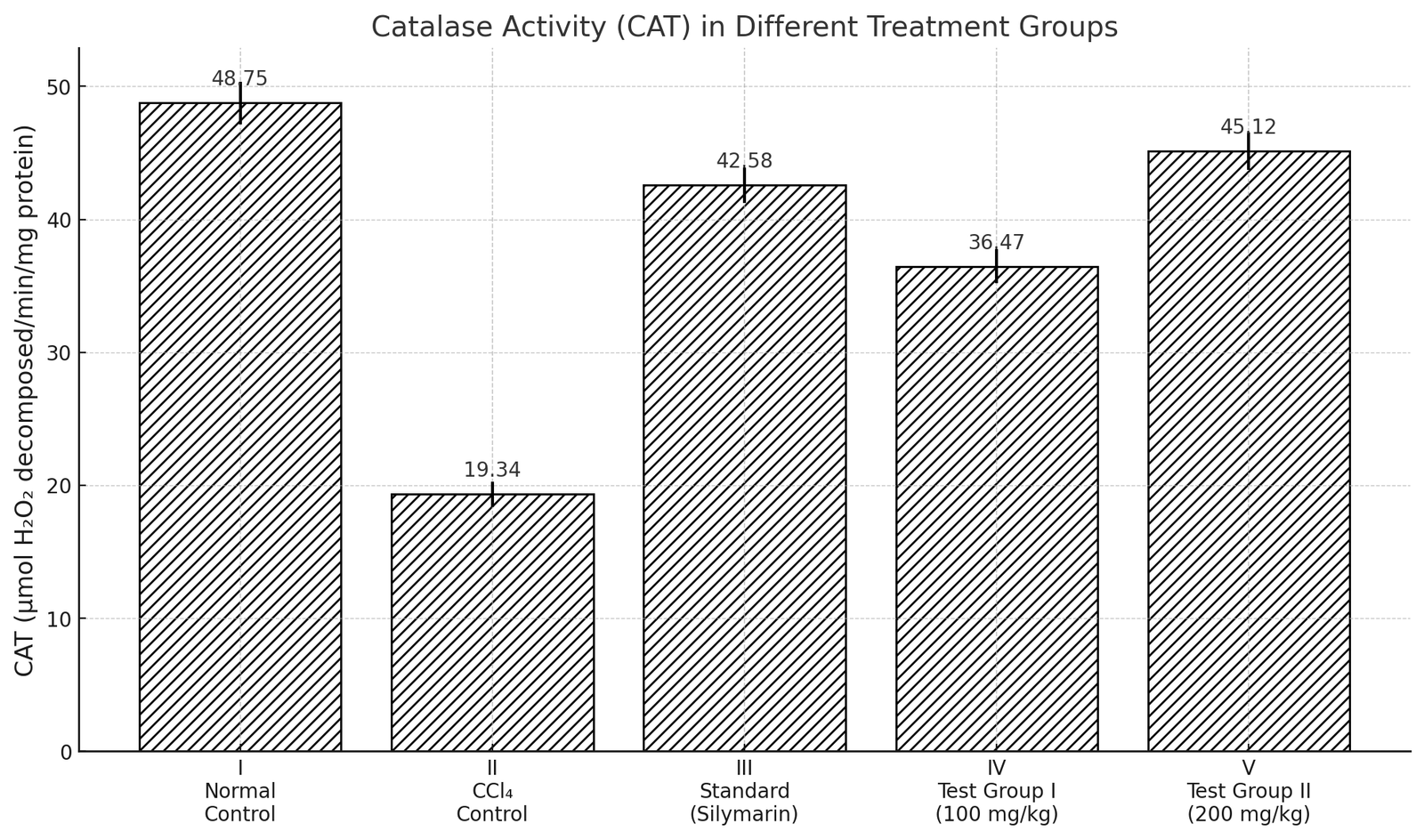
48.75 ± 1.62 |
|
II |
4.91 ± 0.12† |
4.92 ± 0.22† |
19.34 ± 0.95† |
|
III |
1.72 ± 0.09*** |
10.76 ± 0.41*** |
42.58 ± 1.33*** |
|
IV |
2.14 ± 0.11** |
9.14 ± 0.38** |
36.47 ± 1.28** |
|
V |
1.54 ± 0.07*** |
11.42 ± 0.33*** |
45.12 ± 1.41*** |
|
|
|
|
Effect of Ethanolic Extract of Silybum marianum on MDA Levels in CCl₄-Induced Hepatotoxic Rats |
Effect of Ethanolic Extract of Silybum marianum on SOD Levels in CCl₄-Induced Hepatotoxic Rats |
|
|
|
|
Effect of Ethanolic Extract of Silybum marianum on CAT Levels in CCl₄-Induced Hepatotoxic Rats |
|
Figure 2: Effect of Ethanolic Extract of Silybum marianum for Antioxidant activity
Histopathological Evaluation
Histopathological examination of H&E-stained liver sections revealed normal hepatic architecture with intact hepatocytes and no pathological alterations in the normal control group. In contrast, the CCl₄-treated group showed severe hepatocellular necrosis, fatty degeneration, inflammatory cell infiltration, and disruption of normal liver architecture, confirming successful induction of hepatotoxicity. Treatment with silymarin (100 mg/kg) markedly protected the liver, as evidenced by reduced necrosis and inflammation along with preservation of hepatic architecture. Similarly, rats treated with Silybum marianum extract exhibited dose-dependent hepatoprotective effects. The 100 mg/kg dose showed moderate improvement with reduced steatosis and inflammatory changes, whereas the 200 mg/kg dose demonstrated near-normal liver architecture with minimal necrosis and significant hepatocyte regeneration. The hepatoprotective effect of the higher dose was comparable to that of the standard silymarin-treated group.
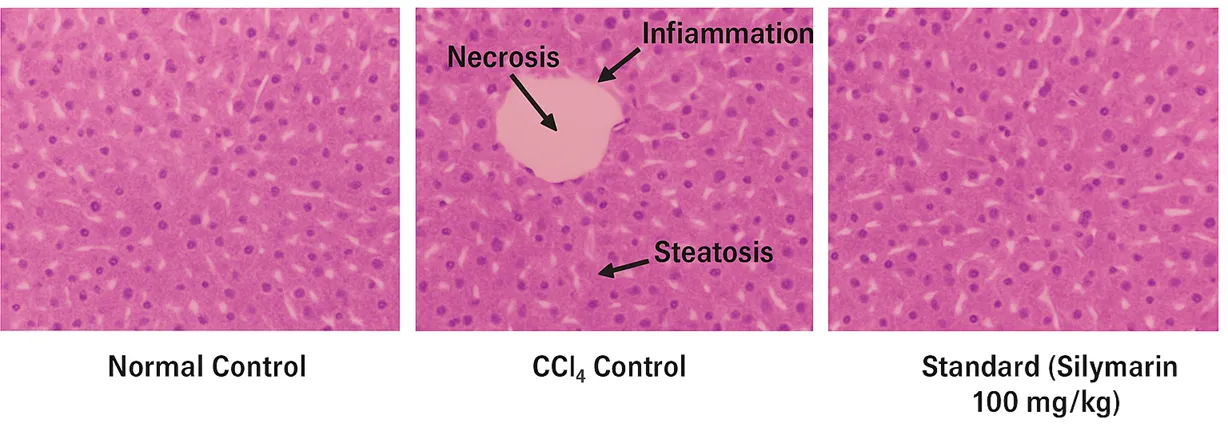
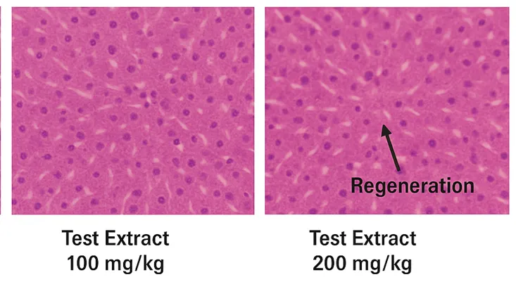
Figure 3: Histopathological Photomicrographs of Liver Tissues from Different Groups (Normal, CCl₄ Control, Standard, Extract 100 mg/kg, Extract 200 mg/kg)
The histopathological findings confirmed the hepatoprotective potential of *Silybum marianum* extract against CCl₄-induced liver injury. Severe hepatic damage observed in the CCl₄ control group was markedly reduced following treatment with the extract. The protective effect was dose-dependent, with the 200 mg/kg dose showing greater restoration of normal liver architecture, reduced necrosis, and decreased inflammatory changes. These effects may be attributed to the antioxidant and membrane-stabilizing properties of silymarin, the principal active constituent of *Silybum marianum*. The presence of regenerating hepatocytes further indicates the liver-restorative potential of the extract, with efficacy comparable to the standard silymarin-treated group.
CONCLUSION
The present study demonstrated that the ethanolic extract of Silybum marianum possesses significant hepatoprotective activity against CCl₄-induced liver damage in Wistar rats. Treatment with the extract effectively reduced elevated liver enzyme levels (ALT, AST, ALP, and bilirubin), restored antioxidant defenses (SOD and CAT), and decreased lipid peroxidation as indicated by reduced MDA levels. Histopathological findings further confirmed the protective effect of the extract, showing improved hepatic architecture, reduced necrosis and inflammation, and enhanced hepatocyte regeneration, particularly at the dose of 200 mg/kg. The hepatoprotective effect was dose-dependent and comparable to that of the standard drug silymarin. These findings suggest that the hepatoprotective activity of Silybum marianum may be attributed to its antioxidant and membrane-stabilizing properties. Therefore, Silybum marianum has potential as a natural therapeutic agent for the prevention and management of liver disorders.
REFERENCES


Yashkirti Vishwakarma, Abhishek Shrivastava, Evaluation of Hepatoprotective Activity of Silybum Marianum on Carbon Tetrachloride Induced Liver Damage in Rats, Int. J. of Pharm. Sci., 2026, Vol 4, Issue 6, 5370-5379, https://doi.org/10.5281/zenodo.20773099
 10.5281/zenodo.20773099