

We use cookies to ensure our website works properly and to personalise your experience. Cookies policy
Dr. J.J. Magdum Pharmacy Collage, Jaysingpur.
Gout is a metabolic and inflammatory condition associated with increased uric acid concentration in the blood, which promotes the formation and accumulation of monosodium urate crystals in the joints. The deposition of these crystals triggers inflammatory reactions that lead to joint pain, swelling, redness, and discomfort. Currently, gout is managed using conventional medications; however, the possibility of adverse effects and limitations associated with long-term treatment has encouraged researchers to explore plant-based therapeutic approaches. This review discusses the potential role of various medicinal plants and their phytochemical constituents in the prevention and management of gout. Their anti-gout properties are mainly associated with the inhibition of xanthine oxidase activity, regulation of uric acid metabolism, improvement in uric acid elimination, and reduction of inflammation. Research findings suggest that several medicinal plants possess promising pharmacological properties that may contribute to gout management. Nevertheless, additional experimental studies and clinical investigations are essential to validate their therapeutic potential, establish appropriate doses, and determine their long-term safety and effectiveness.
Hyperuricemia (HUA) develops due to abnormal purine metabolism, resulting in increased uric acid levels in the blood and the deposition of urate crystals. Acute gouty arthritis (AGA) is an inflammatory disorder that occurs when monosodium urate (MSU) crystals form and accumulate in the joints because uric acid (UA) levels exceed their saturation threshold in the blood or tissue fluids. Elevated serum MSU levels represent the most significant risk factor for the development of gout. Patients with gout are also at an increased risk of associated complications, including hypertension, chronic kidney disease, obesity, diabetes, myocardial infarction, and heart failure. According to the “2021 Trends in Chinese Hyperuricemia and Gout White Paper,” the prevalence of gout in China has continued to increase annually, with the disease increasingly affecting younger individuals. As economic conditions and living standards improve, gout-associated risk factors, including alcohol consumption, the intake of high-fructose beverages, and increased daily consumption of meat and seafood, have also become more prevalent. [1,2]
MSU crystals may accumulate in various tissues, particularly within and around the joints, leading to the formation of tophi. Gout is primarily diagnosed by identifying the characteristic MSU crystals through joint fluid aspiration or aspiration of material from tophi. In the early stages, gout typically presents as acute joint inflammation, which can be rapidly relieved by non-steroidal anti-inflammatory drugs (NSAIDs) or colchicine. Renal stones and tophi generally occur during the later stages of the disease. The primary objective of gout management is to reduce serum uric acid (SUA) levels below the threshold for crystal deposition through dietary modifications and the administration of uric acid-lowering medications. This approach promotes the dissolution of MSU crystals and helps prevent further gout attacks. [3,4]
ETIOLOGY: -
When gout is considered a chronic disease characterized by urate crystal deposition, its underlying cause can be attributed to an imbalance between urate intake and production and its excretion. This imbalance results in urate accumulation and crystal formation in the tissues, creating conditions that trigger an attack by the innate immune system, leading to acute inflammation and the associated clinical manifestations.[5]
GENETICS: -
Most individuals with gout excrete less urate in their urine than people in the general population. Patients with gout also exhibit increased tubular reabsorption of urate. This enhanced reabsorption is influenced by genetic variations in urate transporters. The strongest genetic associations have been identified in SLC2A9 (GLUT9) and SLC22A12 (URAT1), while several other genes, including numerous transporter genes, contribute to a lesser extent. More recent findings have highlighted the role of reduced activity of the ABCG2 transporter in the intestine, which contributes to hyperuricemia. Genetic differences also play a considerable role in explaining variations in gout prevalence among different ethnic groups. Recent advances in gout genetics include the identification of an association between reduced mitochondrial DNA copy number and gout. Although the exact relationship remains uncertain, it may be linked to the functioning of the innate immune system. Several research groups have also identified rare genetic variants associated with gout in transporter genes, such as ABCG2 and SLC22A1, as well as in ALDH16A1, which encodes a member of the aldehyde dehydrogenase superfamily. The association with ALDH16A1 variants is particularly noteworthy because the ALDH16A1 protein may interact with hypoxanthine-guanine phosphoribosyl transferase (HPRT1), a protein involved in urate metabolism. Deficiency of HPRT1 leads to Lesch–Nyhan syndrome and urate accumulation. A recent large-scale genome-wide association study (GWAS) conducted in Japan investigated the genetic factors responsible for hyperuricemia. This study, together with other research, contributes to a better understanding of the genetic associations with serum urate (SU) levels in non-European populations, which have received less attention than those in European populations. Furthermore, the Global Gout Genetics Consortium has planned a large-scale gout GWAS designed to identify a substantial number of additional genetic associations. These recent findings are gradually providing a more comprehensive understanding of the important role genetics plays in the development of gout. [6,7]
ENVIRONMENTAL FACTORS: -
The main sources of urate are (1) dietary purines and food substances that are converted into urate, such as fructose, and (2) metabolic processes involving purine breakdown, including cellular repair and replacement. Large epidemiological studies have shown that the consumption of alcohol, sugar-sweetened beverages, fructose, organ meats, and seafood contributes to hyperuricemia. However, it is important to note that evidence from educational and intervention trials supporting the clinical benefits of restricting these urate sources in the management of established gout remains limited. Dietary modifications alone may produce small reductions in serum urate (SU) levels, but these reductions have not been demonstrated to result in sustained decreases in the frequency of gout flares. For example, one clinical trial provided dietary education to the intervention group on two occasions, while the control group received usual care. Although the intervention improved dietary knowledge and changed eating habits, no reduction in SU levels was observed. A recent study involving 17,000 participants investigated the relative contributions of dietary factors and genetics to SU levels and found that the consumption of 16 different food items was associated with SU levels (2). Variations in the consumption of these food items accounted for 3.3% of the variation in SU levels, whereas genetic factors explained 24% of the variation. This simultaneous assessment of dietary and genetic influences demonstrates the relatively small contribution of diet to variations in SU levels. However, weight loss has been shown to substantially reduce SU levels. For instance, a study evaluating bariatric surgery reported that 83% of patients with gout had SU levels above 0.41 mmol/L before surgery, compared with only 33% one year after the procedure. These findings are supported by evidence from Mendelian randomisation studies indicating that fat mass directly increases SU levels. Such reductions in SU are likely to produce clinically meaningful improvements in gout management. Extensive evidence is available regarding the effects of lifestyle and dietary factors on the incidence of gout. An analysis of the Health Professionals Follow-Up Study cohort, conducted over 26 years, found that adherence to the Dietary Approaches to Stop Hypertension (DASH) diet was associated with a lower incidence of gout. This dietary pattern emphasizes the consumption of fruits, nuts, legumes, vegetables, low-fat dairy products, and whole grains, while limiting sodium, sugar-sweetened beverages, and red and processed meats. Adherence to this dietary pattern was associated with a reduced incidence of gout. Bariatric surgery has also been shown to produce a clinically meaningful reduction in gout incidence. A large Swedish study involving 1,982 obese patients who underwent surgery and 1,999 obese control participants reported an adjusted hazard ratio of 0.60 for the incidence of gout following surgery. [8,9,10]
THE TRANSITION FROM HYPERURICEMIA TO GOUT: -
Only a fraction of individuals with hyperuricemia eventually develop gout. Based on data from the 2007–2008 United States National Health and Nutrition Examination Survey, Choi and colleagues reported that the prevalence of gout was 5.9% in men and 2.0% in women. In contrast, the prevalence of hyperuricemia was 21.2% among men and 21.6% among women. Therefore, within this population, only 28% of hypouricemic men and 9% of hypouricemic women also had gout. The factors that determine why some individuals with hyperuricemia develop gout while others do not are not yet fully understood. However, serum urate (SU) levels and the amount of urate crystals present may be important contributing factors. A strong association has been established between SU levels and gout. Imaging studies have shown that the frequency and volume of urate crystals increase as the condition progresses from asymptomatic hyperuricemia to clinically apparent gout. One possible explanation for the difference between the prevalence of hyperuricemia and gout is that urate crystals gradually accumulate in hyperuricemia individuals until their burden reaches a critical threshold that triggers gout. Nevertheless, the considerable difference between individuals with elevated SU levels and those who have both elevated SU levels and gout suggests that other factors may also be involved. In particular, variations in the innate immune response to urate crystals may contribute to the development of the disease. Evidence supporting this hypothesis comes from a study investigating weight loss following bariatric surgery. In this study, the mean body weight of 20 participants decreased from 123 kg to 98 kg. Significant reductions in white blood cell responses to monosodium urate crystals were observed in relation to interleukin (IL)-1β, IL-8, and IL-6. These findings indicate that factors beyond the mere presence or quantity of urate crystals may influence the development of gout. Furthermore, two studies have reported associations between gout and an important component of the innate immune system. The Toll-like receptor 4 (TLR4) variant rs2149356 has been associated with gout. However, this association was not observed in every cohort investigated and requires further confirmation. [11,12]
RENAL FACTORS: -
Patients with gout and chronic kidney disease (CKD) often experience greater difficulty in maintaining optimal serum urate (SU) levels. Since urate is primarily eliminated through the kidneys, its concentration in the blood increases as renal function declines. Consequently, hyperuricemia is more frequently observed in patients with stage 3–5 CKD Studies using mouse models suggest that hyperuricemia itself may impair kidney function by causing fibrosis of the afferent arterioles, thereby reducing blood flow into the glomeruli. Consistent with these findings, research has demonstrated a five-fold increase in gout prevalence among patients with an estimated glomerular filtration rate (eGFR) of ≤60 mL/min/1.73 m² compared with individuals without kidney disease. Other studies have reported that more than 70% of patients with gout have CKD stage ≥2, while approximately 24% have CKD stage ≥3. Urate crystals may also contribute to renal inflammation and fibrosis by accumulating in the renal medulla and directly impairing kidney function. The relationship between gout and progressive CKD is further complicated by conflicting research findings. Some researchers suggest that persistent, low-grade inflammation in patients with CKD may aggravate gout flares in individuals with gout and hyperuricemia. In contrast, other studies have reported that progression to end-stage kidney disease (ESKD) is associated with a reduction in the frequency of gout flares. [13,14]
DIETARY FACTORS AND RISK OF HYPERURICEMIA AND GOUT: -
RED MEAT: -
Two cross-sectional studies and two prospective cohort studies investigated the risk of gout or hyperuricemia by comparing the highest and lowest categories of red meat consumption. Of these four studies, two examined the association with gout, while the other two focused on hyperuricemia. No heterogeneity was observed among the included studies (p = 0.88, I² = 0%). The pooled data, analysed using a fixed-effect model, demonstrated that high red meat consumption was significantly associated with an increased risk of developing gout and hyperuricemia.[15]
SEAFOODS: -
Choi, Villegas, Poletto, and Teng also investigated the risk of gout or hyperuricemia by comparing the highest and lowest categories of seafood consumption. Among these studies, two focused on gout, while the remaining two examined hyperuricemia. Pooled data analyzed using a random-effects model indicated that high seafood consumption was associated with an increased risk of both gout and hyperuricemia. Considerable heterogeneity was observed between the two studies assessing gout risk (p = 0.06, I² = 73%). This variation may be attributable to ethnic differences, as one study was conducted in the United States, whereas the other was carried out in Singapore. However, sensitivity analyses demonstrated that excluding either of the two studies did not alter the results.[16]
ALCOHOL: -
Alcohol: Three cross-sectional studies and four prospective cohort studies reported odds ratios (ORs) or relative risks (RRs) for the prevalence of gout and hyperuricemia among populations with high alcohol consumption. Choi investigated the risk of gout, whereas the remaining studies examined the risk of hyperuricemia. These studies were conducted in the United States, Korea, China, and Japan. Significant heterogeneity was observed among the included studies (p < 0.00001, I² = 91%). Pooled data analysed using a random-effects model confirmed a positive association between alcohol consumption and an increased risk of gout and hyperuricemia. Sensitivity analysis indicated that ethnic origin might interact with the pooled ORs, potentially accounting for the observed heterogeneity.[17]
PATHOPHYSIOLOGY OF GOUT: -
Necroinflammation plays an important role in the pathophysiology of acute gout. When uric acid becomes supersaturated and crystallizes within the joint, it directly damages synovial epithelial cells. This damage results in the release of damage-associated molecular patterns (DAMPs), also known as alarmins, from dead cells. Mononuclear phagocytes present in the joint engulf monosodium urate (MSU) crystals, triggering activation of the NLR family pyrin domain-containing 3 (NLRP3)/interleukin (IL)-1β inflammasome. As a result, mature IL-1β and IL-1α are secreted locally. Parenchymal cells also release cytokines and chemokines through IL-1 receptor (IL-1R)/nuclear factor kappa B (NF-κB)-mediated pathways. These inflammatory mediators promote the migration of neutrophils into the joint. After entering the joint, neutrophils phagocytose MSU crystals and subsequently undergo necroptosis through the activation of receptor-interacting protein kinases 1 and 3 (RIPK1 and RIPK3) and mixed-lineage kinase domain-like protein (MLKL). This process causes the release of pro-inflammatory substances, including histones and proteases, which further intensify the inflammatory cycle. If this cycle continues, it may ultimately result in a cytokine storm. However, an acute gout attack generally resolves within a few days because of counter-regulatory mechanisms involving macrophages. Macrophages remove apoptotic neutrophils without causing further inflammation. They also release negative-feedback regulators, including pyrin and caspase-containing domains, along with anti-inflammatory cytokines such as transforming growth factor-beta 1 (TGF-β1), which help suppress pro-inflammatory mediators such as TNF I and II and IL-10.[18]
Tophi, which develop in chronic gout, resemble granulomas and consist of three distinct layers. The innermost layer is a central core made up of MSU crystals. Surrounding this core is the second layer, which contains high numbers of innate immune cells, including CD68-positive macrophages, plasma cells, and numerous neutrophil extracellular traps. The outermost, third layer is a fibrovascular region containing smaller numbers of T and B lymphocytes. Tophi may be present without obvious signs of acute gout-related inflammation, even though MSU crystals remain deposited in the center of the lesions. MSU crystals can suppress the viability and differentiation of osteoclasts, thereby reducing the ability to form new bone. As a result, osteoclasts are absent near bone erosions associated with tophi. Furthermore, MSU crystals can reduce the viability and function of chondrocytes, ultimately causing cartilage loss.[19]
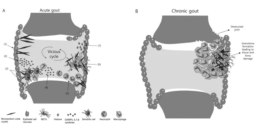
Figure None: 1
DIAGNOSIS:
According to all the included reports, the identification of monosodium urate (MSU) crystals in synovial fluid or tophi is considered the gold standard for the definitive diagnosis of gout. When MSU crystals are not identified, the diagnosis of gout generally requires an assessment of clinical signs, laboratory findings, and imaging results. The serum uric acid concentration used as a threshold for diagnosing hyperuricemia varies across different studies. Generally, serum uric acid levels ranging from 6.8 to 7.0 mg/dL have been reported as the cutoff values.[20]
For gender-specific diagnostic criteria, serum uric acid levels of 6.0 mg/dL (approximately 360 µmol/L) in females and 7.0 mg/dL (approximately 420 µmol/L) in males have been recommended. Several imaging techniques are available for gout diagnosis, including plain radiography, ultrasonography, and dual-energy computed tomography. Since gout progresses slowly, abnormalities on plain radiographs may take several years to become evident. Consequently, although plain radiography has limited diagnostic value during the early stages of gout, it can provide useful evidence in advanced stages of the disease. The characteristic radiographic findings of established gout include bone erosions with overhanging edges and sclerotic margins, bone hyperplasia, narrowing of the joint space, and soft tissue masses known as tophi, which may undergo calcification.[21]
Musculoskeletal ultrasonography is useful in diagnosing gout, particularly when patients present with atypical symptoms or when microscopic confirmation of MSU crystals cannot be obtained. According to the findings of a systematic review, ultrasound features such as the double contour sign, tophi, punctate deposits within the synovial membrane, and hyperechoic spots in the synovial fluid demonstrated good specificity, ranging from 0.65 to 1.00, for gout diagnosis when MSU crystal identification was used as the reference standard. Furthermore, a meta-analysis of diagnostic studies evaluating ultrasonography of joints for gout reported a sensitivity of 0.71 (95% confidence interval [CI] 0.64–0.78), a specificity of 0.62 (95% CI 0.56–0.67), and an area under the curve (AUC) of 0.8549 when the double contour sign was present.[22]
Findings from several meta-analyses indicate that dual-energy computed tomography is an effective diagnostic technique for gout, demonstrating a high sensitivity of 88% (95% CI 84–90), a specificity of 90% (95% CI 85–93), and an area under the curve (AUC) of 0.9565.[23]
Treatment and Prevention:
Colchicine: When administered within 12 hours of the onset of a gout flare, a dose of 1.8 mg of colchicine has been found to be more effective than the traditionally used higher doses.[24]
Non-Steroidal Anti-Inflammatory Drugs (NSAIDs): Non-steroidal anti-inflammatory drugs inhibit cyclooxygenase (COX) enzymes, thereby reducing prostaglandin production throughout the body. Their effectiveness in treating gout is widely accepted. Examples include aspirin and diclofenac.[25]
Allopurinol: Allopurinol is an orally administered xanthine oxidase inhibitor. Allopurinol and other xanthine oxidase inhibitors should not be used in combination with azathioprine and 6-mercaptopurine because xanthine oxidase plays a role in the metabolism of these drugs.[26]
Febuxostat: Febuxostat was the first new urate-lowering medication to receive approval in the United States in more than 40 years. It belongs to the xanthine oxidase inhibitor class and works through the same mechanism as allopurinol. It may be considered as an alternative for patients who experience adverse effects with allopurinol or have kidney disease.[27]
Probenecid: Following the introduction of allopurinol, the use of probenecid declined considerably because it required administration in divided doses, increased fluid intake, and adjustment of urine pH. Probenecid acts on the kidneys to promote the elimination of uric acid from the body. It is taken daily and may be used in combination with antibiotics to enhance their effectiveness.[28]
Lesinurad: Lesinurad is an orally administered medication that promotes the elimination of uric acid from the body. It is used in combination with a xanthine oxidase inhibitor (XOI), such as allopurinol or febuxostat, to enhance uric acid-lowering effects in patients whose gout remains inadequately controlled despite treatment with optimally dosed XOIs alone.[29]
Pegloticase: Pegloticase is used to treat refractory chronic gout when standard medications fail to reduce uric acid levels adequately. It is effective in rapidly reducing uric acid levels and lowering them more quickly than other medications. The drug is administered through an intravenous (IV) infusion once every two weeks.[30]
Serum urate-lowering therapy should be started to prevent recurrent gout attacks in individuals with a previous history of gout who meet any one of the following criteria: two or more flares per year (one flare per year in individuals with stage 2 or higher chronic kidney disease), the presence of tophi, or a history of nephrolithiasis. Serum urate levels should be reduced to a target range of less than 5–6 mg/dL (297–357 µmol/L), depending on the crystal burden and the extent of tophaceous deposits. Serum urate levels should be assessed periodically to evaluate the effectiveness of preventive therapy in patients experiencing recurrent gout. Treatment should be continued for three to six months after a flare if no symptoms persist. However, therapy should be maintained indefinitely if signs or symptoms continue, such as the presence of one or more tophi on physical examination. In men, weight gain is an important risk factor for gout, whereas weight reduction decreases the risk of developing the condition. Consumption of high-fructose corn syrup should be limited, as fructose increases uric acid production as a byproduct of adenosine triphosphate catabolism.[31]
As Conventional allopathic medicine poses severe side effects like-
Hence there is need of Plant based treatments as herbal medicine provide low risk, low side effect therapeutic value. Therefore this study reviewed all available herbal medicaments for management of gout.
METHODS:
The following
keywords were used for the literature search: ‘gout’, ‘gouty arthritis’, ‘hyperuricemia’, ‘uric acid’, ‘XO inhibitor’, ‘uricosuric’, ‘URAT1’, and ‘GLUT9’. The herbal-related keywords included ‘herbal medicine’, ‘medicinal plant’, ‘natural products’, ‘phytomedicine’, and ‘phytotherapy’. The term ‘anti-inflammatory effect’ was combined with the keywords ‘IL-6’, ‘IL-8’, ‘IL-1’, and ‘TNF-’. Furthermore, the reference lists of the identified articles were examined to find additional relevant studies.[32]
RESULT
| Plant Name | Phytocinstituents | Treatment | Study Details | Findings | Comments |
|---|---|---|---|---|---|
| Terminalia bellerica (Bibhitaki) extract | Chebulinic acid, Chebulagic acid, Gallic acid, Ellagic acid Methyl gallate, Galloyl glucose, Termilignan, Thannilignan, Anolignan B, 7-hydroxy flavone, Luteolin, Bellericanin, Belleric acid, β-sitosterol | T. chebula 500 mg twice a day (BID),T. bellerica 250 mg BID, T. bellerica 500 mg BID, placebo BID, and febuxostat 40 mg once dailyplus an identical placebo – for a duration of 24 weeks. Serum uric acid levels were measuredat baseline and at the end of 4, 8, 12, 16, 20, and 24 weeks. Statistical analysis was done usingGraphPad Prism Software 4. | 110 participants randomized across T. bellerica 250 or 500 mg twice daily, T. chebula 500 mg twice daily, placebo, or febuxostat 40 mg daily; 24 weeks, with serial serum urate assessment. [1] | All active arms reduced serum urate versus baseline/placebo; reduction began by week 4. The 500-mg T. bellerica arm reduced mean urate from 8.07 to 5.78 mg/dL at week 24, versus 4.28 mg/dL with febuxostat; no serious adverse effects were reported in this study. [1] | Antiinflamatry Activity Antioxidant Activity Xanthine Oxidase Pathway |
| Terminalia chebula (Haritaki) extract | Chebulinic acid ≥15%, Chebulagic acid ≥10%, Other hydrolysable tannins ≥15% Gallic acid, Ellagic acid, Ethyl gallate, Chebulic acid, Quercetin, Tannic acid, β-sitosterol | T. chebula 500 mg twice a day (BID),T. bellerica 250 mg BID, T. bellerica 500 mg BID, placebo BID, and febuxostat 40 mg once dailyplus an identical placebo – for a duration of 24 weeks. Serum uric acid levels were measuredat baseline and at the end of 4, 8, 12, 16, 20, and 24 weeks. Statistical analysis was done usingGraphPad Prism Software 4. | Randomized, double-blind, placebo- and febuxostat-controlled 24-week study; aqueous extract 500 mg twice daily. [1 | Serum urate fell from baseline; significant reduction was reported from week 4 (P<0.01). The reported urate reduction was smaller than with T. bellerica 500 mg; further confirmation was recommended. [1] | Antiinflamatry Activity Antioxidant Activity Xanthine Oxidase Pathway |
| DKB114: Chrysanthemum indicum flower + Cinnamomum cassia bark extracts | Chrysanthemum indicum Linn flower extractCinnamomum cassia [syn. Cinnamomum cassia] bark extract | Dose: 500 mg / tablet x 4 tablets/day = 2g/day of DKB114 functional ingredient for 12 weeksPreparation: Mixture of Chrysanthemum indicum Linn flower extract + Cinnamomum cassia bark extract in 1:2 ratio w/w | Randomized, double-blind, placebo-controlled, 12-week trial in 80 adults with asymptomatic serum urate 7–9 mg/dL; DKB114 2 g/day of active ingredient versus placebo. [2] | At week 12, serum urate fell 0.58 mg/dL with DKB114 versus 0.02 mg/dL with placebo (between-group P=0.0229); no significant difference was seen at week 6. Small sample and asymptomatic population limit extrapolation to gout patients. [2] | Preclinical work suggests xanthine oxidase inhibition and reduced urate production, plus increased urinary excretion and reduced URAT1/GLUT9-mediated reabsorption; the clinical trial did not show reduced blood xanthine-oxidase activity. [2 |
| Eugenia polyantha (Indonesian bay leaf) + Apium graveolens (celery) + Nigella sativa (black seed) extracts | Quercetin, Quercitrin, Myricitrin, Myricetin | combined extracts 3,000 mg/day versus allopurinol 100 mg/day for four weeks. [3 | Double-blind randomized trial in 44 adults with hyperuricemia; combined extracts 3,000 mg/day versus allopurinol 100 mg/day for four weeks. [ | Both groups showed serum-urate reductions; urinary urate excretion and hsCRP did not show significant reduction in the herbal group. Small sample, short follow-up, and the multi-herb formulation prevent conclusions about individual plants or comparative effectiveness | Studied as a multi-plant urate-lowering intervention in hyperuricemia, not as an isolated plant or acute-flare treatment. [3] |
| Phyllanthus niruri (stonebreaker) | Phyllanthin Hypophyllanthin Phyltetralin Niranthin | Methanol extract of P. niruri leavesMethanol extract - In vitro IC50 39.39 μg/mLIn vivo: IP injection in hyperuricemic ratsLignan Fraction - Fraction 4Most active fraction from resin chromatography - highest plasma uric acid reversalPure Lignans isolatedPhyllanthin, Hypophyllanthin, Phyltetralin, Niranthin, Nirtetralin - Tested at 10 mg/kg IP | In-vitro xanthine-oxidase assay plus uricosuric testing in potassium-oxonate- and uric-acid-induced hyperuricemic rats. [4] | Methanol extract inhibited xanthine oxidase in vitro (IC50 39.39 μg/mL) and increased urinary urate excretion 1.69-fold in hyperuricemic animals; lignans also increased excretion/clearance. No human efficacy trial is described in this source. Pyrazinamide Test: Co-administration of pyrazinamide with phyllanthin exhibited a significant suppression of phyllanthin's uricosuric activity resembling that of pyrazinamide with benzbromarone |
Primarily uricosuric (increased urinary urate excretion), with partial xanthine-oxidase inhibition; isolated lignans appeared mainly uricosuric. Phyllanthin dose-dependently reversed plasma uric acid to normal, comparable to Allopurinol, Benzbromarone, Probenecid |
| Persicaria capitata herba (whole plant) | Quercetin Quercitrin Gallic Acid Trans-p-coumaric acid Myricetrin |
WP - Water extract of P. capitata Aqueous extract - Standardized by UHPLC-HRMS fingerprintHyperuricemia model: 200, 400, 800 mg/kg/day POGouty arthritis model: 400, 800 mg/kgEP - Ethanol extract of P. capitata70% Ethanol extractSame as WP - 200, 400, 800 mg/kg/day POPositive Control DrugsAllopurinol - XO inhibitor10 mg/kg PO5 mg/kg [Gout model]Gout Model InducerMSU crystals - Monosodium urate 1mg/20µL intra-articular ankle injection-Hyperuricemia Model InducerPotassium oxonate 250 mg/kg IP + Hypoxanthine 500 mg/kg PO | Water and 75% ethanol extracts tested in mice with chemically induced hyperuricemia and monosodium-urate-crystal-induced acute gouty arthritis; biochemical and gene/protein-expression analyses. Type: In vivo mice study + In vitro XO assayAnimals: Male Kunming mice, Hyperuricemic model + MSU-induced acute gouty arthritis model Duration: 7 days treatment for hyperuricemia, 3-6h for acute goutParameters measured: Serum uric acid, Urine uric acid, Creatinine, BUN, XO activity [serum & liver], XO expression [Western blot, RT-PCR], URAT1, GLUT9 mRNA & protein, Inflammatory cytokines [IL-1β, IL-6, TNF-α, NLRP3], Ankle swelling, HistopathologySafety: Renal toxicity assessed - BUN, creatinine, histology vs Allopurinol |
Water and 75% ethanol extracts tested in mice with chemically induced hyperuricemia and monosodium-urate-crystal-induced acute gouty arthritis; biochemical and gene/protein-expression analyses. Type: In vivo mice study + In vitro XO assayAnimals: Male Kunming mice, Hyperuricemic model + MSU-induced acute gouty arthritis modelDuration: 7 days treatment for hyperuricemia, 3-6h for acute goutParameters measured: Serum uric acid, Urine uric acid, Creatinine, BUN, XO activity [serum & liver], XO expression [Western blot, RT-PCR], URAT1, GLUT9 mRNA & protein, Inflammatory cytokines [IL-1β, IL-6, TNF-α, NLRP3], Ankle swelling, HistopathologySafety: Renal toxicity assessed - BUN, creatinine, histology vs Allopurinol |
Inhibition of xanthine oxidase and downregulation of renal urate reabsorption transporters URAT1 and GLUT9; anti-inflammatory effects may involve lower proinflammatory-factor expression Anti-inflammatory / Antigouty Arthritis :MSU crystals → Activate NLRP3 inflammasome → Caspase-1 → IL-1β → IL-6, TNF-α → Neutrophil influx → Gout painP. capitata extracts inhibit expression of proinflammatory factors - ↓ NLRP3, ↓ IL-1β, ↓ IL-6, ↓ TNF-α, ↓ NF-κB signalingSimilar to Colchicine mechanism but natural[Joint. |
| Cichorium intybus (chicory) extract / chicoric acid | Chicoric acid [Dicaffeoyltartaric acid] - Specific effective compound - ≥45-55 g/kg extractCaftaric acid [Caffeoyltartaric acid]Chlorogenic acidIsochlorogenic acids A,B,CQuercetin-3-O-glucuronide, Isoquercitrin [Quercetin-3-O-glucoside], Luteolin-7-O-glucuronide, Quercetin, Luteolin, Kaempferol LactonesLactucin, Lactucopicrin, 8-Deoxylactucin, Jacquinelin - Anti-inflammatory Inulin [Polysaccharide 40-60% in root], Cichorioside, Esculetin, Cichoriin, β-sitosterol | Cichorium intybus L. Extract [CIE / CIL]Aqueous-Ethanol extract of aerial partsIn vivo rats: 500 mg/kg, 1000 mg/kg, 2000 mg/kg PO for 7 daysIn vitro macrophages: 50, 100, 200 μg/mLChicoric Acid [CA] - Pure compoundMain active marker of chicory - Specific effective compound in chicoryIn vitro: 25, 50, 100 μMPositive ControlColchicine - 0.3 mg/kg PO [Standard antigout]Disease InducerMSU crystals - 100 μg/mL in vitro100 μL of 50 mg/mL MSU suspension intra-articular into ankle joint [In vivo rats] | Monosodium-urate-injected rat model treated with oral chicory extract or colchicine; ankle edema, inflammatory activity, histology, and signaling readouts assessed, with complementary MSU-stimulated macrophage experiments. In vivo Model: Sprague-Dawley [SD] rats, MSU-induced acute gouty arthritis - MSU injection into ankle jointGroups: Normal, MSU model, MSU + Colchicine 0.3 mg/kg, MSU + CIE low/medium/highIn vitro Model: Primary macrophages isolated from SD rats peritoneal cavity - stimulated with LPS 1 μg/mL + MSU 100 μg/mL |
Extract improved ankle swelling/inflammatory pathology and lowered IL-1β alongside NF-κB/NLRP3 pathway inhibition in rats; chicory extract/chicoric acid also inhibited IL-1β-related signaling in macrophages. Preclinical only. CIE 500-2000 mg/kg significantly improved swelling degree, inflammatory activity score, skin temperature in MSU-injected ankles - Dose dependent In vivo: CIE significantly decreased IL-1β release in serum and ankle joint tissue [p<0.01]In vitro: Both CIE [50-200 μg/mL] and pure Chicoric acid [25-100 μM] inhibited IL-1β release from macrophages dose-dependently without cytotoxicity |
Suppression of NF-κB and NLRP3 signaling, reducing IL-1β release; chicoric acid was examined as an active constituent. |
| Astilbin (plant-derived flavonoid; source plant not specified in the abstract) | Flavanonol / Dihydroflavonol glycoside - Structure: Taxifolin-3-O-rhamnoside | Purified flavonoid - C21H22O11 - Taxifolin-3-O-α-L-rhamnosideHyperuricemia mice: Low, Medium, High dose - Common reported: 5, 10, 20 mg/kg/day PO [Also reported in similar studies as 5.37, 16.11, 48.33 mg/kg]Gouty arthritis rats: 5-20 mg/kg/day POPositive Control - HyperuricemiaFebuxostat [FB] - Xanthine Oxidase Inhibitor - 5-10 mg/kg POPositive Control - Gouty ArthritisColchicine [COL] - 0.3-1 mg/kg POInducer - HyperuricemiaPotassium Oxonate [OXO] 100-250 mg/kg IP + Yeast Extract Powder [YEP] - High purine diet for 7-14 days | Potassium-oxonate/yeast-extract hyperuricemia mouse model and intra-articular monosodium-urate crystal acute-arthritis rat model; febuxostat and colchicine comparators, respectively. | Astilbin lowered serum urate and xanthine oxidase in mice, and reduced ankle swelling, inflammatory-cell infiltration, and several inflammatory mediators in rats. Authors present it as a candidate requiring further evaluation, not established therapy. Anti-hyperuricemia Property - In OXO+YEP mice:AS and Febuxostat reduced serum uric acid [UA] significantly vs ModelAS and FB reduced Xanthine Oxidase [XO] activity [Serum & liver] - XO inhibitory effectAS and FB reduced ROS levels and increased antioxidant enzymes - SOD, GSH-Px, Catalase - Antioxidant effectDose dependent - High dose AS comparable to FebuxostatB. Anti-Gouty Arthritis Property - In MSU rats:AS and Colchicine alleviated ankle joint swelling [Toe volume] vs ModelHistology: Reduced inflammatory cell infiltration in synovium, less edemaAS reduced IL-1β, IL-6, TNF-α, MCP-1 levels in liver [and joint] - Anti-inflammatoryPain threshold improved - less dysfunction |
Reduced xanthine oxidase and oxidative stress markers in hyperuricemic mice; reduced inflammatory cytokines in acute arthritis models. Antioxidant - NRF2 / ROS:↓ Reactive Oxygen Species [ROS] + ↑ SOD, GSH-Px, Catalase → Reduces oxidative stress in hyperuricemia Uric Acid Excretion - URAT1: Inhibits URAT1 expression in kidney proximal tubule → Promotes uric acid excretion → Uricosuric |
| Additional screening-stage plants: Teucrium polium, Prunus avium, Smilax riparia, Rhus coriaria, Foeniculum vulgare, Allium cepa, Camellia japonica, Helianthus annuus | FlavonoidsQuercetin, Luteolin, Apigenin, Astilbin, Kaempferol, Myricetin AlkaloidsColchicine, Berberine, Palmatine Phenolic compoundsCaffeic acid, Chlorogenic acid, Chicoric acid, Gallic acid, Ellagic acid, Mangiferin Essential oilsEucalyptus, Fennel oil, Garlic oil TanninsGeraniin, Corilagin, Chebulagic acid CoumarinsEsculetin, Scopoletin Iridoid glucosidesHarpagoside |
ColchicineAlkaloid - From Colchicum autumnale0.3-1 mg/kg - Anti-inflammatory - Only human studiedQuercetinFlavonoid50-200 mg/kg - XO inhibitor + Uricosuric + Anti-inflammatoryCurcuminPhenolic - Curcuminoid100-400 mg/kg - NLRP3 inhibitorResveratrolStilbene - Phenolic10-40 mg/kgBerberineIsoquinoline Alkaloid50-150 mg/kgLuteolinFlavone50-100 mg/kgApigeninFlavone10-50 mg/kgCaffeic acid / Chlorogenic acidPhenolic acid20-100 mg/kgGallic acid / Ellagic acidTannin50-100 mg/kg | A 2023 review screened literature published 2000–2020 and identified 14 plants and nine phytochemicals with reported antigout properties; plant-specific clinical protocols were not supplied for this grouped list. XO inhibitory activity - IC50Uricosuric action - Urinary uric acid, FEUAAnti-inflammatory - IL-1β, IL-6, TNF-α, CRPSerum urate reduction |
The review highlighted these eight plants for comparatively high xanthine-oxidase inhibition; this is a screening signal, not evidence that they prevent flares or replace urate-lowering therapy. In vitro studies:Flavonoids, alkaloids, essential oils, phenolic compounds, tannins, iridoid glucosides, coumarins show potential anti-gout effects by XO inhibitory actionLignans, triterpenoids, xanthophyll act through anti-inflammatory effects8 plants with highest XO inhibitory: Teucrium polium, Prunus avium, Smilax riparia, Rhus coriaria, Foeniculum vulgare, Allium cepa, Camellia japonica, Helianthus annuus In animal studies:Essential oils, lignans, tannins show dual effects - reduction of uric acid generation + uricosuric actionAlkaloids reveal inhibit uric acid generation, show anti-inflammatory, or combinationPhenolic compounds and flavonoids inhibit uric acid production + show uricosuric + anti-inflammatory - Most promising class In human studies:Only colchicine from Colchicum autumnale showed proven anti-inflammatory in RCTsFor other plant extracts, although revealing anti-gout potential, further phytochemical investigations needed to identify active constituentsPlants with antioxidant activities are much more potent in management of gout |
Review reports xanthine-oxidase inhibitory activity associated with plant phenolics, tannins, coumarins, terpenoids, and alkaloids; mechanisms and potency vary by species/extract. Uricosuric - Increases Excretion - Renal Transporters Anti-inflammatory - Gout Flare Plants with antioxidant activities are much more potent - ROS generated during purine metabolism activates NLRP3 |
| CHERRY | Cyanidin-3-glucoside, Cyanidin-3-glucosyl-rutinoside, Cyanidin-3-rutinoside, Quercetin, Kaempferol, Isorhamnetin-3-rutinoside, Quercetin-3-rutinoside, Catechin, Epicatechin, Chlorogenic acid, Phenolics, Tannins, Coumarins, Terpenoids, Alkaloids |
Cyanidin-3-glucoside + metabolites 0.1, 1, 10 μmol/L Cherry juice concentrate [non-cytotoxic dose] Black soybean anthocyanin 60 mg/kg/day Tart cherry juice 2x8 oz/day - 5 days before, day of, 48h after Purified anthocyanin 320 mg/day |
antioxidant and anti-inflammatory (IL-6, TNF-α, IL-1β, IL-8, COX-I and -II) properties, hypouricemic effects, and the ability to downregulate NFkB-mediated osteoclastogenesis. | ↓ 41-96% production [p<0.03 - p<0.001] - Metabolites strongest effect ↓ 45% secretion at 1:4000 dilution and ↓ 60% secretion at 1:1600 dilution. Treated joints had lower levels [p<0.05] + ↓ Th17 cells, ↓ nitrotyrosine, ↓ iNOS, ↓ osteoclastogenesis. ↓ 49% postrace elevation [41.8 vs 82.1 pg/mL, p<0.001] + ↓ 34% CRP, inhibited urate elevation vs placebo. ↓ 12.8% vs -1.3% placebo [p=0.019] and one marker NS [↓1.8%, p=0.673] + ↑ HDL, ↓ LDL, VCAM-1, hsCRP |
NLRP3 inflammasome → IL-1β pathway, NF-κB → pro-inflammatory cytokine pathway, Purine metabolism → xanthine oxidase (XO) → uric acid pathway, COX-1/COX-2 → prostaglandin pathway, Reactive oxygen species (ROS) → oxidative stress pathway, RANKL → NF-κB → osteoclastogenesis pathway. |
CONCLUSION:
The findings from the reviewed research suggest that several medicinal plants and their phytochemical constituents possess significant potential in managing gout by lowering uric acid concentrations and controlling inflammation. Their therapeutic effects are mainly associated with the suppression of xanthine oxidase activity, enhancement of uric acid elimination, and antioxidant and anti-inflammatory properties. Plants including Terminalia bellerica, Terminalia chebula, Phyllanthus niruri, Persicaria capitata, Cichorium intybus, and cherry have demonstrated promising anti-gout effects. However, the available research is predominantly limited to laboratory investigations and animal experiments, with only a small amount of clinical evidence. Thus, additional clinical investigations are necessary to establish their safety and efficacy in humans. Herbal medicines may provide beneficial support in gout management, but they should not be used as substitutes for currently established treatments.
REFERENCES







Pratiksha Jadhav, Vaidehi Patil, Varad Sutar, Tushar Patil, Omkar Patil, Sammed Lakhane, Abhimanyu Patil, A Review on Anti-Gout Activity by Using Various Herbal Plants, Int. J. of Pharm. Sci., 2026, Vol 4, Issue 11, 3007-3022. https://doi.org/10.5281/zenodo.24334177
 10.5281/zenodo.24334177