

We use cookies to ensure our website works properly and to personalise your experience. Cookies policy
TIT - College of Pharmacy, Bhopal (M.P.) Technocrats Institute of Technology-Pharmacy (M.P.)
Background: Green synthesis of metal nanoparticles using plant extracts has gained considerable attention due to its eco-friendly nature, cost effectiveness, and enhanced biocompatibility. Copper nanoparticles exhibit promising antimicrobial and physicochemical properties, making them suitable for pharmaceutical and biomedical applications. Objective: The present study aimed to formulate and characterize copper nanoparticles using the hydroalcoholic extract of Cordia sinensis and to evaluate their physicochemical characteristics, release behavior, and antimicrobial activity. Methods: Hydroalcoholic extract of C. sinensis was subjected to phytochemical screening and quantitative estimation of total flavonoid and phenolic content. Copper nanoparticles were prepared by green synthesis and formulated into gel preparations. The nanoparticles were evaluated for percentage yield, entrapment efficiency, particle size, zeta potential, and physical characteristics. Gel formulations were assessed for spreadability, viscosity, pH, flavonoid content, and in vitro release kinetics. Antimicrobial activity was evaluated against Streptococcus mutans and Escherichia coli. Results: Phytochemical analysis revealed the presence of flavonoids, phenols, alkaloids, diterpenes, and proteins in the extract. The optimized formulation (F3) showed the highest nanoparticle yield (80.25 ± 0.45%) and entrapment efficiency (0.575 ± 0.074 mg/100 mg). Particle size and zeta potential were found to be 225.65 nm and ?37.45 mV, respectively, indicating good stability. The nanoparticle gel exhibited acceptable physicochemical properties, controlled drug release following zero-order kinetics, and enhanced antimicrobial activity compared to the crude extract. Conclusion: The study confirms the successful green synthesis and formulation of copper nanoparticles using Cordia sinensis extract, demonstrating good stability and superior antimicrobial efficacy, highlighting their potential for topical therapeutic applications.
Nanotechnology has emerged as a transformative field in pharmaceutical and biomedical sciences, enabling the development of metallic nanoparticles with enhanced physical, chemical, and biological properties compared to their bulk counterparts (Rokunuzzaman; 2024). Among these, copper nanoparticles (CuNPs) have attracted significant interest due to their unique optical, electrical, catalytic, and biological activities, including antimicrobial, antioxidant, and anti-inflammatory effects, which make them promising candidates for therapeutic and diagnostic applications. The large surface-to-volume ratio and quantum size effects of CuNPs contribute to their high reactivity and distinctive functionalities in drug delivery systems and biomedical formulations (Yusuf et al., 2023).
Conventional physical and chemical methods of nanoparticle synthesis often involve high energy input and toxic chemicals, which can negatively impact the environment and limit biomedical applications. To address these limitations, green synthesis approaches using plant extracts as reducing and capping agents have been developed (Osman et al., 2024). This eco-friendly method exploits phytochemicals such as phenols, flavonoids, tannins, and other secondary metabolites present in plant extracts, which can both reduce metal ions and stabilize the resulting nanoparticles. Such biologically derived CuNPs demonstrate improved biocompatibility, reduced toxicity, and added therapeutic benefits compared with chemically synthesized nanoparticles. Green synthesized CuNPs have been extensively characterized using techniques such as UV-visible spectroscopy (UV-Vis), Fourier transform infrared spectroscopy (FTIR), X-ray diffraction (XRD), scanning electron microscopy (SEM), and transmission electron microscopy (TEM) to confirm their formation, size, morphology, and surface chemistry (El-Sayyad et al., 2024).
Cordia sinensis Lam. (family Boraginaceae), commonly known as grey-leaved saucer berry, is a medicinal plant traditionally used in various systems of medicine to manage digestive disorders, respiratory ailments, inflammation, and microbial infections, owing to its rich content of bioactive phytochemicals such as flavonoids, phenolics, and tannins (Rawal et al., 2024). Despite the established ethnopharmacological relevance of C. sinensis, scientific investigations into its potential role in the green synthesis and formulation of metal nanoparticles remain limited. Leveraging the intrinsic reducing and stabilizing capacity of C. sinensis phytoconstituents for the biosynthesis of CuNPs could yield a biocompatible nanoparticulate system with enhanced biological activities. Therefore, this study focuses on the formulation and characterization of copper nanoparticles using Cordia sinensis extract, and the evaluation of their physicochemical properties as a foundation for potential biomedical applications.
Material and Methods
Material
Fresh plant material of Cordia sinensis was collected and used for the preparation of a hydroalcoholic extract. Copper sulfate was employed as the copper precursor for the green synthesis of copper nanoparticles. Analytical-grade solvents and reagents required for phytochemical screening, estimation of total flavonoid and phenolic content, and nanoparticle characterization were used. Standard laboratory chemicals were utilized for formulation and evaluation of nanoparticle-loaded gel preparations. All materials and reagents were of analytical grade and used without further purification.
Methods
Extraction procedure
Following procedure was adopted for the preparation of extract from the shade dried and powdered herbs:
Defatting of plant materials
Leaves of Cordia sinensis were shade dried at room temperature. 50 gram dried plant material was coarsely powdered and subjected to extraction with petroleum ether by maceration. The extraction was continued till the defatting of the material had taken place.
Extraction by maceration process
Defatted dried powdered leaves of Cordia sinensis has been extracted with hydroalcoholic solvent (methanol: water: 80:20) using maceration process for 48 hrs, filtered and dried using vacuum evaporator at 40ºC (Mukherjee, 2007; Kokate, 1994).
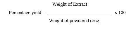
Determination of percentage yield
The percentage yield of each extract was calculated by using following formula:
Phytochemical screening
Phytochemical examinations were carried out for all the extracts as per the standard methods (Audu et al., 2007).
Total flavonoids content estimation
Determination of total flavonoids content was based on aluminium chloride method (Gaur Mishra et al., 2017). 10 mg quercetin was dissolved in 10 ml methanol, and various aliquots of 5- 25μg/ml were prepared in methanol. 10mg of dried extracts of were dissolved in 10 ml methanol and filtered. 3 ml (1mg/ml) of this solution was used for the estimation of flavonoid. 1 ml of 2% AlCl3 methanolic solution was added to 3 ml of extract or standard and allowed to stand for 15 min at room temperature; absorbance was measured at 420 nm.
Total Phenolic content estimation
The total phenolic content of the extract was determined by the modified Folin-Ciocalteu method (Gaur Mishra et al., 2017). 10 mg Gallic acid was dissolved in 10 ml methanol, various aliquots of 5-25µg/ml was prepared in methanol. 10mg of dried extracts of were dissolved in 10 ml methanol and filter. Two ml (1mg/ml) of this solution was used for the estimation of phenol. 2 ml of each extract or standard was mixed with 1 ml of Folin-Ciocalteu reagent (previously diluted with distilled water 1:10 v/v) and 1 ml (7.5g/l) of sodium carbonate. The mixture was vortexed for 15s and allowed to stand for 15 min for colour development. The absorbance was measured at 765 nm using a spectrophotometer.
Biosynthesis of copper nanoparticles
For each formulation, the required quantity of CuSO₄ solution (1–3 mM) was prepared separately. Plant leaf extract (500 mg equivalent) was taken and mixed with the CuSO₄ solution in the specified extract-to-metal salt ratio (1:1 or 1:2). In a typical preparation, 80 mL of CuSO₄ solution was mixed with 20 mL of plant leaf extract under continuous stirring at room temperature.
The reaction mixture was then allowed to stand for 24 hours, during which a gradual change in color of the solution was observed, indicating the formation of copper nanoparticles, as reported earlier (Hariprasad et al., 2016). After completion of the reaction, the mixture was centrifuged at 10,000 rpm for 15 minutes to separate the formed nanoparticles. The obtained pellet was washed and redispersed in double distilled water to remove any unreacted copper ions and unwanted biological materials. The purified copper nanoparticles were then collected and used for further characterization and evaluation.
Table 1: Different formulation of copper nanoparticles of Cordia sinensis
|
Formulation Code |
Extract (mg) |
CuSO4 (mM) |
Ratio |
|
F1 |
500 |
1 |
1:1 |
|
F2 |
500 |
2 |
1:1 |
|
F3 |
500 |
3 |
1:1 |
|
F4 |
500 |
1 |
1:2 |
|
F5 |
500 |
2 |
1:2 |
|
F6 |
500 |
3 |
1:2 |
Characterization of synthesized copper nanoparticles formulations
Percentage yield
The copper nanoparticles, prepared with a size range of 200-300 nm, were gathered and quantified from various formulations. The calculated weight was then divided by the total quantity of all non-volatile components utilized in the microsphere preparation (Saranyaadevi et al., 2014).

Entrapment efficiency
The entrapment efficiency of the drug was defined as the ratio of the mass of the drug associated with the formulations to the total mass of the drug (Moniri et al., 2017). The entrapment efficiency was assessed using the dialysis method, where the copper nanoparticle-entrapped extract was separated from the free drug. For this purpose, the aforementioned formulations were loaded into dialysis bags, and the free drug was dialyzed for 24 hours in 50 ml of buffer at pH 1.2. The absorbance of the dialysate was measured against a blank buffer at pH 1.2, and the absorbance of the corresponding blank was measured under the same conditions. The concentration of free flavonoids was determined based on the absorbance difference using a standard curve.
Surface charge and vesicle size
The particle size, size distribution, and surface charge were determined using the Dynamic Light Scattering method (DLS) with a Malvern Zetamaster, ZEM 5002 instrument from Malvern, UK. Zeta potential measurements for the copper nanoparticles were conducted based on the Helmholtz–Smoluchowsky equation derived from electrophoretic mobility (Usha et al., 2017). For zeta potential measurement, a zetasizer was employed with field strength of 20 V/cm in a large bore measurement cell. Samples were appropriately diluted with 0.9% NaCl and adjusted to a conductivity of 50 µS/cm.
Formulation development of copper nanoparticle gel
Precise quantities of methyl paraben, glycerin, polyethylene glycol, and hydroalcoholic extract of Cordia sinensis were dissolved in approximately 100 ml of water in a beaker. The mixture was vigorously stirred using a mechanical stirrer or sonicator, following the standard method (Pawar et al., 2017).
Subsequently, Carbopol 940 was gradually introduced into the beaker containing the aforementioned liquid while maintaining continuous stirring. The solution was neutralized by slowly adding a triethanolamine solution, stirring constantly, until the gel formation occurred.
Table 2: Formulation of gel
|
Ingredients (mg) |
F1 |
F2 |
F3 |
|
Copper nanoparticle of Cordia sinensis |
500 |
500 |
500 |
|
Carbopol 940 |
250 |
500 |
750 |
|
Polyethylene Glycol 600 |
0.2 |
0.2 |
0.2 |
|
Methyl Paraben |
0.08 |
0.08 |
0.08 |
|
Triethanolamine |
1.0 |
1.0 |
1.0 |
|
Distilled Water |
100 ml |
100ml |
100ml |
Evaluation of gel
Appearance and Consistency: The physical appearance and texture of gel formulations were visually inspected, and observations.
Washability: Formulations were applied to the skin and manually assessed for ease and degree of washing with water (Pawar et al., 2017).
Extrudability Determination: Gel formulations were filled into aluminum collapsible tubes, sealed, and pressed to extrude the material. Extrudability of the formulation was noted.
Determination of Spreadability: Spreadability, a crucial factor for gel formulations, was evaluated using a specially designed apparatus. Two glass slides (6x2) were chosen, and the gel formulation to be tested was placed between them over a length of 6 cm. The time taken for the slides to separate under the application of a 20-gram load was recorded. The experiment was repeated six times for each formulation, and the average was calculated. Two glass slides were selected, and the gel formulation was placed over one slide. The second slide was placed over the formulation, sandwiching it over a length of 6 cm. A 20-gram weight was applied, forming a thin layer. The time taken for the slides to separate under the weight was recorded (Ren et al., 2009).
Spreadability Formula: S=m×lt Where, S = Spreadability (gcm/sec), m = weight tied to the upper slide (20 grams), l = length of the glass slide (6 cm), t = time taken in seconds.
Viscosity: The viscosity of the gel was determined using a Brookfield digital viscometer with spindle no. 6 at 10 rpm and at a room temperature of 25-30°C. Measurements were taken after allowing the gel samples to settle for more than 30 minutes.
Drug Content: The drug content was measured by dissolving 1g of gel in methanol in a 10 ml volumetric flask. A mixture of 3 ml of stock solution and 1 ml AlCl3 solution (2%) was vortexed, and the color production was allowed to stand at 40°C for 30 minutes. Absorbance was measured at 420 nm using a spectrophotometer (Maqusood et al., 2014).
Determination of pH: The pH of the gels was measured using a digital pH meter. One gram of gel was dissolved in 25 ml of purified water, and the electrode was dipped into the gel solution until a steady reading was obtained. pH measurements were repeated twice for each formulation.
In vitro diffusion profile: In vitro diffusion experiments were conducted using Franz diffusion cells. Egg membrane was used as the membrane for dialysis, tied to the diffusion cell. Isotonic phosphate buffer solution (pH 7.4) served as the substrate for receptors. A weighed quantity of the formulation equivalent to 1g of gel was applied to the membrane, and aliquots were withdrawn at different time intervals, measured at 295 nm. The total percent release was calculated for each time period, and the diffusion media were replaced with fresh medium after each withdrawal.
In vitro antimicrobial activity of copper nanoparticle gel
The antimicrobial activity of the copper nanoparticle gel prepared from the Cordia sinensis was determined using the well diffusion method. Three concentrations (25, 50, and 100 mg/ml) of extracted phytochemical were used. Wells containing antibiotics were placed on the agar surface immediately after inoculation with the test organism. Undiluted overnight broth cultures were avoided as inoculums. The plates were then incubated at 37°C for 24 hours and examined for clear zones of inhibition around the wells with specific concentrations of the drug, following the standard procedure by Bauer et al. (1966).
Results and Discussion
The present study was designed to formulate and characterize copper nanoparticles using a green synthesis approach employing the hydroalcoholic extract of Cordia sinensis, followed by evaluation of their physicochemical properties and antimicrobial potential. Green synthesis using plant extracts offers an eco-friendly, cost-effective, and biocompatible alternative to conventional chemical methods due to the involvement of phytoconstituents as reducing and stabilizing agents.
The percentage yield of the hydroalcoholic extract of C. sinensis was found to be 16.5% w/w (Table 3), indicating effective extraction of bioactive constituents using hydroalcoholic solvent. Preliminary phytochemical screening (Table 4) revealed the presence of alkaloids, flavonoids, phenols, proteins, carbohydrates, diterpenes, and tannins, while glycosides and saponins were absent. The presence of flavonoids and phenolic compounds is particularly significant, as these phytoconstituents are known to play a crucial role in the reduction of copper ions and stabilization of nanoparticles during green synthesis.
Quantitative estimation of phytoconstituents (Table 5) demonstrated measurable levels of total flavonoids (0.58 mg/100 mg) and total phenols (0.37 mg/100 mg) in the hydroalcoholic extract. These compounds possess strong antioxidant and metal-chelating properties, which facilitate nanoparticle formation and enhance biological activity.
Copper nanoparticles were successfully formulated using different formulation ratios (F1–F6), with percentage yield ranging from 71.15% to 80.25% (Table 6). Among all formulations, F3 showed the highest yield (80.25 ± 0.45%), indicating optimal conditions for nanoparticle synthesis. Entrapment efficiency studies (Table 7) further supported the superiority of formulation F3, which exhibited the highest flavonoid entrapment (0.575 ± 0.074 mg/100 mg), suggesting effective incorporation of bioactive compounds onto the nanoparticle surface.
Characterization studies of the optimized formulation F3 revealed an average particle size of 225.65 nm with a zeta potential of −37.45 mV (Table 8). The negative zeta potential value indicates good electrostatic stability and reduced tendency for nanoparticle aggregation. Particle size in the nanometer range further supports enhanced surface area and biological interaction potential.
Physical evaluation of the nanoparticle-loaded gel formulations (Table 9) demonstrated acceptable characteristics such as uniform color, absence of clogging, good homogeneity, smooth texture, satisfactory washability, and good to excellent extrudability. These properties are essential for topical formulations and indicate patient acceptability and formulation stability.
Evaluation of gel formulations (Table 10) showed appropriate spreadability, viscosity, flavonoid content, and pH values close to skin physiological pH (6.75–6.85), confirming suitability for topical application. Controlled viscosity and good spreadability ensure ease of application and uniform drug distribution.
In vitro release kinetics analysis of formulation F2 (Table 11) revealed a higher correlation coefficient for the zero-order model (R² = 0.9927) compared to the first-order model, indicating a sustained and controlled release pattern of bioactive constituents from the nanoparticle gel.
Antimicrobial activity studies (Table 12) demonstrated that copper nanoparticle gel exhibited significantly enhanced antimicrobial activity against Streptococcus mutans and Escherichia coli compared to the crude extract alone. The improved antimicrobial efficacy may be attributed to the nanoscale size, increased surface area, and synergistic action of copper ions and plant phytoconstituents. The results confirm that green synthesized copper nanoparticles possess superior antimicrobial potential and can serve as effective antimicrobial agents.
Table 3: % Yield of extract of Cordia sinensis
|
S. No. |
Extract |
% Yield (w/w) |
|
1. |
Hydroalcoholic |
16.5 |
Table 4: Phytochemical screening of extract of Cordia sinensis
|
S. No. |
Constituents |
Hydroalcoholic extract |
|
1. |
Alkaloids Mayer’s Test Wagner’s Test Dragendroff’s Test Hager’s Test |
+ve +ve -ve -ve |
|
2. |
Glycosides Legal’s Test |
-ve |
|
3. |
Flavonoids Lead acetate Alkaline test |
+ve -ve |
|
4. |
Phenol Ferric chloride test |
+ve |
|
5. |
Proteins Xanthoproteic test |
+ve |
|
6. |
Carbohydrates Molisch’s Test Benedict’s Test Fehling’s Test |
-ve -ve +ve |
|
7. |
Saponins Froth Test Foam Test |
-ve -ve |
|
8. |
Diterpenes Copper acetate test |
+ve |
|
9. |
Tannins Gelatin Test |
-ve |
[+ve= positive; -ve= negative]
Table 5: Estimation of total flavonoids and phenol content of Cordia sinensis
|
S. No. |
Extract |
Total flavonoids content (mg/ 100 mg of dried extract) |
Total phenol content (mg/ 100 mg of dried extract) |
|
1. |
Hydroalcoholic |
0.58 |
0.37 |
Table 6: Determination of % yield of prepared copper nanoparticles formulations
|
Formulation code |
% Yield |
|
F1 |
71.15±0.85 |
|
F2 |
73.36±0.36 |
|
F3 |
80.25±0.45 |
|
F4 |
76.65±0.25 |
|
F5 |
73.12±0.88 |
|
F6 |
71.85±0.36 |
Table 7: Determination of entrapment efficiency of prepared formulations
|
Formulation code |
Percentage entrapment efficiency (Flavonoid mg/100mg quercetin equivalent) |
|
F1 |
0.465±0.045 |
|
F2 |
0.482±0.036 |
|
F3 |
0.575±0.074 |
|
F4 |
0.492±0.036 |
|
F5 |
0.547±0.045 |
|
F6 |
0.512±0.074 |
Table 8: Characterization of average particle size and zeta potential of optimized formulation F3
|
Formulation code |
Average Particle size (nm) |
Zeta Potential (mV) |
|
F3 |
225.65 |
-37.45 |
Table 9: Results of physical characteristics
|
Formulation Code |
Colour |
Clogging |
Homogeneity |
Texture |
Washability |
Extrudability |
|
F1 |
Light brown |
Absent |
Good |
Smooth |
Good |
Good |
|
F2 |
Brown |
Absent |
Very good |
Smooth |
Very good |
Excellent |
|
F3 |
Dark brown |
Absent |
Good |
Smooth |
Good |
Very good |
Table 10: Results of evaluation of gel formulations
|
Formulation code |
Spreadability* (gcm/sec) |
Viscosity* (cp) |
Flavonoid Content (mg/100mg) |
pH |
|
F1 |
11.25±0.85 |
3265±18 |
0.512±0.018 |
6.85±0.05 |
|
F2 |
10.32±0.92 |
3155±15 |
0.575±0.016 |
6.80±0.02 |
|
F3 |
9.85±0.74 |
3045±13 |
0.563±0.012 |
6.75±0.03 |
*Average of three determinations (n=3 ±SD)
Table 11: Release kinetics regression values of formulation F2
|
Formulation code |
Zero order |
First order |
|
F2 |
0.9927 |
0.8105 |
Table 12: Antimicrobial activity against selected microbes
|
S. No. |
Microbes |
25 mg/ml |
50 mg/ml |
100 mg/ml |
|
Extract |
||||
|
1 |
S. mutans |
7.2 ± 0.48 |
9.6 ± 0.52 |
11.2 ± 0.45 |
|
2 |
E. coli |
9.5 ± 0.88 |
11.4 ± 0.82 |
12.5 ± 0.46 |
|
Copper nanoparticles gel (F2) |
||||
|
1 |
S. mutans |
9.5 ± 0.18 |
11.3 ± 0.70 |
12.8 ± 0.54 |
|
2 |
E. coli |
10.2 ± 0.48 |
12.0 ± 0.80 |
15.1 ± 0.30 |
CONCLUSION
The present study successfully demonstrated the green synthesis, formulation, and characterization of copper nanoparticles using the hydroalcoholic extract of Cordia sinensis. The extract, rich in flavonoids and phenolic compounds, effectively acted as a reducing and stabilizing agent, leading to the formation of stable copper nanoparticles with desirable physicochemical properties. The optimized nanoparticle formulation exhibited nanoscale particle size, good zeta potential, satisfactory entrapment efficiency, and acceptable gel characteristics suitable for topical application. Furthermore, the copper nanoparticle gel showed enhanced and dose-dependent antimicrobial activity against selected microbial strains compared to the crude extract. The findings highlight the potential of C. sinensis-mediated copper nanoparticles as a promising, eco-friendly, and effective nanotechnological platform for antimicrobial and pharmaceutical applications.
REFERENCES


Devshankar Dwivedi*, Sunil Kumar Shah1, B. K. Dubey, Deepak Basedia, Formulation And Characterization Of Copper Nanoparticles Of Cordia Sinensis, Int. J. of Pharm. Sci., 2026, Vol 4, Issue 8, 3059-3069. https://doi.org/10.5281/zenodo.22029083
 10.5281/zenodo.22029083