

We use cookies to ensure our website works properly and to personalise your experience. Cookies policy
Department of Chemistry, St. Xavier’s College (Autonomous), Ahmedabad -380009, Gujarat
Herpes Zoster (HZ), commonly known as shingles, is a painful viral disorder caused by the reactivation of latent Varicella-Zoster Virus (VZV). The disease is frequently observed in elderly and immunocompromised individuals and may lead to serious complications such as postherpetic neuralgia, ophthalmic involvement, hearing impairment, long-term neuropathic pain, and even facial palsy. Acyclovir is one of the most widely prescribed antiviral drugs for treating HZ and related viral infections. However, the greatest disadvantage of the drug is its poor aqueous solubility and low oral bioavailability, which often limit its therapeutic potential. The present experimental work aims to prepare acyclovir co-crystals via three different methods: solvent evaporation, motor dry grinding, and microwave-assisted solvent evaporation (MASE). Among the methods utilized, the (MASE) produced the best result with the highest number of co-crystals. The microwave method not only provided a fast result but also remained a green method, producing 390 mg of Acyclovir: Tartaric acid (ACV:TA) 1:1 co-crystals with 30% net aqueous solubility enhancement. Apart from the ACV:TA other sets such as; Acyclovir: Citric acid, Acyclovir:Succinic acid The formation of co-crystals were characterized by Fourier transform infrared spectroscopy (FTIR), Ultraviolet (UV) spectroscopy and meting point, confirming the solubility enhancement of the formed co-crystals compared to the pure acyclovir.
Herpes zoster (HZ), also known as shingles, is a nerve tissue based painful outburst of lesions on the skin of an individual, caused by the activation of a dormant neurotrophic virus called Varicella Zoster. The pain associated with this disease can be fatal, so much so that sometimes it may lead to a burdensome life with disabilities. The eruption of the rash is mostly observed on the private parts, sex organs, under the armpits, chest, back, and even on the eyes and face. The virus persists asymptomatically in the dorsal root ganglia of anyone who has had chicken pox[1] or any other forms or episodes of Varicella Zoster in their lifetime. The virus is host-specific meaning to say that, occurring only in humans. [2]
Prey to this virus are those, who have a mediated immunity or have immunosuppression, due to either old age or due to any physiological conditions. People affected by diabetes malignancy, HIV, psychological trauma, etc., can also be attacked by HZ. Individuals who are older have a major risk of developing herpes zoster.[3,4] In more than 90% of individuals, herpes zoster begins with a prodrome of itching, tingling, tenderness, and hyperesthesia, which is soon followed by painful eruption of vesicles in groups on an erythematous base in various dermatomal areas. It also causes fever, headache, numbness, chills, and fatigue. Once the virus is transmitted and replicated, it manifests its cardinal features of throbbing pain and paresthesia in the affected regions.[5] Moreover, if the virus involves the mucocutaneous division of the VII Carnial nerve, the ear and side tongue, or the VIII Carnial nerve, there is a very high risk of development of lesions in the ear, facial paralysis, hearing defect, and vestibulary (imbalance) symptoms. This phenomenon is known as Ramsay Hunt syndrome. Herpes zoster may also cause facial nerve palsy without vesicles (rash) in the external meatus. [6]
The most interesting fact about the virus is; Patients with Herpes Zoster can easily transmit Vericella Zoster Virus even to their sero-negative contacts, who may develop varicella (Chicken pox) but not Herpes Zoster.[7,8] The rate of transmission of the aliment to weak immune contacts remains about 15% for zoster, whereas, it bounces up to 80-90% for varicella.[9] Moreover, the lifetime risk to developing Herpes Zoster remains between 25% to 30%, which rises to 50% in those aged 80 years or more.[10,11]
HZ can be diagnosed clinically only after the rash becomes visible, nonetheless, prior to the rash appearing for atypical cases, diagnosis can be done using by PCR (Polymerase Chain Reaction) analysis, which is highly capable of detecting the DNA of the Herpes Zoster Virus rapidly as well as accurately.[12,13] Varicella in immunocompetent children can be treated with antipyretic, antihistamines, calamine lotion, and tepid baths according to the severity. If treatment is started within 72 h of rash onset, the time period and severity of varicella may be controlled. The FDA has approved oral acyclovir and valacyclovir for the treatment of varicella in children (2-17 years of age), whereas acyclovir is approved for adults. [14] The treatment of HZ with an appropriate antiviral drug and analgesics reduces acute rash and pain thus preventing some complications.[13,15]
ACV has been proven to be the most effective antiviral drug for various viral infections; such as Herpes and Varicella.[16,17] However, according to the Biopharmaceuticus Classification System (BCS) the given drug Acyclovir has a very minimal bio-availability leading to just 15 to 30% absorption through the gut wall with a peak plasma concentration.[18,19]
Hence, Co-crystal engineering has been recognized as one of the most effective methods to enhance the kinetic solubility, dissolution rate, stability, and subsequent bioavailability of an active pharmaceutical ingredient. [20,21]
Thus, the objective of this study was to enhance the solubility of pure acyclovir via the formulation of co-crystals. Lets now see The chemistry of co-crystallization is briefly described as follows: the API is mixed in equimolar amounts with a suitable pharmaceutical molecule, also known as co-formers, to form a crystal lattice through a non-covalent adhesive interaction, generally known as hydrogen bonding. This lattice changes the physical and chemical properties of the parent drug while maintaining its actual activity.[22] The following co-formers can be made use of for the formulation of the co-crystals, they are, Tartaric acid (TA), Citric acid (CA), Succinic acid (SA), Glutaric acid (GA), Fumaric acid (FA) etc.[23] However, in our experiment, we used only TA, SA, and CA along with Glacial Acetic acid (GAA) and Ethyl Alcohol (Eth) to fulfil the aim of the experiment.
In our study, we used three methods of co-crystal formation: solvent evaporation, dry motor grinding, and microwave-assisted methods. The latter produced the most effective result, yielding 390 mg of ACV: TA co-crystals with the highest solubility, as confirmed by UV, FTIR, and melting point analyses.
MATERIALS AND METHODS
Standard stock preparation and UV-Visible analysis
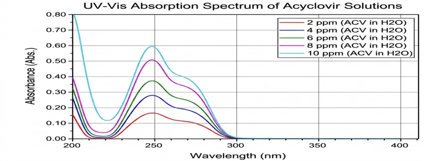
Precisely 1 g of API was weighed on a digital weighting balance and transferred to a 250 ml glass beaker with 60 ml of distilled water. The bulk was sonicated for 5 min to dissolve as much substance as possible. Then, remaining water was added to obtain a 1000ppm stock solution. The solution was then further diluted to get a 100ppm solution. Once the stock was ready, different ppm solutions were prepared, such as; 2ppm, 4ppm, 6ppm, 8ppm and 10ppm of Acyclovir. The solutions were further used to examine their solubility characteristics by taking their UV (UV - 1800 SHIMADZU) spectrograph at 252 nm. As the concentration of the solutions increased from lower to higher, the curing rate increased. Thus, we obtained a straight line graph of concentration (on the x-axis) and absorbance (on the y-axis).
CO-CRYSTAL FORMATION: UV, FTIR ANALYSIS
SOLVENT EVAPORATION METHOD (SE)
Four sets of samples were prepared for analysis using the solvent evaporation method. The API and co-formers were calculated and weighed on a digital balance to prepare an equimolar (1:1) mixture. This mixture was then transferred to a beaker, and co-solvents were added to dissolve them. The solution was stirred manually for approximately 30 min and then left to dry until no solvent remained in the beaker. UV analysis was performed, which showed the solubility enhancement of the drug. FTIR was also conducted to analyze the formation of co-crystals via new bond formation. Based on the spectral analysis and observed changes in peak position, broadening, and %Transmittance, Hydrogen bonding was altered due to the interaction, and the reduced free C=O indicated bonding with ACV. The fingerprint region showed a new lattice structure; thus, it was concluded that a co-crystal between acyclovir and the co-formers had been formed. The changes observed in the O–H, C=O, and N–H regions indicate strong hydrogen bonding and molecular interactions, which confirm the formation of a new molecular structure.
Table 1: solvent evaporation (SE) method for different Co-formers
|
Sr. No. |
API: Co-formers (1:1) |
Co-Solvents |
Method |
|
1) |
ACV:TA |
Ethanol |
SE |
|
2) |
ACV:SA |
Ethanol |
SE |
|
3) |
ACV:CA |
Ethanol |
SE |
DRY GRINDING METHOD
Some traditional methods which were proposed by the chemists for crystal synthesis could be solution and grinding based.[24] To perform this method of co-crystallization, we first calculated the concentrations of the API and co-formers. The compounds were then weighed on a digital balance. As only 1 mol of ACV and the co-formers were used to make an equimolar mixture, we preferred a crucible to a mortar for grinding. The crucibles were washed with absolute alcohol before use and weighed. The drug, along with the co-formers TA, SA, and CA (1:1), was thoroughly ground in three different crucibles. The grinding process was performed manually. Then they were characterized using FTIR spectroscopy at wave number 4000 to 400 cm.-1
FTIR analysis of the ACV:TA co-crystals revealed the presence of intermolecular hydrogen bonding. When the FTIR spectra of pure ACV, TA, and ACV:TA’s FTIR was compared, the co-crystals indicated broadening and shifting in O-H/N-H stretching (3600–2800 cm-1), and COOH stretching was found at (1750-1600 cm-1), showing H bonding. Moreover, the fingerprint regions (1500-800 cm-1) indicate molecular rearrangement and supramolecular crystalline phase formation.
Table 2: Dry motor grinding method for different Co-formers
|
SR. NO |
NAME |
METHOD |
AMOUNT (mg) |
|
01. |
ACV:TA |
Grinding |
225.21:150.09 |
|
02. |
ACV:SA |
Grinding |
225.21:118.09 |
|
03. |
ACV:CA |
Grinding |
225.21:192.12 |
MICROWAVE ASSISTED SOLVENT EVAPORATION METHOD (MASE)
Microwave-assisted co-crystallization is a green method and modern technique used to enhance the formation of co-crystals with efficacy in a few seconds using microwave rations. The instrument relies solely on dielectric heating, where microwave energy interacts with polar molecules and solvents, generating rapid internal heat. The heat generated enhances molecular mobility and accelerates co-crystallization by disrupting crystal lattice energies, leading to better solute-solvent interactions. [25,26]
Table 3: Microwave assisted method for different Co-formers
|
Sr. No. |
API: Co-formers (1:1) |
Co-Solvents ( 3 ml ) |
Method |
|
1) |
ACV:TA |
GAA |
MASE |
|
2) |
ACV:SA |
GAA |
MASE |
|
3) |
ACV:CA |
GAA |
MASE |
Like the other two methods, the API (active pharmaceutical ingredient) and Co-former were weighed accurately on the digital balance. Equimolar mixtures were prepared, as shown in Table 3. The mixtures were then transferred into 10 ml glass vials, and 3 ml of glacial acetic acid was added to the vials. The solution was mixed thoroughly after covering it with a lid. The vial was placed into the instrument by following various parameters such as temperature 800C and voltage 100 W, reaction time 1 min and the cooling temp 50 OC.
To obtain a clear solution, we followed the same procedure for 2 times until the solution became highly saturated. The solutions were then removed from the instrument and poured into previously washed and weighed watch glasses. The solutions on the watch glasses were left standing to obtain Co crystals at room temperature. Among the given sets, the highest experimental yield was obtained from the ACV:TA Co-Crystal, which was then characterized by UV–visible analysis, FTIR analysis, and melting point. The latter was observed at 150 °C, confirming the formation of a co-crystal.
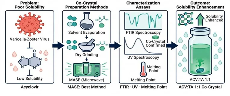
Figure 1. Various methods of solubility enhancement
Table 4. Optimization of Microwave Synthesis
|
Sr. No. |
Name |
Amount |
Solvent |
Power |
Time (min) |
Obtained crystal |
|
01 |
ACV:TA |
1:1 |
GAA |
100w |
2 |
390mg |
|
02 |
ACV:SA |
1:1 |
GAA |
100w |
2 |
270mg |
|
03 |
ACV:CA |
1:1 |
GAA |
100w |
2 |
360mg |
UV analysis of the Co-Crystals
To perform the UV analysis of the co-crystals obtained via the MASE method, we prepared solutions of various concentrations for all three sets. After Co-Crystallization, the weighed compound was added to distilled water, and the drug was immediately soluble without any effort compared to the solubility before co-crystallization, implying enhanced solubility. We prepared the solutions of 2ppm, 4ppm, 6ppm, 8ppm and 10ppm of ACV:TA, ACV:SA, and ACV:CA, respectively. The relationship between the ACV concentration and UV absorption was validated by drawing a standard calibration curve. Linear regression analysis was performed using the standard Beer-Lambert Law relationship, Y= mXC, Where, Y is the absorbance at lambda max =252 nm, X is the concentration of the solution in ppm, m represents the calibration slope, and C is the concentration in the Y-axis. Thus, the resulting calibration was Y= 0.0775X, establishing a direct proportionality between concentration and absorbance within the said ppm solutions. According to Beer Lambert's law, higher absorption is directly proportional to greater solubility. Moreover, the pure ACV UV-visible spectra were compared with the spectrum of the co-crystals, and an enhancement in the solubility was observed.
Table 5. comparative solubility and enhancement ratio of ACV formulations at maximum nominal concentration (10 ppm)
|
Formulation |
Absorbance (10ppm) |
Apparent solubility enhancement factor (Ef)a |
Relative intensity / Hyperchromic factor % |
Net dissolution growth % |
|
ACV |
0.60 |
1.00x |
100% |
- |
|
ACV:CA |
0.40 |
0.67x |
67% |
-33% |
|
ACV:SA |
0.46 |
0.77x |
77% |
-23% |
|
ACV:TA |
0.78 |
1.30x |
130% |
+30% |
FTIR analysis of the Co-Crystals
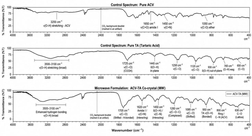
Although FTIR analyses were performed for the other two sets, the ACV:TA ratio gave the best result, manifested through the highest co-crystal yield obtained via the MASE method. The above co-crystal exhibited significant spectral changes compared to the parent drug, indicating intermolecular hydrogen bonding. A broad absorption band was observed in the 3500-3100 nm-1corresponding to the overlapping of O-H stretching of tartaric acid and N-H/O-H stretching of ACV ultimately showing the new hydrogen bonding. 1690-1075 cm-1 a downshift suggests the participation of the carboxyl group in forming intermolecular hydrogen bonds. An overlap of amide and C=N was observed at approximately 1610-1620 cm-1 indicating the interaction between the heterocyclic nitrogen atom of acyclovir and the carboxylic acid groups of tartaric acid. The rearrangement of crystal packing and hydrogen bond formation disturbed the oxygen-containing functional groups seen at 1280 cm-1, which corresponded to C-O-C/C-O vibrations. Supramolecular assembly was observed in the fingerprint region of 1000-600 cm-1 exhibiting changes in the position, intensity, and shape of the band. Thus, the formation of a co-crystal was confirmed, and a faster yield was obtained through the MASE method.
RESULTS AND DISCUSSION
Co-crystal formation resulted in enhanced aqueous solubility. The altered dissolution and solubility of the modified co-crystals were quantitatively verified at a maximum concentration threshold of 10.0 ppm. The 1:1 ACV:TA mixture with glacial acetic acid produced 390 mg of co-crystals via the MASE method, yielding an increased regression slope (m=0.0775) and a relative absorbance intensity of 130% compared to that of pure ACV. The calculated hyperchromic shift of 130% indicates the direct modification of the electronic transition probabilities of the ACV chromophore, caused by the structural rearrangement of tartaric acid in the new microenvironment. This structural change provides a clear idea of a 1.30 fold boost in apparent solubility, where ACV:TA’s UV absorption reached 0.78 compared to pure ACV.
In contrast, the negative dissolution of ACV:CA and ACV:SA can be attributed to steric and thermodynamic limitations. The bulky nature of CA causes steric hindrance within the upcoming crystal lattice. The generated spatial mismatch could restrict the formation of hydrogen bonds with pure ACV, resulting in limited dissolution. SA, a flexible dicarboxylic acid, can form tight and symmetric hydrogen bonds. When ACV was treated with SA, an interlocking network might have formed, which might have increased the overall thermodynamic stability of the crystal. This could have caused the drug to be locked inside the stable lattice, restricting dissolution.
The UV spectrum of pure ACV was compared with that of the co-crystals, and solubility enhancement was observed, indicating that a suitable co-former could change the solubility through structural changes. FTIR showed the formation of new intermolecular bonds between the pure drug and co-former, along with the formation of hydrogen bonds. The melting of the crystals occurred at 150 °C, indicating the formation of stable co-crystals. Moreover, when a preliminary qualitative analysis test for aqueous solubility was performed, the co-crystals were found to be completely soluble without any external force, confirming the solubility enhancement.
ACKNOWLEDGEMENT
I would like to express my sincere gratitude to Dr. Devang R. Pandya, an assistant professor in the Department of Chemistry at St. Xavier’s College, Ahmedabad, for his invaluable guidance and support. Moreover, I am indebted to him for providing ACV as a gift.
REFERENCES

Ralph Rajesh, Microwave Assisted Synthesis and Characterization of Acyclovir Co-crystals to Enhance Apparent Aqueous Solubility and Bioavailability in Herpes Zoster’s Treatment, Int. J. of Pharm. Sci., 2026, Vol 4, Issue 9, 4160-4167. https://doi.org/10.5281/zenodo.23058979
 10.5281/zenodo.23058979