

We use cookies to ensure our website works properly and to personalise your experience. Cookies policy
Department of Pharmaceutics, Dayanand Education Society's Dayanand College of Pharmacy, SRTMU - Latur 413512.
Background: Skin, hair, and nails may all be affected by superficial fungal infections, which are frequent dermatological diseases. Prolonged treatment, low patient compliance, recurrence, and insufficient drug retention at the infection site may all be linked to conventional antifungal therapy. Because of their natural nature and possible therapeutic benefits, herbal topical preparations may provide a viable alternative. Numerous phytoconstituents linked to antibacterial, anti-inflammatory, and antioxidant properties are present in Clitoria ternatea L. The goal of the current research was to improve the formulation using a design of experiments method and to create and assess a herbal emulgel containing Clitoria ternatea flower extract for possible topical antifungal treatment.Methods: An emulgel comprising Tween 80 and Span 80 as emulsifiers and Carbopol 934 and HPMC K100M as gelling agents was made using an ethanolic extract of Clitoria ternatea flowers. A design of experiments technique was used to create nine formulations (F1–F9). Physical appearance, homogeneity, pH, viscosity, spreadability, drug content, in vitro drug release, stability, and antifungal effectiveness against Candida albicans were all assessed for the produced formulations.Results: With a viscosity of 2948 cP, pH of 6.5, drug content of 86%, and 88.3% in-vitro drug release at 8 hours, the improved formulation, F5, demonstrated good physical features. In comparison to 13 mm for 1% clotrimazole, the formulation showed a 10 mm zone of inhibition against Candida albicans. During a month of accelerated storage at 40°C and 75% relative humidity, the improved formulation demonstrated adequate stability with very slight variations in pH, viscosity, drug content, and drug release.
Applying a pharmaceutical formulation directly to the skin to achieve a targeted therapeutic impact is known as topical medication delivery. Easy administration, better patient compliance, avoidance of gastrointestinal degradation and first-pass metabolism, and potential reduction of systemic exposure are just a few of its many benefits. However, the stratum corneum is the main barrier to medication entry through the skin, and topical formulations may be less effective due to low permeability or skin irritation. [1, 2] Keratinized tissues including the skin, hair, and nails are susceptible to superficial fungal infections, often known as superficial mycoses. Itching, scaling, erythema, irritation, swelling, and blister development are typical clinical symptoms. Topical or systemic antifungal medications may be used in therapy, depending on the extent and location of the infection. [3, 4] The majority of antifungal drugs work by disrupting vital elements of fungal cell membranes, including ergosterol production. Fungal membrane production and proliferation are disrupted by azole antifungals, which inhibit lanosterol 14α-demethylase, whereas allylamines block squalene epoxidase.[4,5] Important types of azole antifungals used to treat superficial fungal infections include imidazoles and triazoles. [5, 6]
Classification of Topical Drug Delivery System:
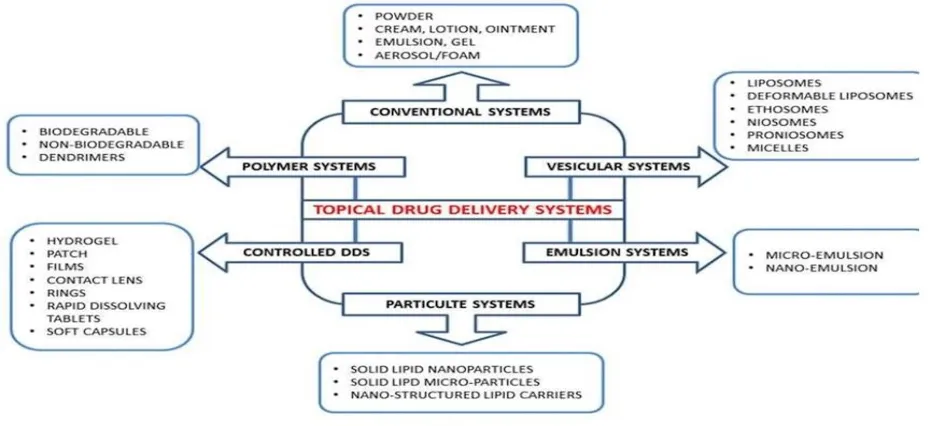
Figure No. 1. Topical Drug Delivery System
Long-term treatment, poor adherence, recurrence, and insufficient drug retention or penetration into hyperkeratotic lesions are still significant drawbacks of topical antifungal therapy, even with the availability of potent antifungal drugs. As a result, the perfect topical antifungal formulation should provide sufficient drug concentration at the site of infection, extended skin retention, efficient antifungal action, easy dosage, high tolerability, and enhanced patient acceptance. [7-19]
An emulsion is mixed with a gel matrix to create Emulgel, a topical administration method. It is especially helpful for combining poorly water-soluble or lipophilic medicines because it combines the drug-loading capacity of an emulsion with the advantageous application characteristics of a gel. When compared to certain traditional semisolid formulations, emulgels may provide better spreadability, stability, skin retention, controlled drug release, and patient acceptance. [15-26]
An aqueous vehicle, oily phase, emulsifier, gelling agent, permeation enhancer, preservative, antioxidant, and humectant are the main ingredients of an emulgel. To achieve appropriate viscosity, physical stability, drug release, skin compatibility, and therapeutic effectiveness, these excipients must be chosen and concentrated appropriately.[8,9] While surfactants like Tween 80 and Span 80 are often employed as emulsifiers, common gelling agents include carbomers, hydroxypropyl methylcellulose, xanthan gum, poloxamers, and hydroxyethyl cellulose. [8, 9]
There is growing interest in medicinal plants as possible sources of bioactive chemicals for the treatment of inflammatory and infectious diseases. Butterfly pea, or *Clitoria ternatea* L. (Fabaceae), is a medicinal plant that is said to possess physiologically active components. Its promise as a plant-derived antifungal candidate is supported by extracts from several sections of *C. ternatea*, such as seeds, leaves, and callus, which have shown antifungal efficacy against certain fungal strains.[11]
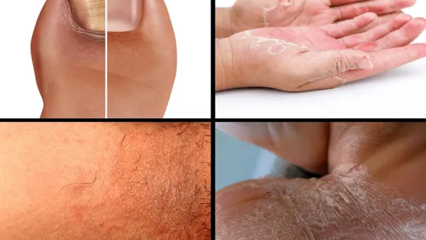
Figure No. 2: fungal infection
Based on these factors, the creation of a herbal antifungal emulgel containing *C. ternatea* extract may offer a promising method for topical treatment of superficial fungal infections by fusing the beneficial delivery properties of an emulgel system with the plant extract's potential antifungal activity.[23-29]
2. MATERIALS AND METHODS:
2.1 Material:
The herbal active ingredient was Clitoria ternatea flowers; The gelling agents were Carbopol 934 and HPMC K100M; the preservatives were methyl and propyl paraben; the penetration enhancer was propylene glycol; the pH-adjusting agent was triethanolamine; and the emulsifying agents were Tween 80 and Span 80. The materials were purchased from the appropriate sources and were of analytical/reagent or pharmaceutical grade.[14-31]
2.2 Tools and Equipment: A digital weighing balance (Contech CAH-223), hot-air oven (Lap Hosp), rotary vacuum evaporator (Buchi type), FTIR spectrometer (Agilent Cary 630), UV-visible spectrophotometer (Shimadzu UV-1900), digital pH meter (Equiptronics-610A), Franz diffusion cell, homogenizer (REMI), probe sonicator, bath sonicator (Toshniwal), magnetic stirrer (REMI), Brookfield digital viscometer (DV-E), heating mantle, and hot plate were the main tools utilized during formulation development and evaluation. [29-43]
2.3 Clitoria ternatea Plant Profile: Clitoria ternatea L., sometimes referred to as blue pea or butterfly pea, is a member of the Fabaceae family. Several kinds of phytoconstituents, including as flavonoids, phenolic compounds, tannins, saponins, triterpenoids, alkaloids, glycosides, anthocyanins, steroids, and other secondary metabolites, have been found in the plant, which has long been utilized as a medicinal herb. Numerous biological actions, such as antioxidant, antibacterial, anti-inflammatory, antidiabetic, analgesic, and other pharmacological properties, have been linked to these components. [1-3]
2.4 Purchasing and Verifying Plant Material: Fresh C. ternatea flowers were gathered from a nearby farm in the Latur area and the botanical garden on the college campus. The gathered flowers were carefully chosen, cleaned, and given a water wash to get rid of any dust or other objects that could have stuck to them. After being shade-dried at room temperature, the material was ground into a coarse powder using a grinder. Dr. C. S. Swami, a professor and head of the botany department at Dayanand Science College in Latur, verified the authenticity of the plant sample. Until it was needed again, the verified plant material was kept in an airtight container.
2.5 C. ternatea Flower Extract Preparation: The dried flowers were ground into a powder and put through an appropriate sieve. Maceration was used to extract around 50 g of powdered plant material using 200 mL of ethanol. For seven days, the extraction process was carried out with sporadic shaking. A rotating vacuum evaporator was used to concentrate the extract under low pressure after it had been filtered using Whatman No. 1 filter paper. Before being used again, the resultant C. ternatea flower extract (CTFE) was gathered in a sterile container and kept chilled. [33-37]
2.6 Plant Extract Characterization: Preliminary characterisation of the produced CTFE included organoleptic assessment, FTIR analysis, UV-visible spectrophotometric analysis, and preliminary phytochemical screening.[37-41]
2.6.1 Organoleptic Assessment: Visual and sensory observation were used to assess the extract's color, smell, physical characteristics, and overall nature.
2.6.2 Calculating λmax: A UV-Visible spectrophotometer was used to scan an acceptable concentration of CTFE in the chosen solvent across a suitable wavelength range against a comparable blank. The wavelength that exhibited the highest absorption was chosen as the λmax. The reported λmax of CTFE in this investigation was 266 nm. [41-49]
2.6.3 Analysis of FTIR: The extract's distinctive functional groups were identified using FTIR spectroscopy. An FTIR spectrometer was used to capture the CTFE spectra, and the absorption bands that were seen were interpreted based on their distinctive functional groups.
2.6.4 Initial Screening for Phytochemicals: Using conventional phytochemical techniques, the ethanolic extract was tested qualitatively for the presence of carbohydrates, alkaloids, glycosides, saponins, terpenoids, phenolic compounds, steroids, tannins, proteins, anthocyanins, and reducing sugars.[4,5]
2.7 Plant Material Standardization: Standard pharmacognostic measures were used to evaluate the quality of the herbal raw material. Total ash, acid-insoluble ash, water-soluble ash, loss on drying/moisture content, and foreign matter were all determined as part of standardization. These criteria are often used to evaluate the identification, quality, and purity of herbal products. [6]
2.7.1 Calculating Total Ash: A crucible that had already been lit was filled with precisely 1-2 g of powdered plant material, which was then burned until a carbon-free ash was produced. After cooling in a desiccator, the crucible was weighed. The weight of the ash in relation to the air-dried sample was used to compute the percentage of total ash.[6]% Total ash is equal to (ash weight divided by sample weight) times 100. [51-61]
2.7.2 Ash That Is Insoluble in Acid: The resulting ash was heated for about five minutes after being treated with diluted hydrochloric acid. After gathering and cleaning with hot water, the insoluble residue was burned, cooled, and weighed. The air-dried material was used to determine the proportion of acid-insoluble ash.[6]
2.7.3 Ash Soluble in Water: The quantity of ash that remained insoluble after filtering, igniting, and weighing was calculated by treating the whole amount of ash with water.[6]
2.7.4 Drying Loss: A predetermined amount of powdered plant material was weighed and dried in an oven set to between 100 and 105 degrees Celsius until the weight remained consistent. Moisture/loss on drying was used to compute the % weight loss.[6]% Drying loss is equal to [(Initial weight − Final weight) / Initial weight] x 100.
2.7.5 International Issues: The presence of extraneous items, such as dirt, stones, insects, plant debris, and other foreign objects, was visually inspected in the powdered plant material. The proportion of foreign matter was calculated using established quality-control techniques for herbal materials. [6]
2.8 Herbal Emulgel Formulation and Development: A two-step process was used to make the herbal emulgel: first, the gel base and emulsion were made, and then the emulsion was incorporated into the gel matrix. Because they combine the properties of gels and emulsions and may enhance the application and distribution of poorly water-soluble substances, emulgel systems are often utilized for topical administration. [7–9]
2.8.1 Gel Phase Preparation: To create homogeneous polymeric dispersions, the necessary amounts of Carbopol 934 and HPMC K100M were dispersed independently in purified water while being continuously stirred. The polymer dispersion was given enough time to hydrate. Triethanolamine was used to bring the gel's pH down to around 6.0–6.5
2.8.2 Oil Phase Preparation: To create the oil phase, liquid paraffin and Span 80 were combined while being constantly stirred.
2.8.3 Aqueous Phase Preparation: The aqueous phase was created by adding Tween 80 to filtered water. Propylene glycol was used to dissolve methyl and propyl parabens. The chosen solvent was used to integrate the CTFE into the proper phase.
2.8.4 Emulsion and Emulgel Preparation: To create a homogenous emulsion, the oil and aqueous phases were heated separately to between 70 and 80 degrees Celsius before being blended while being continuously stirred. After cooling to room temperature, the emulsion was left. To create a uniform herbal emulgel, the created emulsion was then progressively added to the previously made gel basis while being continuously stirred. According to the formulation technique, the emulsion and gel were mixed in a ratio of around 1:1.[7]
2.9 Herbal Emulgel's Physicochemical Assessment: Physical characteristics, color, odor, greasiness, consistency, pH, viscosity, spreadability, homogeneity, washability, drug content, and in-vitro drug diffusion were assessed for the developed emulgel formulations.
2.9.1 Appearance: The color, appearance, homogeneity, consistency, odor, presence of any visible particles, and phase separation of the formulations were all visually inspected.
2.9.2 pH: After dispersing around 1 g of each emulgel formulation in 100 mL of distilled water, the mixture was left to equilibrate for about two hours. A digital pH meter that had been calibrated was used to test the pH. [65-69]
2.9.3 Viscosity: A Brookfield digital viscometer was used to measure the compositions' viscosity. Using the designated spindle, around 5 g of formulation was assessed at 50 rpm for 60 seconds, and the viscosity measurement was noted.
2.9.4 Spreadability: The parallel-plate approach was used to assess spreadability. Two glass slides were sandwiched with around 1 g of emulgel. The top slide was given a preset weight, which was left there for around five minutes. The formulation's spreading was measured, and the spreadability was computed using: S is spreadability, M is the weight supplied to the higher slide, L is the distance the slide travels, and T is the time it takes.[10]
2.9.5 Uniformity: The regularity of the manufactured formulations and the existence of lumps, aggregates, or other particle matter were visually examined.
2.9.6 Washability: Applying a little amount of formulation to the skin and then washing the treated area with water was how washability was assessed. The presence of leftover formulation and its ease of removal were evaluated visually.
2.9.7 Substance Content: After precisely weighing 100 mg of emulgel, it was diluted in 100 mL of phosphate buffer (pH 7.4). To guaranty full extraction of the active ingredients, the dispersion was shook for around two hours. After filtering the solution, the absorbance was measured at 266 nm using phosphate buffer pH 7.4 as a blank. The mean drug content was determined after the analysis was carried out in triplicate. [69-73]
2.10 Drug Diffusion Study in Vitro: A Franz diffusion cell was used for the in-vitro diffusion analysis of the herbal emulgel compositions. The temperature was maintained at 37 ± 1°C, and the receptor media was phosphate buffer pH 7.4. The described experimental approach called for the use of an egg membrane as the diffusion membrane.
The membrane of the donor compartment was covered with around 1 g of emulgel. At around 50 rpm, the receptor media was constantly agitated. To maintain sink conditions, samples were taken out at prearranged intervals for up to eight hours and replaced with an equivalent amount of new receptor media. The extracted materials were spectrophotometrically measured at 266 nm after being appropriately diluted. At every sample interval, the cumulative proportion of medication released was computed. [71-73]
2.11 Analysis of Stability: Stability tests were conducted on the improved emulgel formulation under various storage conditions. For the duration of the investigation, samples were kept at room temperature, 37 ± 2°C, and refrigerated (4°C). To determine formulation stability, samples were routinely assessed for physical properties and in vitro drug release.
2.12 Activity Against Fungi: An agar diffusion technique was used to assess the antifungal activity of CTFE and the created herbal emulgel. The research used Candida albicans ATCC 9027, a fungus strain that was acquired from NCIM. The reference antifungal medication was clotrimazole. [61-73] Before being used, the necessary culture media was sanitized and prepared in accordance with the manufacturer's instructions. Under aseptic circumstances, the fungal culture was injected into the proper agar medium. Predetermined amounts and concentrations. [61-73] of CTFE and herbal emulgel formulations were added to wells that had been created using a sterile cork borer. For a whole day, the plates were incubated at around 37°C. The diameter of the zone of inhibition was measured in millimeters to assess antifungal activity. [61-73]
2.13 Experiment Design and Formulation Optimization: The impact of formulation factors on the herbal emulgel's performance was examined using Design of Experiments (DoE). Design-Expert® software version 13 was used to optimize the formulation utilizing the response surface approach.
At three levels, HPMC K100M (X?) and Carbopol 934 (X?) were chosen as the independent formulation variables. The primary dependent response was chosen to be the percentage medication release (Y?). The chosen design was used to create nine trial formulations. To determine the ideal formulation and assess how the formulation factors affected drug release, response-surface and three-dimensional plots were used.. [61-73]
Table No.01: The experimental formulation combinations were.
|
Run |
Carbopol 934 (mg) |
HPMC K100M (mg) |
Drug Release (%) |
|
1 |
550 |
450 |
70.4 |
|
2 |
500 |
550 |
78.9 |
|
3 |
500 |
450 |
68.0 |
|
4 |
450 |
450 |
65.9 |
|
5 |
550 |
550 |
88.3 |
|
6 |
550 |
500 |
81.9 |
|
7 |
450 |
500 |
66.0 |
|
8 |
500 |
500 |
72.2 |
|
9 |
450 |
550 |
69.4 |
The optimized formulation was selected based on the desired drug-release response and overall formulation characteristics
Collection and authentication of plant material:
In the month of January, leaves and flower of clitoria ternatea were collected in the Latur area. Authentication was done by Dr.C.S Swami, Head of the department of Botany, Dayanand Science College, Latur.
3.1 Preparation of ethanolic extract:
3.1.1 Percentage yield:
Table No.02: Percentage yield of plant extract
|
Sr. no |
Solvents |
colour |
odour |
consistency |
% yield w/w |
|
1 |
Clitoria ternatea flower |
Blue |
No |
Semisolid sticky mass |
88.3% |
3.2 Preliminary phytochemical screening of extract:
Result of phytochemical screening depends upon chemical constituents in extract. Either results are positive or negative. Results are given in table no. 03
Table No.03: Phytochemical test of extract
|
Sr. no |
Test |
Clitoria ternatea |
|
|
Flavonoids |
_ |
|
2 |
Alkaloids |
+ |
|
3 |
Glycoside |
+ |
|
4 |
Saponin |
+ |
|
5 |
Tannin |
+ |
|
6 |
Terpeoids |
+ |
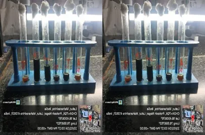
Figure No. 03: phytochemical screening of plant extract
3.3.1 Estimation of extract by U.V Spectrophotometer Clitoria ternatea: Estimation of standard Calibration curve for clitoria ternatea ethanol extract in Buffer pH 7.4 Estimated Value at Max 266nm.
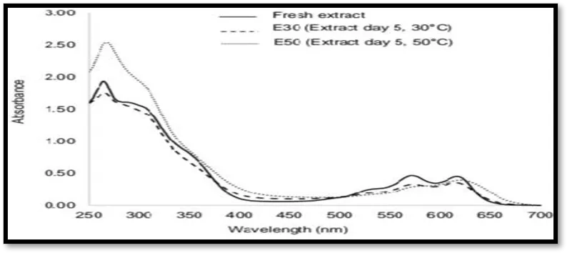
Figure No. 04: UV spectrum of clitoria ternatea flower extract
Table No.04: Calibration of Clitoria ternatea flower extract
|
Sr. no |
Concentration in µgm/ml |
Absorbance 1 |
|
1 |
0 |
O |
|
2 |
5 |
0.01426 |
|
3 |
10 |
0.0237 |
|
4 |
15 |
0.0326 |
|
5 |
20 |
0.0414 |
|
6 |
25 |
0.0513 |
|
7 |
30 |
0.0598 |
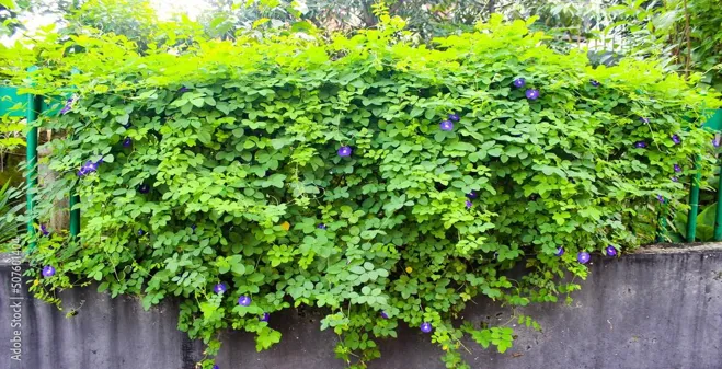
Figure No. 05: calibration of clitoria ternatea flower extract
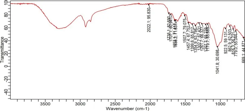
Figure No. 06: FTIR graph of clitoria ternatia flower extract
Table No.05: frequency of functional group of clitoria ternatea flower extract dissolve in ethanol.
|
Sr.no |
Type of vibration |
Peak cm1 |
Peak observed |
|
1 |
C=O stretching (Aldehydes or ketones) |
1700 - 1750 |
1735 |
|
2 |
C=C stretching (Aromatic rings or alkenes) |
1500 - 1600 |
1507 |
|
3 |
C-O stretching (Alcohols ,esters, ethers) |
1000 - 1300 |
1041 |
3.4.2 FTIR of polymers:
CARBOPOL 934:
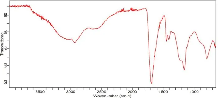
Figure No. 07: FTIR graph of carbopol 934
Table No. 06: Frequency of functional group of carbopol 934
|
Sr.no |
Type of vibration |
Peak cm1 |
Peak observed |
|
1 |
O-H stretching (Carboxylic acid) |
1400 - 1448 |
1435 |
|
2 |
C-O stretching (Ester or carboxylate groups) |
1000 - 1130 |
1125 |
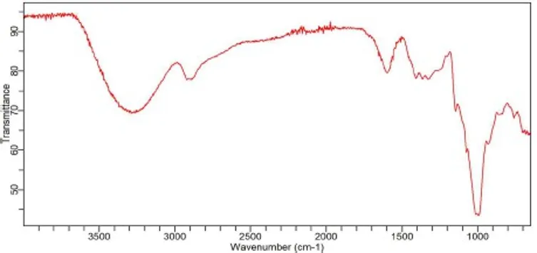
Figure No. 08: FTIR graph of HPMC K100M
Table No. 07: Frequency of functional group of HPMC K100M
|
Sr.no |
Type of vibration |
Peak cm1 |
Peak observed |
|
1 |
C-O-C stretching |
1000 - 1050 |
1033 |
|
2 |
C-H stretching |
1300 - 1450 |
1407 |
|
3 |
C-O stretching |
1300 - 1400 |
1364 |
|
4 |
C=H stretching |
700 - 900 |
861 |
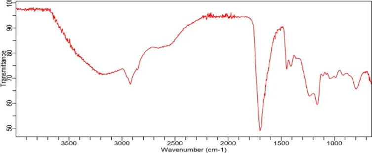
Figure No. 09: FTIR for mixing in clitoria ternatea, carbopol and HPMC K100m
Table No. 08: frequency of functional group of HPMC K100M
|
Sr.no |
Type of vibration |
Peak cm1 |
Peak observed |
|
1 |
C-O-C stretching |
1000 - 1050 |
1033 |
|
2 |
C-H stretching |
1300 - 1450 |
1407 |
|
3 |
C-O stretching |
1300 - 1400 |
1364 |
|
4 |
C=H stretching |
700 - 900 |
861 |
Table No. 09: Optimization Analysis
|
Runs |
Factor 1 A Carbopol 934 |
Factor 2 B HPMC K100M |
Response 1 in vitro drug release % |
|
1 |
550 |
450 |
70.4 |
|
2 |
500 |
550 |
78.9 |
|
3 |
500 |
450 |
68 |
|
4 |
450 |
450 |
65.9 |
|
5 |
550 |
550 |
88.3 |
|
6 |
550 |
500 |
81.9 |
|
7 |
450 |
500 |
66 |
|
8 |
500 |
500 |
72.2 |
|
9 |
450 |
550 |
69.4 |
3.5. Optimization analysis:
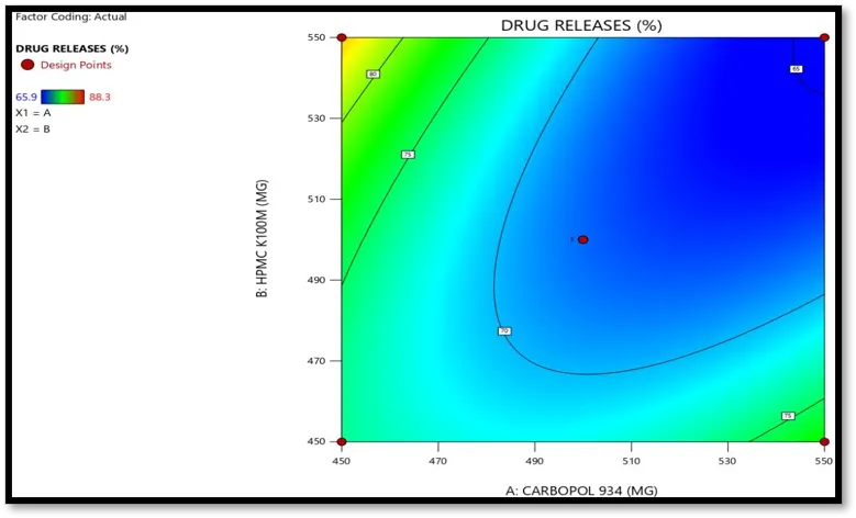
Figure No. 10: 3D response surface plot showing effect of polymer concentration on drug release.
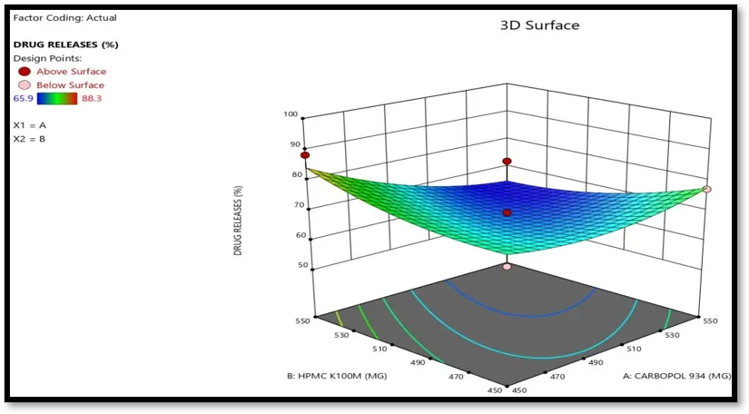
Figure No. 11: Contour graph showing effect of % drug release
With the help design of expert get the optimized batch F5 show the highest percentage drug release. Design of experiment also shows different four batches also shows good release. it It also predicted the Anova result dignostics result and model graphs. It also give information about point prediction. In that predicted mean, standard deviation, SE mean 95% cl low mean.Design of experiment predict itself and tell this batch also good. With the help of different equation such as standard deviation and there mean.
Emulgel formulations were viscous creamy preparations with a smooth homogeneous texture and glossy appearance. The color of formulation was checked against white the consistency of emulgel was checked by applying on skin. Results have been discussed in Table no 10.
Table No. 10: Physical parameters of formulation batches
|
Batch code |
Colour |
Homogeneity |
|
F1 |
Greenish white |
Homogenous |
|
F2 |
Greenish white |
Homogenous |
|
F3 |
Greenish white |
Homogenous |
|
F4 |
Greenish white |
Homogenous |
|
F5 |
Greenish white |
Homogenous |
|
F6 |
Greenish white |
Homogenous |
|
F7 |
Greenish white |
Homogenous |
|
F8 |
Greenish white |
Homogenous |
|
F9 |
Greenish white |
Homogenous |
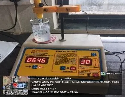
Figure No. 12: photograph showing determination PH of formulation by using Digital PH meter.
Table No. 11: Determination of PH
|
Batch code |
pH. meter |
|
F1 |
6.3 |
|
F2 |
6.46 |
|
F3 |
6.2 |
|
F4 |
6.1 |
|
F5 |
6.5 |
|
F6 |
6.46 |
|
F7 |
6.2 |
|
F8 |
6.42 |
|
F9 |
6.3 |
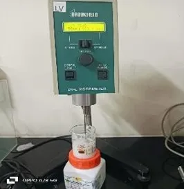
Figure No. 13: viscosity of herbal emulgel
Table No. 12: viscosity of herbal emulgel
|
Formulation code |
Viscosity (cps ) |
|
F1 |
2726 |
|
F2 |
2768 |
|
F3 |
2279 |
|
F4 |
2167 |
|
F5 |
2948 |
|
F6 |
2879 |
|
F7 |
2179 |
|
F8 |
2645 |
|
F9 |
2334 |
Table No. 13: Spreadability of formulations F1-F9
|
Sr.no |
Formulation |
spreadability |
|
1 |
F1 |
27.26 |
|
2 |
F2 |
28.56 |
|
3 |
F3 |
23.54 |
|
4 |
F4 |
21.56 |
|
5 |
F5 |
31.56 |
|
6 |
F6 |
30.46 |
|
7 |
F7 |
22.66 |
|
8 |
F8 |
27.36 |
|
9 |
F9 |
26.25 |
Table No. 14: % drug content
|
Formulation |
% drug content |
|
F1 |
68.2 |
|
F2 |
76 |
|
F3 |
66.3 |
|
F4 |
63 |
|
F5 |
86 |
|
F6 |
78.2 |
|
F7 |
65 |
|
F8 |
70 |
|
F9 |
68.3 |
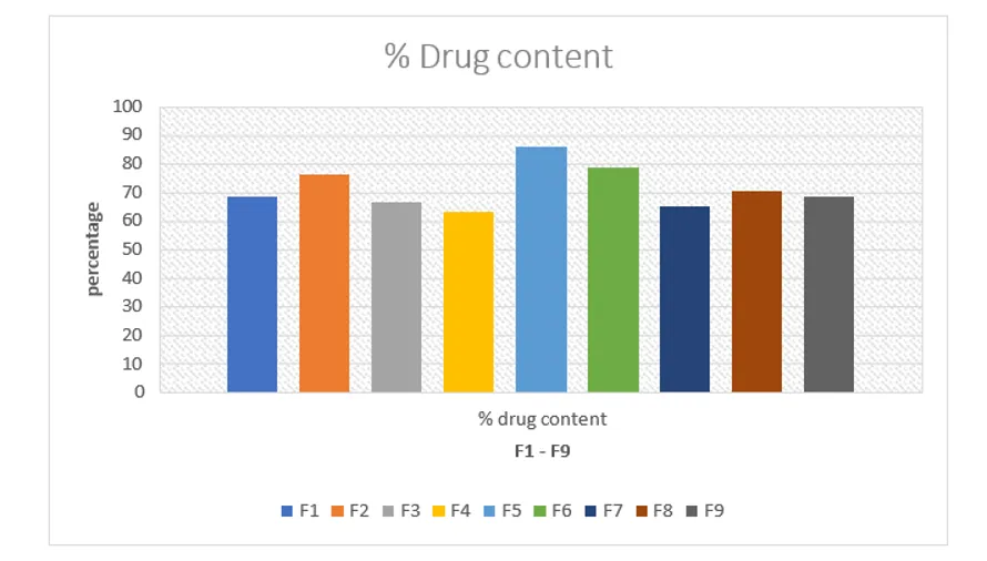
Figure No. 14: drug content of clitoria ternatea
The receptor compartment's 5ml of sample was removed at specified intervals and refilled with an equal volume of phosphate buffer at pH 7.4 by using a UV spectrophotometer at 266 nm, the aliquots were examined. The F6 batch's total drug release after 4 hours was determined to be 46.45 which is a better percentage of drug release when compared to other emulgel formulations.
Table No. 15: % drug release
|
Time (hrs.’) |
F1% |
F2% |
F3% |
F4% |
F5% |
F6% |
F7% |
F8% |
F9% |
|
0 |
0 |
0 |
0 |
0 |
0 |
0 |
0 |
0 |
0 |
|
1 |
3.4 |
1.2 |
1.5 |
2.2 |
1.5 |
15.2 |
8.1 |
2.4 |
3.7 |
|
2 |
12.5 |
8.8 |
10.6 |
8.6 |
11.1 |
23.5 |
17.6 |
10.2 |
11.3 |
|
3 |
25.2 |
24.5 |
18.7 |
20.8 |
23.3 |
39.2 |
30.2 |
26.5 |
27.6 |
|
4 |
43.8 |
40.5 |
29.3 |
33.5 |
32.9 |
46.45 |
38.8 |
45.3 |
46.4 |
|
5 |
53.6 |
50.9 |
35.6 |
46.5 |
41.5 |
58.41 |
41.5 |
55.8 |
56.1 |
|
6 |
60.4 |
60.3 |
42.1 |
55.2 |
60.7 |
60.16 |
48.2 |
60.5 |
61.8 |
|
7 |
65.5 |
72.4 |
60.6 |
61.9 |
78.2 |
75.01 |
55.8 |
67.5 |
67.1 |
|
8 |
70.4 |
78.9 |
68 |
65.9 |
88.3 |
81.9 |
66 |
72.2 |
69.4 |
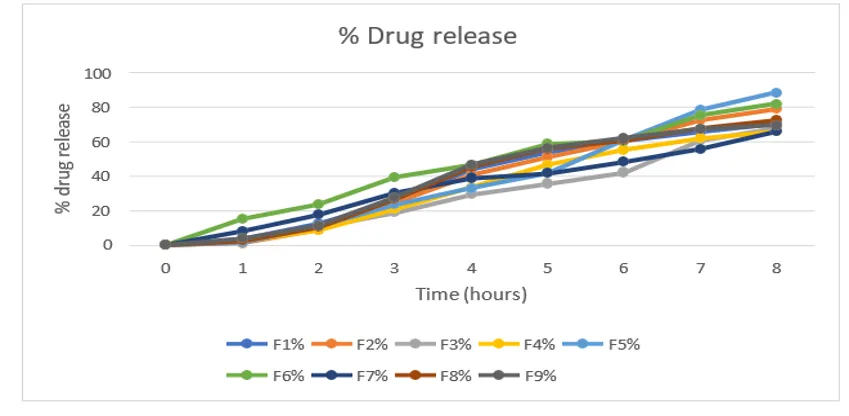
Figure No. 15: Percentage drug release curve of batch F1 to F9
To evaluate the stability of the drug formulation, stability experiments were conducted in accordance with ICH. A packaging made of aluminium and polyethylene was laminated and used to seal the optimised F5 formulation. For one month, samples were stored at 40° c and 75% RH. At the conclusion of the trial period, the formulation's physical features, colour, drug content, and characteristics of drug release observe only little change.
Table No. 16: Stability study of emulgel
|
Parameters |
Initial |
After 1 month |
|
Viscosity |
2948 |
2948 |
|
PH |
6.5 |
6.5 |
|
Drug content |
86 |
85 |
|
% drug release |
88.3 |
86 |
After 24 hours, the plates were examined. By measuring the smallest dimension of the area around the patch where no fungal growth occurred, the zone of inhibition is estimated.
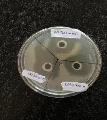
Figure No. 16: Antifungal activity against candida albicans
Table No. 17: Antifungal activity against candida albicans
|
Sr.no |
Microbial strain |
Zone of inhibition |
|
1 |
Candida albicans |
10 mm |
|
2 |
1% clotrimazole |
13 mm |
CONCLUSION
Due of their medicinal qualities, medicinal plants are quite valuable. These plants can be found in untamed areas throughout India. Traditional methods for discovering herbal products include extraction, phytochemical screening, biological activity testing, and lastly formulation into a suitable dosage form. The productivity, therapeutic effectiveness, and competitiveness of medicinal plants can be affected by accumulating scientific information about them and turning them into formulations.
The antimicrobial and anti-inflammatory properties of medicinal plants, which have been found to be effective in treating diseases by inhibiting the growth of microorganisms or significantly reducing the burden of microorganisms, are provided by traditional systems of medicine. This has a positive impact on fungal infection. Specific microbes, including fungi like candida albicans, Staphylococcus aureus, and E-coil, can cause or worsen fungal infection. Only a small percentage of chronic wound patients can be treated with antibiotics since these germs are adaptable and resistant to them. A number of medicinal plants have been tested for their antifungal activity as a result of the increasing failure of therapeutic agents due to antibiotic resistance by pathogenic microbes. To fulfil the demand for new antimicrobial medicines with improved efficacy and minimal or no adverse effects going forward, the development of new medications derived from plants may be helpful. Therefore, it is fairly conceivable to design safe, efficient, affordable, and simple-to-apply emulgel compositions beneficial in treating chronic wounds using the existing literature and instructions provided in the traditional system of medicine. A survey of the literature lists a number of plants with a reputation for having antifungal and anti-bacterial properties. For the purpose of this investigation a plants were chosen Freshly dried plant materials were gathered from reliable market sources and authenticated at Dayanand Science College in Latur by renowned botanist Dr. C.S. Swami. Drug standardization was done in accordance with WHO recommendations. Following standardization, ethanol was used as a solvent to extract the plant material. The preliminary phytochemical screening of every prepared ethanol extract revealed that it contains a variety of phytoconstituents. Plant extracts were used to prepare a topical herbal emulgel formulation. With varied amounts of plant extract, herbal emulgel formulations were made using solvent dispersion method from ethanolic extracts. The physicochemical characteristics of the herbal emulgelgel formulations, including appearance, pH, homogeneity, and
REFERENCES




Govind Mise, Pritam Navagire, Siddhant Bedre, Rohit Sarda, Formulation And Evaluation of Clitoria Ternatea Emulgel for Treatment of Fungal Infection., Int. J. of Pharm. Sci., 2026, Vol 4, Issue 9, 972-992, https://doi.org/10.5281/zenodo.22678089
 10.5281/zenodo.22678089