

We use cookies to ensure our website works properly and to personalise your experience. Cookies policy
1Associate professor, Department of Pharmacognosy, Bojjam Narasimhulu Pharmacy College, Vinay nagar, Saidabad, Hyderabad, Telangana 500059.
2,3,4,5,6Student, Bojjam Narasimhulu Pharmacy College, Saidabad, Vinay nagar, Hyderabad, Telangana - 500059, India.
The present study aimed to formulate and evaluate Camellia sinensis extract-loaded ethosomes for enhanced antioxidant and anti-aging activity through topical delivery. Ethosomes were prepared using varying concentrations of phosphatidylcholine and ethanol and evaluated for their physicochemical characteristics, drug entrapment efficiency, in vitro drug release, and stability. FTIR studies indicated the compatibility between the extract and formulation excipients without significant interactions.The prepared ethosomes exhibited nanosized vesicles with particle sizes ranging from 186 to 208 nm and zeta potential values between -24 and -28 mV, suggesting good physical stability. Entrapment efficiency of the formulations ranged from 78.51% to 81.25%, with formulation F2 showing the highest entrapment efficiency (81.25%). In vitro drug release studies demonstrated sustained release behavior over 8 hours, with formulation F2 exhibiting the maximum cumulative drug release of 97.17%. Stability studies performed under different storage conditions for three months revealed no significant changes in the formulation characteristics, confirming the stability of the optimized formulation.The findings suggest that Camellia sinensis extract-loaded ethosomes can serve as an effective topical delivery system to enhance the antioxidant and anti-aging potential of green tea polyphenols. Among the developed formulations, F2 was identified as the optimized formulation due to its superior entrapment efficiency, drug release profile, and stability. Therefore, ethosomal delivery of Camellia sinensis extract represents a promising strategy for the development of novel anti-aging and antioxidant skincare formulations.
Defination of Ethosomes
“Ethosomes are ethanolic liposomes”. Ethosomes can be defined as noninvasive delivery carriers that enable drugs to reach deep into the skin layers and/or the systemic circulation. These are soft, malleable vesicles tailored for enhanced delivery of active agents. The vesicles have been well known for their importance in cellular communication and particle transportation for many years. Vesicles would also allow controlling the release rate of drug over an extended time, keeping the drug shielded from immune response or other removal systems and thus be able to release just the right amount of drug and keep that concentration constant for longer periods of time. One of the major advances in vesicle research was the finding of a vesicle derivative, known as an Ethosomes. Ethosomes are the slight modification of well established drug carrier liposome. Ethosomes are lipid vesicles containing phospholipids, alcohol (ethanol and isopropyl alcohol) in relatively high concentration and water. Ethosomes are soft vesicles made of phospholipids and ethanol (in higher quantity) and water. The size range of ethosomes may vary from tens of nanometers (nm) to microns (µ) ethosomes permeate through the skin layers more rapidly and possess significantly higher transdermal flux.
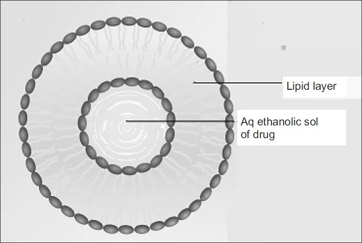
Figure 1: Structure of Ethosomes
Advantages of Ethosomal Drug Delivery
1. Delivery of large molecules (peptides, protein molecules) is possible.
2. It contains non-toxic raw material in formulation.
3. Enhanced permeation of drug through skin for transdermal drug delivery.
4. Ethosomal drug delivery system can be applied widely in Pharmaceutical, Veterinary, Cosmetic fields.
5. High patient compliance: The ethosomal drug is administrated in semisolid form (gel or cream) hence producing high patient compliance.
6. Simple method for drug delivery in comparison to Iontophoresis and Phonophoresis and other complicated methods.
7. The Ethosomal system is passive, non-invasive and is available for immediate commercialization.
8. The main advantage of ethosomes over liposomes is the increased permeation of the drug.
Disadvantages
Chemical instability
High ethanol content can lead to oxidation and degradation of phospholipids over time.
Ethanol is volatile, so long-term storage may cause vesicle shrinkage or leakage.
Skin irritation potential
The high percentage of ethanol (20–45%) may cause skin dryness, redness, or irritation in sensitive individuals.
Limited loading capacity for hydrophilic drugs
Ethosomes are more suitable for lipophilic or amphiphilic molecules; hydrophilic drugs may show lower encapsulation efficiency.
Scale-up and manufacturing challenges
Maintaining uniform vesicle size and stability during large-scale production can be difficult.
Ethanol handling requires safety measures and specialized equipment.
Storage and stability issues
Sensitive to temperature fluctuations and light, which may destabilize the vesicles.
Possible aggregation or fusion of vesicles during long-term storage.
Limited systemic delivery
While ethosomes enhance skin penetration, they may not provide high systemic bioavailability for drugs requiring precise systemic dosing.
Regulatory and formulation concerns
High ethanol concentration may limit their use in pediatric or sensitive patient populations.
Regulatory approval for ethanol-rich topical formulations can be more complex.
MATERIALS
Green tea extract was procured from Synpharma Research Labs, Hyd.Soya lecithin, Ethanol, Span 80 were obtained from Synpharma Research Labs, Hyderabad. Other chemicals and the reagents used were of analytical grade.
METHODOLOGY
Extraction of Green Tea Extract
Fresh green tea leaves were collected, washed, and shade-dried at room temperature. The dried leaves were pulverized into a coarse powder using a mechanical grinder. About 50 g of the powdered material was subjected to Soxhlet extraction using ethanol as the extraction solvent for 6–8 hours. The extraction was continued until the solvent in the siphon tube became colorless. The obtained extract was filtered through Whatman filter paper and concentrated under reduced pressure using a rotary evaporator. The concentrated extract was further dried to obtain a semisolid mass. The dried extract was weighed, stored in an airtight container, and used for further formulation studies.
Formulation development
Table 1: Preparation of Camellia Sinensis Extract
| S.No | Ingredients | F1 | F2 | F3 | F4 |
|---|---|---|---|---|---|
| 1 | Camellia sinensis extract | 50 | 50 | 50 | 50 |
| 2 | Phosphotidyl choline | 100 | 200 | 300 | 400 |
| 3 | Ethanol | 10 | 20 | 30 | 40 |
| 4 | Water | q.s | q.s | q.s | q.s |
Procedure
Using the cold technique, ethosomal formulations were created. When preparing ethosomal material, this is the approach that is most frequently utilised. In a covered jar, the phospholipids, medication, and other pharmaceutical ingredients mentioned in the table were dissolved in ethanol while vigorously stirring at room temperature. A closed container was used to gradually add a fine stream of distilled water while stirring continuously at 100 rpm. The liquid was then heated to 300C ± 10C. The mixing process was kept going for another 5 minutes with the system maintained at a temperature of 300 C ± 10C. Following a 30-minute cooling period at ambient temperature, the mixture was subjected to five cycles of sonication at 40 C, with a one-minute interval between each cycle, using a probe sonicator.
CHARACTERIZATION
Particle size:
The size of each batch of ethosomes was examined under a microscope. The average size of the ethosomes was calculated by collecting a little drop of ethosomes from each batch and measuring them at several locations on the slide.
SEM analysis
Using a scanning electron microscope, the shape of ethosomes was examined. A sputter coater was used to apply a layer of gold particles to the surface of the lyophilised sample that had been deposited on aluminium stubs. A scanning electron microscope (SEM) (XL30, Philips, the Netherlands) operating at 15 kV and 750 mA was used to ascertain the Ethosomes' form.
Zeta Potential:
The charges on the surface of ethosomes are referred to as the zeta potential. How frequently an electron is present on an ethosome's surface. The component or ingredient employed in the production process is the source of this charge. All of the ethosomes in the formulation must have an electrical charge on their surfaces so that the Ethosomes can repel each other and prevent the particles from clumping together. We used a zeta sizer equipment with Malvern software to measure the zeta potential of ethosomes. The sample was analysed at 25oC with a detection angle of 90o. A zeta potential value between +30 and -30 mV is good. Within these limits, Ethosomal particles are unable to clump together.
Entrapment efficiency
If you want to know how effective the entrapment process is, you have to divide a known quantity of the created formulation (such a nanoparticle or vesicular suspension) into two parts: the unentrapped drug and the drug linked with the carrier. Centrifugation, ultrafiltration, or dialysis are the primary methods for separation; high-speed centrifugation is often used for vesicular systems such as liposomes or ethosomes. It is common practice to use UV-visible spectrophotometry to determine the amount of free drug after meticulously collecting and analysing the supernatant, which includes the unentrapped or free drug. The entire amount of drug in the formulation can be found by dissolving or breaking down the particles or vesicles and then measuring the amount of drug that remains. The formula is then used to calculate entrapment efficiency:
Entrapment efficiency (%)=Total drug−Free drug/Total drug×100
As a measure of the loading capacity and overall effectiveness of the formulation, this percentage indicates the amount of the initial medication dose that is really trapped within the delivery system.
In-vitro Drug release studies:
Utilising a modified Franz type glass diffusion cell, an in-vitro investigation of drug penetration through the Dialysis membrane was conducted. To keep the sink state, the modified cell is used, which has a higher capacity of 10 ml. The spectrophotometric analysis was used to determine the drug release in the samples. Refilling the receptor phase with phosphate buffer of the same volume was done with each sample removal.
Stability studies:
To define an appropriate shelf life and suggested storage conditions, stability studies are conducted to ascertain the quality variations of a medicinal substance or medication product over time in response to environmental elements including light, humidity, and temperature. Preparation of a stability protocol outlining the quantity of batches, type of packaging, storage conditions, testing intervals, and acceptability criteria is the first step in the study, as per ICH Q1A (R2) recommendations. The batches that will be sold or used as a pilot are sealed in the designated commercial container and kept in stability chambers under specific conditions. These conditions can be long-term (25 ± 2 °C / 60 ± 5% RH for 12-24 months), accelerated (40 ± 2 °C / 75 ± 5% RH for 6 months), or intermediate (30 ± 2 °C / 65 ± 5% RH) in nature.
RESULTS ANDDISCUSSION
FTIR Studies
Compatibility study (IR spectroscopy)
FTIR analysis was performed in order to study the compatibility of ingredients used in the preparation of nanoparticles, using a Shimadzu FTIR spectrophotometer (Prestige21, Shimadzu Corporation, Kyoto, Japan). Camellia sinensis extract and Excipients their mixture with ratio (1:1) was evaluated using FTIR spectrophotometer using potassium bromide disc technique where 1mg of the sample is mixed with 100 mg of dry powdered KBr, the mixture is pressed into a transparent disc and was inserted in the apparatus for IR scan.
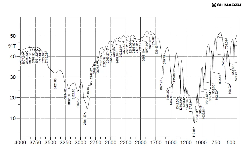
Figure 2: FT-IR Sample for Camellia Sinensis Extract
Table 2: Characteristic Peaks and Frequency of Camellia Sinensis Extract
| S. No. | Characteristic Peaks | Frequency range (cm-1) |
Frequency (cm-1) |
|---|---|---|---|
| 1 | OH stretching | 4000-3500 | 3855.83 |
| 2 | OH Bending | 3000-2750 | 2891.39 |
| 3 | C-H stretching | 1750-1250 | 1342.50 |
| 4 | C-N stretching | 1250-1000 | 1112.96 |
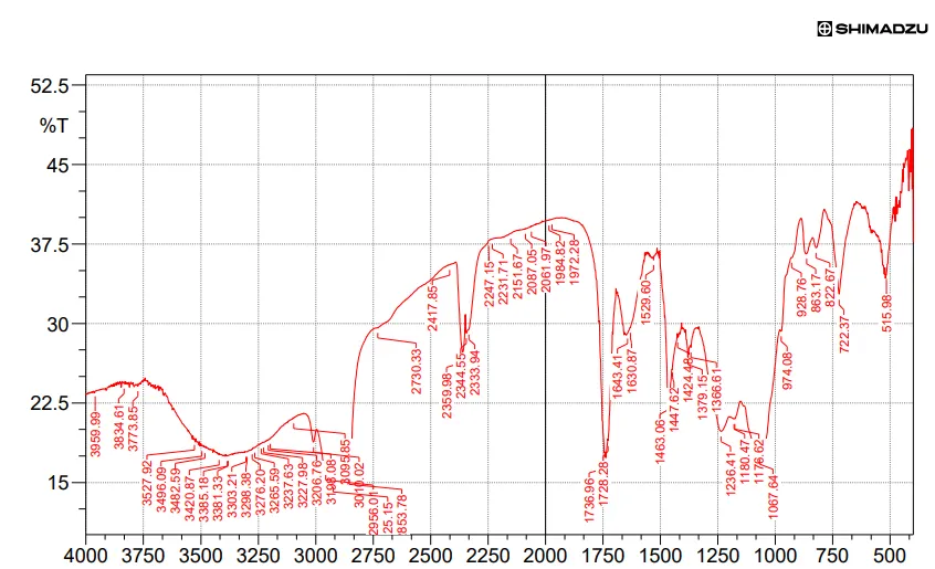
Figure 3: FT-IR Sample for Optimized Formulation
Table 3: Characteristic Peaks and Frequency of Optimized Formulation
| S. No. | Characteristic Peaks | Frequency range (cm-1) |
Frequency (cm-1) |
|---|---|---|---|
| 1 | OH stretching | 4000-3500 | 3834.61 |
| 2 | OH Bending | 3500-2750 | 3227.98 |
| 3 | C-H stretching | 1750-1250 | 1463.06 |
| 4 | C-N stretching | 1250-1000 | 1180.47 |
An infrared spectrophotometer was used to conduct the compatibility studies. Researchers looked at the infrared spectra of both the pure drug and a physical mixture of the drug and excipients. Since the aforementioned peaks' typical absorption was within the statutory limits (±100 cm-1), the medication can be safely used with excipients.
Determination of Vesicle morphology and Size
The morphological features of the ethosomes that were created were examined using scanning electron microscopy (SEM). A sample holder made of gold-plated copper with a tiny drop of Ethosomes sandwiched between two rivets. Liquid nitrogen was used to slush the entire apparatus. The surface moisture of the sample was sublimed by heating it to -850C for 30 minutes. Once the sample was coated with gold, it was subjected to a temperature of -1200c and a voltage of 5kV so that the scanning electron microscope (SEM) could take photographs.
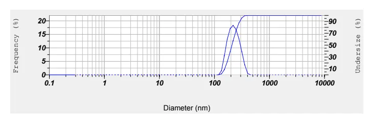
Figure 4: Particle Size Analysis of Optimized Ethosomes
SEM Analysis
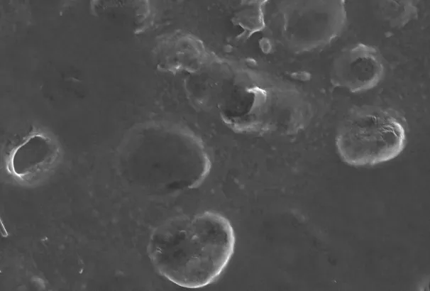
Figure 5: SEM Analysis of Ethosomes
Zeta potential
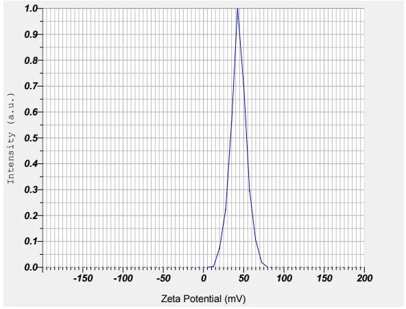
Figure 6: Zeta Potential of Ethosomes
Table 4: Evaluation Studies of Particle Size and Zeta Potential Ethosomes
| F. No | Particle size (nm) | Zeta potential(mV) |
|---|---|---|
| F1 | 197 | -26 |
| F2 | 186 | -24 |
| F3 | 208 | -28 |
| F4 | 199 | -25 |
Entrapment Efficiency:
Entrapment efficiency was determined by measuring the concentration of unentrapped free drug in aqueous medium. About 1 ml of the drug loaded ethosomes dispersion was placed in the Eppendorf tubes and centrifuged at 17000 rpm for 30 min. The ethosomes along with encapsulated drug were separated at the bottom of the tubes. Plain ethosomes without drug was used as blank sample and centrifuged in the same manner. In order to measure the free drug concentration, the UV absorbance of the supernatant was determined.
Table 5: Drug Entrapment Efficiency
| F. code | Drug entrapment efficiency |
|---|---|
| F1 | 79.68 |
| F2 | 81.25 |
| F3 | 78.51 |
| F4 | 80.17 |
[Figure - see the formatted PDF]
Figure 7: Drug Entrapment Efficiency of All Formulation
In vitro drug release studies:
Figure 6: In Vitro Drug Release Studies of (F1-F4) Formulation
| Time | F1 | F2 | F3 | F4 |
|---|---|---|---|---|
| 0 | 0 | 0 | 0 | 0 |
| 1 | 25.68 | 24.17 | 21.08 | 23.07 |
| 2 | 34.76 | 36.89 | 37.69 | 38.20 |
| 3 | 47.80 | 46.58 | 47.11 | 45.21 |
| 4 | 59.14 | 56.80 | 58.80 | 59.81 |
| 5 | 68.10 | 67.15 | 65.15 | 69.10 |
| 6 | 76.81 | 78.91 | 76.17 | 77.81 |
| 7 | 83.56 | 84.82 | 82.33 | 85.24 |
| 8 | 93.28 | 97.17 | 94.08 | 91.02 |
[Figure - see the formatted PDF]
Figure 8: Drug Release for All Formulations
The drug release studies of all formulations of Camellia Sinensis Extract Ethosomes were conducted by means of diffusion apparatus for a time period of 8 hrs. From the drug release studies as depicted in Figure, the results showed that F2 formulation showed maximum drug release rate of 97.17 % within 8 hrs.
Stability studies
There was no significant change in physical and chemical properties of the solid lipid nanoparticles of formulation F-2 after 90 days. Parameters quantified at various time intervals were shown.
Table 7: Stability Studies of All Formulations
| F.no | Parameters | Initial | 1st Month | 2nd Month | 3rd Month | Limits as per Specifications |
|---|---|---|---|---|---|---|
| F-2 | 250C/60%RH % Release |
97.17 | 95.55 | 94.58 | 93.68 | Not less than 85 % |
| F-2 | 300C/75% RH % Release |
97.17 | 95.26 | 94.30 | 93.53 | Not less than 85 % |
| F-2 | 400C/75% RH % Release |
97.17 | 95.18 | 94.16 | 93.18 | Not less than 85 % |
CONCLUSION
The present study successfully formulated and evaluated Camellia sinensis extract-loaded ethosomes for potential antioxidant and anti-aging applications. Preformulation studies confirmed the suitability of the extract for ectosome formulation development.FTIR compatibility studies confirmed the absence of significant interactions between Camellia sinensis extract and the excipients used in the formulation, indicating good compatibility and stability of the developed ethosomal system. Morphological analysis by SEM demonstrated the formation of discrete, spherical vesicles with smooth surfaces. The particle size analysis revealed that all formulations possessed nanosized vesicles ranging from 186 to 208 nm with zeta potential values between -24 and -28 mV, indicating good physical stability.The entrapment efficiency of the formulations ranged from 78.51% to 81.25%, with formulation F2 exhibiting the highest entrapment efficiency (81.25%). In vitro drug release studies showed sustained release characteristics for all formulations over 8 hours. Among the developed formulations, F2 demonstrated the highest cumulative drug release of 97.17% at the end of 8 hours, indicating its superior release profile. Stability studies conducted under different storage conditions for three months revealed no significant changes in drug release characteristics, with all values remaining within acceptable limits, confirming the stability of the optimized formulation.Based on the overall evaluation parameters, formulation F2 was identified as the optimized formulation due to its favorable vesicle size, satisfactory zeta potential, highest entrapment efficiency, enhanced drug release profile, and excellent stability. Therefore, Camellia sinensis extract-loaded ethosomes represent a promising vesicular delivery system for improving the topical delivery of green tea polyphenols, thereby enhancing their antioxidant and anti-aging efficacy. Further in vivo and clinical studies are warranted to establish the therapeutic potential and safety of the developed formulation for cosmetic and dermatological applications.
REFERENCES

B. Kusuma Pravallika, Tarannum Sultana, Iram Usva, Javeriya Firdous, Mariyam Mirza, Hafsa Idrees, Formulation and Evaluation of Green Tea Extract Loaded Ethosomes for Antioxidant and Anti Aging Effect, Int. J. of Pharm. Sci., 2026, Vol 4, Issue 11, 1241-1249. https://doi.org/10.5281/zenodo.24000734
 10.5281/zenodo.24000734